Hospital de Clínicas de Porto Alegre, Universidade Federal do Rio Grande do Sul, Porto Alegre RS, Brazil (UFRS); 1Pediatric Immunologist; 2Associate Pro f e s s o r, Social Medicine, UFRS, PhD in Epidemiology, London University; 3Biochemist, Immunology Unit; 4A s s o c i a t e P ro f e s s o r, Internal Medicine Department, UFRS, Chief, Immunology Unit, Hospital de Clínicas de Porto Alegre, Scientific Dire c t o r, Laboratório DNA Reference, Brazil. Financial support was provided by Fundação de Amparo à Pesquisa do Rio Grande do Sul (FAPERGS).
Received 28 February 2005, received in final form 14 July 2005. Accepted 1 September 2005.
Dra. Luciane Failace - Rua Carlos Von Koseritz 1353/201 - 90540-031 Porto Alegre RS - Brasil. Acute bacterial meningitis is responsible for m o s t
infections affecting the central nervous system ( C N S ) . Close to two-thirds of all cases occur among chil-d ren, anchil-d Haemophilus influenzae, Neisseria me-ningitidis, and Streptococcus pneumoniae are the pathogens responsible for 80 to 90% of the
cas-e s1 , 2. A rapid and precise etiologic diagnosis of bac-terial meningitis is essential to determine adequate t reatment, and it can also significantly reduce mor-tality and the risk for long-term sequelae3.In epi-demiological terms, the diagnosis of bacterial me-ningitis may indicate the need for selective
immu-SIMULTANEOUS DETECTION OF
NEISSERIA
MENINGITIDIS, HAEMOPHILUS INFLUENZAE
AND
STREPTOCOCCUS
SP. BY POLYMERASE
CHAIN REACTION FOR THE DIAGNOSIS OF
BACTERIAL MENINGITIS
Luciane Failace
1, Mario Wagner
2, Marisa Chesky
3,
Rosana Scalco
3, Luiz Fernando Jobim
4ABSTRACT - The simultaneous detection of Neisseria meningitidis, Haemophilus influenzae, and S t re p t o c o c c u s s p .was assessed by polymerase chain reaction (PCR) for the diagnosis of bacterial meningitis, as well as t h e applicability of PCR as a routine test. A cohort study was carried out with 182 children (2 months to 12 years of age) with suspicion of bacterial meningitis. Routine tests identified the etiologic agent in 65/84 childre n whose clinical status and laboratory findings suggested the presence of bacterial meningitis. Bacterial me-ningitis was ruled out in 98 children. In 19 children, the etiologic diagnosis was not possible using stand a rd methods; in 14 of these patients, the etiologic agent was identified by PCR (N. meningitidis=12; H.i n f l u e n -z a e=1; S t reptococcus sp.=1). The sensitivity of PCR was 88.1%; specificity, 99.0%; positive predictive value, 98.7%; and negative predictive, 90.1%. PCR is a useful complementary diagnostic technique, especially w h e n Gram stain, culture, or antigenic detection are negative or inconclusive.
KEY WORDS: polymerase chain reaction (PCR), bacterial meningitis, cerebrospinal fluid (CSF).
Detecção simultânea da Neisseria meningitidis, Haemophilus influenzae e S t reptococcus sp. pela reação em cadeia da polimerase no diagnóstico das meningites bacterianas
RESUMO - Avaliamos o desempenho da reação em cadeia da polimerase (PCR) para detecção simultânea da Neisseria meningitidis, Haemophilus influenzae eS t reptococcus sp. no diagnóstico das meningites bacte-rianas e sua aplicabilidade na rotina diagnóstica. Foi realizado um estudo de coorte com 182 crianças apre-sentando suspeita de meningite bacteriana. Em 84, havia alterações clínicas e laboratoriais sugestivas de meningite bacteriana. Destas, 65 tiveram o agente etiológico identificado pelos métodos laboratoriais de rotina e 19 ficaram sem diagnóstico etiológico. Em 98 pacientes foi excluído o diagnóstico de meningite bacteriana. Analisando o desempenho da PCR encontramos sensibilidade de 88,1%, especificidade de 99,0% e valores preditivos positivo e negativo de 98,7% e 90,1% respectivamente. Nos 19 pacientes com meningite bacteriana mas sem diagnóstico etiológico a PCR detectou microrganismos em 14, sendo 12 N. m e n i n g i t i d i s ,um H. influenzaee um S t reptococcus sp. A PCR possui o potencial de poder aumentar os ín-dices de identificação das técnicas tradicionais, principalmente nas situações onde a microscopia dire t a , cultura ou identificação antigênica são negativos ou inconclusivos.
nization of a community, such as by chemopre v e n-tion and vaccinan-tion of the contacts of patients c a r-rying meningococcal disease4.
Among the routine tests used for the etiolog-ic diagnosis of bacterial meningitis, bacterial cultu-re is considecultu-red the gold standard. However, this is a time-consuming technique, which re q u i res via-ble micro o rganisms for cultivation, and its sensiti-vity is directly affected by the start of antibiotic t re-atment before spinal tap5. Other faster tests, such as latex agglutination, have a lower sensitivity and p resent reliable results only with samples contain-ing more than 105 CFU/mL6. Currently, molecular biology techniques, such as PCR, are being wide-ly used for the etiologic diagnosis of CNS infect i o n s7. In 1991, Kristiansen et al. detected the pre s e n c e of N. meningitidis DNA in the culture-negative ce-re b rospinal fluid (CSF) specimen of a patient with meningococcal disease after she had received an-tibiotic tre a t m e n t8. In 1992, Ni et al. studied the u s e of PCR in diagnosing meningococcal meningitis; results indicated specificity and sensitivity of 91%. Neither sensitivity nor specificity were affected by p revious antibiotic tre a t m e n t9. In 1994, Radstro m et al. described a nested PCR strategy for the simul-taneous detection of Neisseria meningitidis, Hae
-mophilus influenzae, and Streptococcus sp.1 0i n
C S F. That study showed that it is possible to simul-taneously and efficiently amplify the three main pathogens that cause bacterial meningitis. Ho-w e v e r, it also revealed certain difficulties associat-ed with the technique, such as false-positive re s u l t s due to contamination, and false-negative re s u l t s due to the presence of PCR inhibitors in CSF, which still compromise diagnostic re l i a b i l i t y1 0 - 1 3. Additio-nal studies correlating clinical and laboratory find-ings to PCR results are essential to confirm the d i a g-nostic value of PCR.
The present study was carried out in order to assess the simultaneous detection of Neisseria me
-ningitidis, Haemophilus influenzae,and S t re p t o
-coccus sp.by PCR for the diagnosis of bacterial
me-ningitis. The study also evaluated the applicabili-ty of PCR as a routine laboratory test.
METHOD
A cohort study was carried out with children betwe-en 2 months and 12 years of age, with suspicion of bac-terial meningitis at three public hospitals in the city of P o rto Alegre, State of Rio Grande do Sul, Brazil: Hospital de Clínicas de Porto Alegre, Hospital da Criança Santo Antônio, and Hospital da Criança Conceição. Most pedi-atric cases in the region are treated at these hospitals.
The study was submitted to and approved by the Re-s e a rch EthicRe-s CommitteeRe-s of the participating hoRe-spitalRe-s. C h i l d ren with suspected bacterial meningitis were includ-ed in our study after an informinclud-ed consent form was sig-ned by a parent or guardian. A 0.5 mL aliquot of CSF w a s separated during spinal tap or after the CSF was re c e i v e d in the laboratory. Specimens were stored at - 20oC until
PCR was performed.
The signs and symptoms of bacterial meningitis vary depending on the age group. In preschool and school age children, the predominant symptoms include menin-geal irritation and increase of intracranial pressure ex-p ressed as neck stiffness (Kern i g ’s and Bru d z i n s k i ’s signs). In younger children, these symptoms may be absent, wi-th predominance of unspecific signs of infection. In wi-these cases, meningitis was considered in the presence of fever, l e t h a rg y, irritability and sleepiness, especially if associa-ted with signs of meningeal irritation or increased intra-cranial pressure.
Alterations in CSF, such as leukocytosis with pre d o m-inance of polymorphonuclears, CSF glucose concentra-tion below 40 mg/dL and increased CSF protein (100-500 mg/dL) are considered to be suggestive of bacteria l meningitis. Diagnostic confirmation is based on the iden-tification of the etiological agent through direct micro s-copy (Gram), culture, latex agglutination or counterim-m u n o e l e c t ro p h o resis. Leukocytosis with pre d o counterim-m i n a n c e of lymphocytes, normal or increased CSF glucose and in-creased proteins suggest viral meningitis.
Clinical history and results of physical examination w e re reviewed. Patients were followed until discharg e f rom the hospital. The results of routine CSF analysis ( d i f-f e rential cell count; glucose; protein; Gram stain; bacte-rial culture; and latex agglutination) were obtained fro m the medical chart during hospitalization, or directly fro m the laboratory. Counterimmunoelectro p h o resis was car-ried out at a state-run laboratory (Laboratório Central do Estado do Rio Grande do Sul); these results were ob-tained from the State Department of Health.
In the present study, the gold standard for diagnos-ing bacterial mendiagnos-ingitis included clinical signs and symp-toms (as previously described), course of disease (antibi-otic therapy, length of admission, neurological seque-lae), and routine laboratory results (CSF analysis: diff e r-ential cell count; glucose; protein; Gram stain; bacteri-al culture; latex agglutination and counterimmunoelec-t ro p h o resis). According counterimmunoelec-to counterimmunoelec-these cricounterimmunoelec-teria, pacounterimmunoelec-tiencounterimmunoelec-ts were divided into three groups:
G roup II – Clinical indication of bacterial meningitis, positive laboratory results, and unsuccessful diagnosis of etiologic agent by routine methods;
G roup III – Clinical indication of bacterial meningi-tis, diagnosis of bacterial meningitis ruled out by course of disease, and negative laboratory results.
PCR sensitivity, specificity, and predictive values were calculated by comparing the results for groups I+II ver-sus group III. It is important to stress that the diagnosis of meningitis and the distribution in the groups was in-dependent, i.e., blinded for the result of PCR.
We employed the seminested multiplex PCR techni-que for the simultaneous detection of N. meningitidis, H. i n f l u e n z a e ,and S t reptococcus sp. A region of bacterial gene 16S rRNA was amplified. Species-specific primers we-re used along with primers complementary to universal regions U3 and U8 of gene 16S rRNA, in a two-stage PCR a s s a y. In the first stage, amplification was perf o rmed using e x t e rnal universal primers (u3, ru8). A generic bacterial amplicon was generated, not specific to the type of bacte-rium present in the specimen. The second stage combined t h ree specific primers one for each type of bacterium -with the ru8 universal primer, generating species-specif-ic amplspecies-specif-icons with diff e rent molecular weights.
In the first stage, the PCR reaction contained (NH4)2S O4 (16 mM); Tris-HCl (67 mM; pH 8.8 at 25oC); 0.01 % Tw e e n
-20 (w/v); MgCl2 (1.5 mM); triphosphate deoxyribonucleo-side mixture (0.25 mM); external primers (at 0.1 µM ) syn-thesized according to previously published sequences1 0;
and polymerase DNA enzyme (0.625 U). During this first step, 10 µL of CSF were added. DNA extraction from hem-o rrhagic hem-or xanthhem-ochrhem-omic CSF specimens, and frhem-om speci-mens with high leukocyte concentration (causing
turbidi-ty) was carried out using the QIAamp kit (70 µL CSF speci-men). Other CSF specimens were submitted to PCR dire c t-l y, without any treatment. The finat-l reaction vot-lume was 50 µL. Amplification was done on a 9600 Perkin Elmer t h e rm o c y c l e r. The initial denaturation period was of 1 mi-nute and 40 seconds at 94o C, followed by 33 incubation
cycles (20 seconds each) at 94oC, 60oC and 72oC.
The second amplification stage employed species-specific primers and the universal ru8 primer in a mix-t u re idenmix-tical mix-to mix-thamix-t of mix-the firsmix-t smix-tage. The final re a c-tion volume was 25 µL, and we used 2 µL from the first reaction as specimen. Denaturation was perf o rmed for 45 seconds at 94oC, followed by 33 (15-second cycles)
at 94oC, 15 seconds of annealing at 55oC, and 20
sec-onds of polymerization at 72oC. Some pro c e d u res were
followed to avoid contamination, such as using aero s o l -resistant tips, working in four distinct enviro n m e n t s , including negative controls, and submitting the re a g e n t m i x t u re to UV radiation (254 nm) for 15 minutes before adding CSF. All positive specimens were retested for con-firmation of the results.
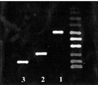
A 10 µL specimen from the second PCR reaction was run in an ethidium bromide-stained 2% agarose gel. A m-plification products were identified following electro-p h o resis. Selectro-pecimens were visualized using UV transillumi-nation. Specific N. meningitidis(700 bp), H. influenzae
(500 bp), and S t reptococcus sp.(300 bp) bands were c o m-p a red to the resm-pective controls or to molecular weight markers (Figure).
M e a s u rements of PCR perf o rmance were expre s s e d as percentages with their respective 95% confidence in-t e rvals (95% CI). Confidence inin-tervals were calculain-ted by binomial distribution. Due to the asymmetric distribu-tion of the data, the Kru s k a l - Wallis test was used to ana-lyze the comparisons between groups I, II and III. The n o n-parametric multiple comparison test proposed by Dunn was used to assess diff e rences between the groups.
RESULTS
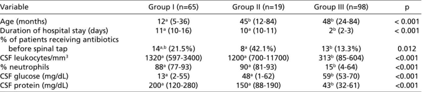
During the 18 months of the study, we analyzed 182 patients with suspected bacterial meningitis. In 65 patients, the diagnosis of bacterial meningi-tis was confirmed by identification of the etiolog-ic agent using routine laboratory tests (group I). N i-neteen patients presented clinical and laboratory indication of bacterial meningitis without identi-fication of the etiologic agent by standard tests ( g roup II). In the remaining 98 patients, the course of the disease and laboratory results ruled out a diagnosis of bacterial meningitis (group III). The c l i-nical and laboratory characteristics of the thre e groups are presented in Table 1.
Among the 65 patients from group I, 29 cases (44.6%) were caused by H. influenzae,21 (32.3%) by N. meningitidis,and 15 (23.1%) by S t re p t o c oc c u s
s p .PCR identified the correct pathogen in 60 of t h e
65 cases. However, PCR did not identify the cor-rect pathogen in three cases of N. meningitidis a n d in two cases of Streptococcus sp. Among the 98 p a-tients from group III, PCR was negative in 97 CSF specimens. There was one false-positive result indi-cating N. meningitidisDNA.
Among the 19 patients from group II, PCR iden-tified pathogens in 14 specimens. Twelve speci-mens contained N. meningitidis DNA, one speci-men contained H. influenzaeDNA, and one speci-men contained S t reptococcus sp.DNA. In group II, 42.1% of the patients had started antibiotic tre a t-ment before spinal tap, compared to 21.5% in group I and 13.3% in group III.
The sensitivity of PCR in groups I and III was 92.3% (95% CI: 82.2-97.1), and the specificity was 99.0% (95% CI: 93.6-99.9). The positive pre d i c t i v e value was 98.4% (95% CI: 90.0-99.9), and the neg-ative predictive value was 95.1% (95% CI: 88.4-98.2). When group II patients were included in the assessment of PCR perf o rmance, sensitivity dro pp e d to 88.1% (95% CI: 78.8-93.8), and the negative pre-dictive value was reduced to 90.1% (95% CI: 83.1-95.2). Nevertheless, both the specificity and the p o s i-tive predici-tive value remained practically the same: 99.0% (95% CI: 93.6-99.0) and 98.7% (95% CI: 91.8-99.9), respectively.
When assessing groups I and III, there was no statistically significant diff e rence between sensiti-vity of PCR, Gram stain, culture, and latex aggluti-nation. When group II patients were included, the sensitivity of PCR was significantly higher than that of the other tests (Table 2).
DISCUSSION
In general, the number of cases of bacterial me-ningitis whose etiologic agent is not identified is still significant. Several factors interf e re with the re c o v e ry of micro o rganisms from CSF, such as pre-vious antibiotic treatment, inadequate collection and storage of culture specimens, and re d u c e d number of bacteria in CSF. Tests for detecting bac-terial antigens, such as latex agglutination and c o u n-t e r i m m u n o e l e c n-t ro p h o resis, may assisn-t in n-the diag-nosis of bacterial meningitis. However, although these tests are fast and not affected by pre v i o u s antibiotic treatment, their sensitivity is limited. T h e use of molecular biology techniques, such as PCR, for the etiologic diagnosis of CNS infections is un-der study. It is expected that such techniques will have a higher sensitivity and specificity for diagno-sing meningitis.
We assessed the accuracy of the seminested PCR technique for the simultaneous detection of the t h ree main pathogens which cause bacterial me-n i me-n g i t i s :N. meningitidis, H. influenzae, and S t re p
-tococcus sp. Due to the absence of an absolute g o l d
s t a n d a rd, in the present study clinical status, course of disease, CSF analysis, and identification of eti-ologic agent by routine tests (Gram stain, culture , latex agglutination, and counterimmunoelectro-p h o resis) were established as the gold standard for the diagnosis of bacterial meningitis. Since the etiologic agent is not identified in many patients (especially in those receiving antibiotics before spi-nal tap), we later decided to consider another gold Table 1. Clinical and laboratory characteristics of groups I, II, and III.
Variable Group I (n=65) Group II (n=19) Group III (n=98) p
Age (months) 12a(5-36) 45b(12-84) 48b(24-84) < 0.001
Duration of hospital stay (days) 11a(10-16) 10a(10-11) 2b(2-3) < 0.001 % of patients receiving antibiotics
before spinal tap 14a,b(21.5%) 8a(42.1%) 13b(13.3%) 0.012
CSF leukocytes/mm3 1320a(597-3400) 1200a(700-11700) 313b(85-604) <0.001
% neutrophils 88a(77-93) 90a(81-93) 15b(4-64) <0.001
CSF glucose (mg/dL) 13a(2-55) 48a(1-62) 59b(53-70) <0.001 CSF protein (mg/dL) 200a(120-280) 150a(88-190) 43b(32-61) <0.001
All values are expressed as medians (interq u a rtile range), except for percentage of patients taking antibiotics. Superscript letters indicate statistically significant differences.
Table 2. Sensitivity of routine laboratory tests for the etiologic diagnosis of patients with bacterial meningitis.
Method No. tests Sensitivity CI 95% No. tests Sensitivity CI 95%
(groups I and III) (groups I, II, and III)
PCR 65 92.3% 82.2-97.1 84 88.1% 78.8–93.8
Gram stain 65 81.5% 70.0-90.1 84 63.3% 52.4–72.9
Culture 65 81.5% 70.0-90.1 84 63.3% 54.4–72.9
Latex agglutination 40 75.0% 58.8-87.3 50 60.0% 46.0–72.8
CI* 36 52.8% 35.5-69.6 46 41.3% 27.8–55.9
standard, i.e., the clinical and laboratory diagno-sis of bacterial meningitis, regardless of etiologic identification.
When the etiologic diagnosis of bacterial m e n i n-gitis was used as the gold standard, group II pa-tients (clinical and laboratory indication of bacte-rial meningitis; unsuccessful etiologic identificat i o n by routine tests) were excluded from the calcula-tion of PCR accuracy, and consequently these re s u l t s may be overestimated. The inclusion of patients with clinical and laboratory indication of bacteria l meningitis whose etiologic identification pro v e d unsuccessful through routine laboratory tests re-veals a PCR accuracy that is closer to what would be expected if this assay were used routinely for the diagnosis of bacterial meningitis; there is a sta-tistically nonsignificant drop in sensitivity and neg-ative predictive value, whereas specificity and posi-tive predicposi-tive value remain practically unchanged.
PCR was the only test that was capable of iden-tifying the etiologic agent in 14 patients fro m g roup II. In 63% of these patients, N. meningitidis
DNA was detected in CSF. The notion that over half of the bacterial meningitis cases without etiolog-ic identifetiolog-ication were caused by N. meningitidisi s i m p o rtant both clinically and epidemiologically, a n d allows a more adequate management of patients and contacts. The fact that antibiotic use before spinal tap was more frequent in group II unders-cores the importance of PCR in the identification of the etiologic agent in meningitis.
PCR did not identify pathogens in five cases of bacterial meningitis with etiologic diagnosis by routine tests, a fact that points to the presence of PCR inhibitors in CSF. PCR inhibitors had been de-tected in some hemorrhagic, xanthochromic, and high leukocyte-concentration CSF specimens, and DNA extraction techniques were used in an at-tempt to remove the inhibitors. In these five spec-imens, there had been no CSF alterations indicat-ing the need for extraction, and, there f o re, CSF w a s d i rectly submitted to PCR. At the end of the study, these specimens were submitted to DNA extrac-tion and resubmitted to PCR. After extracextrac-tion, PCR identified pathogens in three of the five specimens with previously negative results. This suggests that purification and concentration (70 µL) of bacteri-al DNA in CSF using extraction techniques may im-p rove the sensitivity of the method. Extraction te-chniques should be applied to all CSF specimens, regardless of their macroscopic aspect. While the detection of PCR inhibitors could indicate the need
for an internal control, the use of a multiplex re a c-tion could lead to difficulties in the interpre t a t i o n of results.
The high specificity and the positive pre d i c t i v e values found in our study indicate that the pro ba-bility of false-positive PCR results is low, and that PCR detection of pathogens is a strong indicator o f bacterial meningitis. In turn, the sensitivity and n e-gative predictive values found in the present study suggest that a diagnosis of bacterial meningitis should not be ruled out when PCR results are neg-ative. Our results suggest that PCR may enhance t h e diagnostic accuracy of traditional techniques, espe-cially when Gram stain, culture, and antigenic de-tection tests are negative or inconclusive.
Molecular biology techniques, such as PCR, have a wide range of applications for diagnosis. Howe-v e r, before incorporating PCR into a diagnostic routine, it is necessary to standardize this pro c e d u-re, to establish strict laboratory quality control m e a-s u rea-s, and to carry out further a-studiea-s aimed at ea-s- es-tablishing PCR accuracy. Finally, PCR should not re-place standard tests, rather, it should contribute to more precise and rapid diagnoses.
REFERENCES
1. Bell WE. Bacterial meningitis in children: selected aspects. Pediatr Clin N Am 1992;39:651-658.
2 . Schelch WF III, Wa rd JI, Band JD, Hightower A, Fraser DW, Broome CV. Bacterial meningitis in the United States, 1978 through 1981: the nation-al bacterination-al meningitis surveillance study. JAMA 1 9 8 5 ; 2 5 3 : 1 7 4 9 - 1 7 5 4 . 3. Salih MA, Khaleefa OH, Bushara M, et al. Long-term sequelae of
child-hood acute bacterial meningitis in a developing country: a study fro m the Sudan. Scand J Infect Dis 1991;166:177-182.
4 . G reenwood BM, Wali SS. Control of meningococcal infection in the A f r i c a n meningitis belt by selective vaccination. Lancet 1980;8171:729-732. 5. Gray LD, Fedorko DP. Laboratory diagnosis of bacterial meningitis.
Clin Microbiol Rev 1992;2:130-145.
6 . Olcén P. Serological methods for rapid diagnosis of Haemophilus in-fluenzae, Neisseria meningitidis and Streptococcus pneumoniae in cere-b rospinal fluid: a comparison of coagglutination, immunofluore s c e n c e and immunoelectro o s m o p h o resis. Scand J Infect Dis 1978;10:283-289. 7 . J e ffery KJM, Bangham CRM. Recent advances in the laboratory diagnosis
of central nervous system infections. Curr Opin Infec Dis 1996;9:132-137. 8. Kristiansen B, Ask E, Jenkins A, Fremer C, Radstrom P, Skold O. Rapid
diagnosis of meningococcal meningitis by polymerase chain re a c t i o n . Lancet 1991;337:1568-1569.
9. Ni H, Knight A, Cartwright K, Palmer WH, McFadden J. Polymerase chain reaction for diagnosis of meningococcal meningitis. Lancet 1992; 340:1432-1434.
10. R a d s t rom P, Backman A, Qian N, Kragsjberg P, Pahlson C, Olcen P. De-tection of bacterial DNA in cere b rospinal fluid by an assay for simulta-neous detection of Neisseria meningitidis, Haemophilus influenzae, and Streptococci using a seminested PCR strategy. J Clin Micro b i o l 1994;32:2738-2744.
11. Olcen P, Lantz P, Backman A, Radstrom P. Rapid diagnosis of bacteri-al meningitis by a seminested PCR strategy. Scand J Infect Dis 1995; 27:537-539.
12. Backman A, Lantz P, Radstrom P, Olcen P. Evaluation of an extended diagnostic PCR assay for detection and verification of the common cau-ses of bacterial meningitis in CSF and other biological samples. Mol Cell Probes 1999;1:49-60.