INTRODUCTION
Article/Artigo
1. Enfermaria de Clínica Médica, Hospital Barão de Lucena, Recife, PE. 2. Serviço de Doenças Infecciosas e Parasitárias, Hospital Universitário Oswaldo Cruz, Recife, PE. 3. Setor de Bacteriologia, Laboratório Central de Saúde Pública Dr. Milton Bezerra Sobral, Recife, PE. 4. Programa de Pós Graduação em Medicina Tropical, Universidade Federal de Pernambuco, Recife, PE. 5. Departamento de Medicina Clínica, Universidade de Pernambuco, Recife, PE.
Address to: Dr. Alfredo Pereira Leite de Albuquerque Filho.Enfermaria de Clínica Médica/Hospital Barão
de Lucena. Avenida Caxangá 3860, 50731-000 Recife, PE, Brasil. Phone: 55 81 3184-6606
e-mail: [email protected]
Received in 16/06/2010
Accepted in 28/07/2011
Validation of a case deinition for leptospirosis diagnosis in patients with
acute severe febrile disease admited in reference hospitals at the state of
Pernambuco, Brazil
Validação de uma deinição de caso para diagnóstico de leptospirose em pacientes com doença febril
aguda grave, admitidos em hospitais de referência do Estado de Pernambuco, Brasil
Alfredo Pereira Leite de Albuquerque Filho
1, Jéssica Guido de Araújo
1, Inacelli Queiroz de Souza
1,
Luciana Cardoso Martins
2, Marta Iglis de Oliveira
2, Maria Jesuíta Bezerra da Silva
3, Ulisses Ramos Montarroyos
4and Demócrito de Barros Miranda Filho
2,5ABSTACT
Introduction: Leptospirosis is oten mistaken for other acute febrile illnesses because of its nonspeciic presentation. Bacteriologic, serologic, and molecular methods have several limitations for early diagnosis: technical complexity, low availability, low sensitivity in early disease, or high cost. his study aimed to validate a case deinition, based on simple clinical and laboratory tests, that is intended for bedside diagnosis of leptospirosis among hospitalized patients. Methods: Adult patients, admited to two reference hospitals in Recife, Brazil, with a febrile illness of less than 21 days and with a clinical suspicion of leptospirosis, were included to test a case deinition comprising ten clinical and laboratory criteria. Leptospirosis was conirmed or excluded by a composite reference standard (microscopic agglutination test, ELISA, and blood culture). Test properties were determined for each cutof number of the criteria from the case deinition. Results: Ninety seven patients were included; 75 had conirmed leptospirosis and 22 did not. Mean number of criteria from the case deinition that were fulilled was 7.8±1.2 for conirmed leptospirosis and 5.9±1.5 for non-leptospirosis patients (p<0.0001). Best sensitivity (85.3%) and speciicity (68.2%) combination was found with a cutof of 7 or more criteria, reaching positive and negative predictive values of 90.1% and 57.7%, respectively; accuracy was 81.4%. Conclusions: he case deinition, for a cutof of at least 7 criteria, reached average sensitivity and speciicity, but with a high positive predictive value. Its simplicity and low cost make it useful for rapid bedside leptospirosis diagnosis in Brazilian hospitalized patients with acute severe febrile disease.
Keywords: Leptospirosis. Case deinition. Clinical diagnosis. Validation study.
RESUMO
Introdução: Por sua apresentação clínica inespecífica, a leptospirose é frequentemente confundida com outras doenças febris agudas. Métodos bacteriológicos, sorológicos e moleculares apresentam limitações para o diagnóstico precoce: complexidade técnica, baixa disponibilidade, insensibilidade na doença precoce, ou alto custo. Este estudo objetivou validar uma deinição de caso, baseada em dados clínicos e laboratoriais simples, destinada ao diagnóstico da leptospirose em pacientes hospitalizados. Métodos: Foram incluídos pacientes adultos, admitidos em 2 hospitais de referência no Recife, com doença febril de até 21 dias e suspeita clínica de leptospirose, para testar uma deinição de caso contendo 10 critérios clínico-laboratoriais. Leptospirose foi conirmada ou afastada por uma combinação de teste de aglutinação microscópica, ELISA e hemoculturas. Foram determinadas as propriedades do teste, para cada número de critérios da deinição de caso preenchidos. Resultados: Incluíram-se 97 pacientes, 75 com leptospirose e 22 negativos para a doença. O número médio de critérios da deinição de caso preenchidos foi 7,8±1,2 e 5,9±1,5, respectivamente (p < 0,0001). A melhor combinação de sensibilidade (85,3%) e especiiciidade (68,2%) foi obtida com a presença de 7 ou mais critérios, atingindo valores preditivos positivo de 90,1% e negativo de 57,7%, e acurácia 81,4%. Conclusões: A deinição de caso proposta, com um ponto de corte de pelo menos 7 critérios presentes, alcançou sensibilidade e especiicidade moderadas, mas um elevado valor preditivo positivo. Sua simplicidade e o baixo custo tornam-na útil para o diagnóstico rápido da leptospirose à beira do leito, em pacientes brasileiros hospitalizados com doença aguda febril grave.
Palavras-chaves: Leptospirose. Deinição de caso. Diagnóstico clínico. Estudo de validação.
Leptospirosis is an endemic disease that occurs mainly in tropical regions. It is associated with high morbidity and still causes many deaths, predominantly among previously health young people. As the symptoms and signs of leptospirosis are commonly found in other acute febrile illnesses, it is oten mistaken for other infectious (dengue, viral hepatitis, pneumonia, cholangitis, and others) and noninfectious diseases¹. To initiate appropriate therapeutical measures, such as antibiotics and directed supportive care, it is important to diagnose leptospirosis as soon as possible after hospital admission. Speciic diagnostic tools for the disease include blood cultures for leptospiras, serologic methods [microscopic agglutination test (MAT) and enzyme-linked immunosorbent assay (ELISA)], as well as molecular methods [such as polymerase chain reaction (PCR)]. However, all these methods have serious limitations for early and immediate diagnosis. For instance, blood cultures are time-consuming, technically demanding, and have very low sensitivity. Serological methods and PCR are also insensitive during the irst week of symptoms, when many patients come to the hospital; MAT is technically complex. All of these methods are expensive and available only at reference laboratories, especially in developing countries such as Brazil. It is estimated that, in this country, diagnosis is confirmed in no more than 25% of the cases². herefore, there is still a demand for an accurate, quickly available, and cheap diagnostic test for leptospirosis.
Although the clinical picture of the disease is considered nonspeciic, some authors have tried to validate clinical criteria for diagnosis3-6. hree of
METHODS
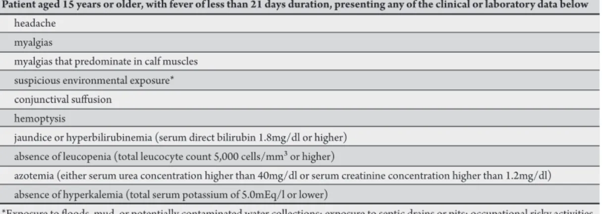
TABLE 1 - Case deinition to be validated in the present study.
Patient aged 15 years or older, with fever of less than 21 days duration, presenting any of the clinical or laboratory data below
headache myalgias
myalgias that predominate in calf muscles suspicious environmental exposure* conjunctival sufusion
hemoptysis
jaundice or hyperbilirubinemia (serum direct bilirubin 1.8mg/dl or higher) absence of leucopenia (total leucocyte count 5,000 cells/mm³ or higher)
azotemia (either serum urea concentration higher than 40mg/dl or serum creatinine concentration higher than 1.2mg/dl) absence of hyperkalemia (total serum potassium of 5.0mEq/l or lower)
*Exposure to loods, mud, or potentially contaminated water collections; exposure to septic drains or pits; occupational risky activities such as rubbish handling, unblocking of water streams, animal handling or farming in looded areas.
(84.9% to 98.6%)3-5. he low positive predictive values put in doubt
the utility of these criteria to assist in decisions taken at the bedside. Other authors validated Sri Lanka’s Ministry of Health criteria (slightly modiied from the World Health Organization (WHO) criteria), obtaining a beter positive predictive value (78%)6. None
of these criteria have been validated for use in the epidemiologic context of a Brazilian hospital.
he main objective of the present study is to validate a case deinition, based on simple clinical data and routine laboratory tests, which can be applied for bedside diagnosis of leptospirosis in reference hospitals of an endemic region in Brazil.
Patients were consecutively admited to two reference hospitals [Hospital Barão de Lucena (HBL) and Hospital Universitário Oswaldo Cruz (HUOC)] in Recife, a great city in Northeastern Brazil. They were recruited from February to December 2009, irrespective of the rainy season (that occurs from March to September). Informed consent was obtained from all participants or from their relatives, when patients were unconscious or were aged younger than 18 years. Subjects of either sex, aged 15 years or older, were included if they had a history of fever with a duration of 21 days or less and had leptospirosis as one of the diagnostic possibilities listed by the admiting physician.
Exclusion criteria were the inability to provide clinical information at hospital admission, as well as the awareness of a conirmed diagnosis for the febrile disease before hospital entry. Patients were considered study losses if they (or their relatives, when appropriate) refused to participate in the study; if data pertaining to either the clinical or laboratorial items that comprised the case deinition were unavailable; or if there were missing laboratory data that prevented conirmation or exclusion of leptospirosis diagnosis, including cases in which there was discordance between ELISA (positive) and MAT (negative) test results.
All patients had clinical data, and a single blood sample was collected at admission. The presence of headache, generalized myalgias, myalgias that predominated in calf muscles, suspicious environmental exposure, conjunctival sufusion, hemoptysis, and jaundice was evaluated; serum bilirubin, urea, creatinine, and potassium concentrations as well as total and diferential leukocyte count were tested. All patients admited up to the seventh day of symptoms had samples for blood cultures for leptospiras collected,
unless they had already taken antimicrobials. Cultures were performed in Ellinghausen-McCullough-Johnson-Harris (EMJH) media. After the seventh day of symptoms, patients had blood samples collected for ELISA-IgM and irst MAT testing. Second samples were collected for convalescent MAT testing two weeks or more ater the irst sample. ELISA tests were performed at Laboratório Central de Saúde Pública Dr. Milton Bezerra Sobral (LACEN), the local reference laboratory in Recife. MAT tests included antigens for the search of 22 serovars and were performed in the national reference laboratory of the Fundação Oswaldo Cruz, in the City of Rio de Janeiro.
Patients were considered as having confirmed leptospirosis (cases) if they fulilled at least one of the following criteria: a positive blood culture; a positive ELISA test; a positive MAT with title equal or above 1:800; or the demonstration of a negative (or a lower than 1:800 titer) MAT that became positive (or increased at least four times) ater a minimal 2-week interval, respectively.
Patients were considered without leptospirosis (non-cases) if they fulilled all the following conditions: negative blood culture, when it had been done; negative ELISA, with sample collected at least 7 days ater the beginning of symptoms; negative results for the two paired samples of the MAT; or low titer (less than 1:800) irst sample positive MAT that did not increase at least four times in the paired sample.
he case deinition designed for this study comprised ten criteria, all of them equally valued (Table 1).
RESULTS
TABLE 2 -Demographic data and duration of symptoms before admission from cases, non-cases, and lost patients. Cases Non-cases Cases and non-cases Lost patients
(n=75) (n=22) (n=97) (n=23)
Age (years, ± SD) 32.3 (12.7) 34.8 (15.0) 32.9 (13.2) 41.4 (18.8)*
Sex, male (%) 93.3 63.6** 86.6 82.6
Duration of symptoms before admission (days, ± SD) 6.4 (2.5) 5.2 (2.7) 6.1 (2.6) 5.0 (2.6) *p=0.01 for diference between included (cases and non-cases) and lost patients; **p=0.002 for diference between cases and non-cases; SD: Standard deinition.
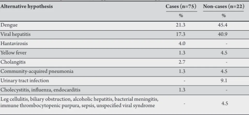
TABLE 3 -Alternative diagnoses initially suggested by the admiting physician for cases and non-cases.
Alternative hypothesis Cases (n=75) Non-cases (n=22)
% %
Dengue 21.3 45.4
Viral hepatitis 17.3 40.9
Hantavirosis 4.0
-Yellow fever 1.3 4.5
Cholangitis 2.7
-Community-acquired pneumonia 1.3 4.5
Urinary tract infection - 9.1
Cholecystitis, inluenza, endocarditis 1.3 -Leg cellulitis, biliary obstruction, alcoholic hepatitis, bacterial meningitis,
immune thrombocytopenic purpura, sepsis, unspeciied viral syndrome - 4.5
From February to December 2009, 120 patients were recruited — 64 from HBL and 56 from HUOC. Of them, 5 were lost because they had missing information on one of the laboratorial criteria from the case deinition. No patients had missing clinical criteria.
Another 18 patients were also considered study losses: 14 patients did not have convalescent serum MAT samples collected (4 died before the seventh day ater the beginning of symptoms, and 10 did not atend follow-up visits), whereas 4 patients had discordant results from conirmatory tests (positive ELISA but negative convalescent phase MAT results). Therefore, the final analysis included the remaining 97 patients: 75 had a diagnosis of leptospirosis as conirmed by the gold standard (cases), and 22 had that diagnosis denied (non-cases). For the 75 cases, diagnosis was conirmed by a combination of positive MAT, ELISA, and blood culture in 2 (2.7%) subjects; by both MAT and ELISA in 51 (68%) subjects; by MAT alone in 3 (4%) subjects; and by ELISA alone in 19 (25.3%) subjects. Serovars most oten identiied by MAT were L. copenhageni (61.6% of patients), L. icterohaemorragiae (27.5%), and L. tarassovi (4.5%).
Cases were more oten male (93.3%) than non-cases (63.6%), p=0.002. Cases were aged 32.3±12.7 years old and not statistically speciicity combination of the test; the area under the ROC curve was calculated. In addition, we calculated the likelihood ratios for each cutof number of the criteria.
Ethical considerations
he study protocol is in accordance with the revised Declaration of Helsinki and was approved by the local ethics research commitee (Comitê de Ética em Pesquisa em Seres Humanos do Complexo Hospitalar HUOC/PROCAPE), under number 139/2008.
diferent from non-cases (34.8±15.0 years old, p=1.0). Patients lost from the study were older (43.6±19.3 years, p=0.009 when compared to cases), with a gender distribution that was intermediate between the study groups (Table 2).
From the whole group of 97 patients included, 24 were initially admited to the intensive care unit (ICU), whereas the remaining 73 (75.3%) patients were admited to the wards. In groups CL and NL, 30.7% and 4.6% were admited to the ICU, respectively (p=0.04).
Among the cases, 72% had leptospirosis as the single diagnosis suggested by the physician responsible for hospital admission, while this happened for only 13.6% of non-cases (p<0.001). he diseases more oten suggested as alternative diagnoses in the 75 cases were dengue (16 citations), viral hepatitis (13 citations), and hantavirosis (3 citations). In the 22 non-cases, the alternative diagnoses more oten suggested were dengue (10 citations) and viral hepatitis (9 citations). Other diseases mentioned as alternative diagnoses are listed in Table 3.
Cases had environmental exposures suggestive of potential contact with leptospiras in 90.7% of cases; this happened to 77.3% of non-cases (non-signiicant diference, p=0.09).
Most patients admited before the seventh day of symptoms had received antibiotics prior to having their blood culture samples collected. Only 12 patients had blood cultures for leptospiras performed, of whom 4 were non-cases (with negative results) and 8 were samples from cases. Two of these last 8 blood cultures (25%) showed positive results; both of these patients had also positive ELISA and MAT results.
he mean number of criteria of the case deinition that were fulilled was 7.8±1.2 for cases and 5.9±1.5 for non-cases, showing a statistically signiicant diference (p<0.0001).
and accuracy obtained for each cutof number of criteria (from 5 or more to 9 or more) are shown in Table 4. he remaining cutof levels (0 to 4 or more, and 10 criteria) were not shown because sensitivity or speciicity levels obtained were clearly unacceptable. he best sensitivity (85.3%) and speciicity (68.2%) combination was found when having 7 or more criteria as cutoff
(Figure 1). For this cutoff level, a positive predictive value of 90.1%, a negative predictive value of 57.7%, and an accuracy of 81.4% were reached. he area under the curve (AUC) was 0.83 (95% conidence interval, 0.72 to 0.94).
TABLE 4 -Values obtained for Se, Sp, PPV, NPV, Ac, and LR for diferent cutof levels of the number of case deinition criteria fulilled.
Cutof Se (%) Sp (%) PPV (%) NPV (%) Ac (%) LR
(number of criteria) (95% CI) (95% CI) (95% CI) (95% CI)
At least 5 100.0 13.6 79.8 100.0 80.4 1.16 (93.9-100.0) (3.6-36.0) (70.0-87.1) (31.0-100)
At least 6 96.0 54.5 87.8 80.0 86.6 2.11
(88.0-99.0) (32.7-74.9) (78.3-93.7) (51.4-94.7)
At least 7 85.3 68.2 90.1 57.7 81.4 2.68
(74.8-92.1) (45.1-85.3) (80.2-95.6) (37.2-76.0)
At least 8 62.7 86.4 94.0 40.4 68.0 4.61
(50.7-73.3) (64.0-96.4) (82.5-98.4) (26.7-55.7)
At least 9 29.3 90.9 91.7 27.4 43.3 3.22
(19.7-41.1) (69.4-98.4) (71.5-98.5) (17.9-39.3)
Se: sensitivity; Sp: specificity; PPV: positive predictive value; NPV: negative predictive value; Ac: accuracy;
LR: likelihood ratios; CI: conidence interval.
FIGURE 1 - Receiver operating characteristic curve. he number of criteria from the case deinition fulilled is shown on the boxes.
DISCUSSION
he present study validated a case deinition of leptospirosis, based on clinical indings and simple laboratory tests, aimed for patients with acute severe febrile disease admited to reference tertiary-care hospitals in Northeastern Brazil. he best sensitivity and speciicity values were obtained for a cutof of 7 or more criteria. Although levels of sensitivity and speciicity were a litle lower than those observed with other deinitions, such as Faine’s and the modiied WHO criteria, the present study’s deinition atained a high positive predictive value, making it atractive for usage in the speciic population evaluated. Furthermore, interpreting the likelihood ratios obtained suggests that the case deinition can signiicantly raise the pretest probability of leptospirosis diagnosis7.
he sample studied seems to be representative of the average patients usually admited to our reference hospitals with suspected leptospirosis: predominantly young adult male subjects. he subjects were admited to the hospital 5 to 6 days ater the beginning of symptoms, a time point in which serologic methods have low sensitivity; this reinforces the demand for a test that can make an early diagnosis of the disease. As expected, non-cases had proportionately
more alternative diagnoses suggested by the admiting physicians. Dengue and viral hepatitis were, by far, the more cited ones for both cases and non-cases, although 15 more diseases were thought about. It is interesting to note that in 3 (13.6%) patients who were non-cases, leptospirosis was the sole diagnosis proposed upon admission, leading probably to equivocal therapeutic decisions. he epidemiologic component of the case deinition (exposure to potentially contaminated environment) was present almost as oten in cases as in non-cases, with a non-signiicant diference. It suggests that the discriminative potential of this information should not be overvalued when diferentiating leptospirosis from other diseases in our epidemiologic context. his assumption would argue against the utilization of Faine’s criteria in our hospitals, as that case deinition atributes a heavy weight to suspicious exposure8.
Direct comparisons of the present study with previous work on the subject demand some considerations. First, one of the studies that validated Faine’s criteria included outpatients as well as inpatients3.
In contrast, only hospitalized subjects were included in the present study, suggesting they had more severe disease. In fact, it should be noted that one quarter of our patients were admited directly to the ICU. he other two studies validating Faine’s criteria4-5, as well
as the study validating the modiied WHO criteria6, included only
hospitalized patients. However, they elected to include all patients who were admited with fever. In contrast, the present study only included subjects in whom leptospirosis was speciically suspected by the admiting physician. his restriction puts these patients in a situation of higher pretest probability of having leptospirosis, qualifying the present study as a Phase III validation study9. his
category of validation studies evaluates the capability of the test in distinguishing patients with and without the disease among patients in whom it is clinically reasonable to suspect that the disease is present9. In such context, test properties can be reduced, and the test
can appear less useful7. he three studies validating Faine’s criteria
obtained varying sensitivities (41.9% to 88.9%), speciicities (72.9% to 84.9%), positive predictive values (30.8% to 41.9%), and negative predictive values (84.9% to 98.6%)3-5. he study conducted in Sri
Lanka, which evaluated the modiied WHO criteria, showed 91.9%, 73.8%, 78.1%, and 90% for the same parameters above, respectively, with an accuracy of 82.9%6. he comparably high positive predictive
value of the present case deinition (90.1%) makes it atractive for use at the bedside, assisting in therapeutic decisions.
100
90
80
70
60
50
40
30
20
10
0
1 - specificity
Sensi�vity
0 10 20 30 40 50 60 70 80 90 100 >9
>8
>7
he authors declare that there is no conlict of interest. CONFLICT OF INTEREST
REFERENCES he present study fulills the main principles for the validity
of diagnostic test studies10. First, we performed an independent,
blind comparison of our test (the case deinition) with a reference standard. It should be emphasized that we used MAT test (usually considered the reference standard for leptospirosis diagnosis) for conirmation of 75% of cases. he remaining 25% of patients were conirmed by ELISA-IgM, a test with high sensitivity and speciicity, when compared with MAT11-13. In fact, the Brazilian Health Ministry
recommends the use of either ELISA or MAT for speciic diagnosis of leptospirosis14. The US Food and Drug Administration has
also approved the commercial utilization of ELISA15. Second, the
spectrum of patients included was precisely the same in which the case deinition is to be applied in practice, in particular, those with acute and severe febrile disease, admited to reference hospitals with a clinical suspicion of having leptospirosis. hird, the reference standard was performed in all patients who fulilled the inclusion criteria, independently of the test (case deinition) results. his minimized the so-called veriication bias16.
On the other hand, the present study has some limitations. First, the number of subjects was small, notably in non-cases. he stringent inclusion criteria, limiting entry to patients with a previous suspicion of leptospirosis diagnosis at hospital admission, contributed to it. However, 14 patients who had negative ELISA and irst MAT samples did not have the convalescent phase serum collected, either because they died or did not atend follow-up visits. As several of these subjects had ELISA samples collected ater the seventh day of symptoms (data not shown), it is reasonable to presume that many of them, notably the non-atendants, would have been non-cases. Hence, these omissions (lost cases) reduced the power of the validation parameters, enlarging its conidence intervals. Second, we did not pursue the alternative diagnoses in the patients who were assumed not to have leptospirosis. It was not pursued because of local resource limitations. However, our deinition of non-leptospirosis cases was very stringent and was based on the best practice in serological diagnosis of the disease. he small number of blood cultures performed is not a real limitation, if we consider the low sensitivity of this test17.
Lastly, it should be considered that the case deinition was tested in a speciic epidemiologic context, namely, the state of Pernambuco, Brazil, in 2009, among hospitalized patients with acute severe febrile disease. Diferences in the nosological proile at other regions, as well as an option to evaluate non-hospitalized patients, would be a demand for a new validation of the diagnostic tool.
he present study designed and validated a case deinition for leptospirosis diagnosis, based on simple clinical and laboratorial tests, to be used in hospitalized patients with acute severe febrile disease in Northeastern Brazil. he deinition reached an average sensitivity and speciicity but presented with a high positive predictive value. his inding, allied to its simplicity and low cost, makes it useful for rapid bedside diagnosis and for assistance in therapeutic decisions, before a rapid and early sensitive laboratory test becomes available.
1. Terpstra WJ. International leptospirosis Society. Human Leptospirosis: Guidance for diagnosis, surveillance and control. Geneva: World Health Organization; 2003.
2. Mcbride AJA, Santos BL, Queiroz A, Santos AC, Hartskeerl A, Reis MG, et al. Evaluation of four whole-cells leptospira-based serological tests for diagnosis of human leptospirosis. Clin Vaccine Immunol 2007; 14:1245-1248.
3. Rao P, Sethi S, Sud A, Banga SS, Sharma M. Screening of patients with acute febrile illness for leptospirosis using clinical criteria and sorology. Natl Med J India 2005; 18:244-246.
4. Bal AM, Kakrani AL, Bharadwaj RS, Kagal AS, Joshi AS, Arjunwadkar VP. Evaluation of clinical criteria for the diagnosis of leptospirosis. J Assoc Physicians India 2002; 50:394-396.
5. Shivakumar S, Shareek PS. Diagnosis of leptospirosis utilizing modiied Faine’s criteria (leter). J Assoc Physicians India 2004; 52:678-679.
6. Dassanayake DLB, Wimalaratna H, Agampodi SB, Liyanapathirana VC, Piyarathna TACL, Goonapienuwala BL. Evaluation of surveillance case deinition in the diagnosis of leptospirosis, using the microscopic agglutination test: a validation study. BMC Infect Dis 2009; 9:48.
7. Jaeschke R, Guyat GH, Sacket DL. User’s guide to the medical literature. III. How to use an article about a diagnostic test. B. What are the results and will they help me in caring for my patients? JAMA 1994; 271:703-707. 8. Faine S. Guidelines for the control of leptospirosis. Geneva: World Health
Organization; 1982.
9. Sacket DL, Haynes RB. Evidence base of clinical diagnosis: the architecture of diagnostic research. BMJ 2002; 324:539-541.
10. Flahault A, Cadilhac M, homas G. Sample size calculation should be performed for design accuracy in diagnostic test studies. J Clin Epidemiol 2005; 58:859-862. 11. Mcbride AJA, Pereira FA, Silva ED, Matos RB, Silva ED, Ferreira AGP, et al. Evaluation of the EIE-IgM Leptospirose assay for the serodiagnosis of leptospirosis. Acta Trop 2007; 102:206-211.
12. Dey S, Mohan M, Ramadass P, Nachimuthu K. Diagnosis of leptospirosis by recombinant antigen based single serum dilution ELISA. Indian J Med Res 2008; 128:172-177.
13. Mulla S, Chakraborty T, Patel M, Pandya HP, Dadhaniya V, Vaghela G. Diagnosis of leptospirosis and comparison of ELISA and MAT techniques. Indian J Pathol Microbiol 2006; 49:468-470.
14. Ministério da Saúde. Doenças infecciosas e parasitárias: guia de bolso. 6th ed.
rev. Brasília: Secretaria de Vigilância em Saúde. Departamento de Vigilância Epidemiológica; 2005.
15. Mathur M, De A, Turbadkar D. Leptospirosis outbreak in 2005: L.T.M.G. hospital experience. Indian J Med Microbiol 2009; 27:153-155.
16. Jaeschke R, Guyat GH, Sacket DL. User’s guide to the medical literature. III. How to use an article about a diagnostic test. A. Are the results of the study valid? JAMA 1994; 271:389-391.