RBCCV 44205-1442 DOI: 10.5935/1678-9741.20130009
Beneicial effects of hyperosmotic perfusion
in the myocardium after ischemia/reperfusion
injury in isolated rat hearts
Efeitos benéicos da perfusão hiperosmótica no miocárdio após lesão isquemia/reperfusão em corações
isolados de ratos
Yong Cao
1, Lie Wang
2, Hong Chen
3, Zhiqian Lv
41. Doctor - No 6th People's Hospital, Shanghai Jiaotong University School of Medicine, Shanghai, China. Participated the experiment and drafted the manuscript.
2. Doctor - Research Institute of General Surgery, General Hospital, Fuzhou Military Area Command of PLA, Shanghai, China. Participated the experiment and drafted the manuscript.
3. Doctor - Department of Pharmacology, Shanghai Jiaotong University School of Medicine, Shanghai, China. Designed the experiment and participated in the experiment.
4. Doctor - No 6th People's Hospital, Shanghai Jiaotong University School of Medicine, Shanghai, China. Provided the research grant and research plan.
The work was carried out at No 6th People's Hospital and Department of Pharmacology, Shanghai Jiaotong University School of Medicine, Shanghai, China.
Correspondence address Zhiqian Lv and Hong Chen
Department of Cardiosurgery, No 6th People's Hospital, Shanghai Jiaotong University School of Medicine, 200233, Shanghai, P R. China E-mail: [email protected] and [email protected]
Article received on September 18th, 2012 Article accepted on October 1st, 2012
Abstract
Objective: A simple method to reduce the ischemia/ reperfusion injury that can accompany cardiac surgery would have great clinical value. This study was to investigate the effect of hyperosmotic perfusion on ischemia/reperfusion injury in isolated perfused rat hearts.
Method: Forty male Sprague-Dawley rats were randomly divided either to have their isolated hearts perfused with normal osmotic buffer or buffer made hyperosmotic by addition of glucose. Hearts were then subjected to 30 min
ischemia followed by 30 min reperfusion. Coronary low, time to ischemic arrest, reperfusion arrhythmia, and ventricular
function were recorded. Creatine phosphokinase leakage into
the coronary artery, and myocardial content and activity of
superoxide dismutase and catalase were also examined.
Results: Rat hearts with hyperosmotic perfusion showed
higher coronary low, a prolonged time to ischemic arrest (10.60 vs. 5.63 min, P<0.005), a lower reperfusion arrthythmia score (3.2 vs. 5.3, P<0.001), better ventricular function, and less creatine phosphokinase leakage (340.1 vs. 861.9, P<0.001) than normal osmotic controls. Myocardial catalase content
and activity were increased signiicantly (1435 vs. 917 U/g
wet weight, P<0.001) in hearts perfused with hyperosmotic solution in comparison to the normal osmotic controls.
Conclusion: Pretreatment with hyperosmotic perfusion in
normal rat hearts, which is attributed partly to the increased antioxidative activity, could provide beneicial effects from
ischemia and reperfusion-induced injury by increasing
coronary low, and decreasing reperfusion arrhythmia.
Resumo
Objetivo: Um método simples para reduzir a lesão de isquemia/reperfusão que pode acompanhar a cirurgia cardíaca teria grande valor clínico. O objetivo deste estudo foi investigar o efeito da perfusão hiperosmótica na isquemia/ reperfusão em corações isolados de ratos perfundidos.
Métodos: Quarenta ratos machos Sprague-Dawley foram divididos aleatoriamente e tiveram os seus corações isolados perfundidos com tampão osmótico normal ou tampão hiperosmótico com a adição de glucose. Os corações foram
então submetidos a 30 minutos de isquemia, seguida de 30
min de reperfusão. O luxo coronariano, tempo de parada isquêmica, arritmia de reperfusão e da função ventricular
foram registrados. Vazamento creatinofosfoquinase na
artéria coronária, o miocárdio e atividade de
superóxido-dismutase e catalase foram também examinados.
Resultados: Crações de ratos com perfusão hiperosmótica
apresentaram maior luxo coronariano, tempo prolongado de parada isquêmica (10,60 vs. 5,63 min, P<0,005), menor pontuação de reperfusão arritmica (3,2 vs. 5,3, P<0,001),
melhor função ventricular e menos vazamento de creatina
fosfoquinase (340,1 vs. 861,9, P<0,001) do que controles
normais osmóticos. Teor de catalase e atividade do miocárdio
também tiveram aumento signiicativo (1435 vs. 917 peso
U/g de peso fresco, P<0,001) em corações perfundidos com
solução hiperosmótica em comparação com os controles normais osmóticos.
Conclusão: O pré-tratamento com perfusão hiperosmótica
em corações de ratos normais, o que é atribuído, em parte, ao aumento da atividade antioxidante, pode oferecer efeitos benéicos de isquemia e reperfusão induzida por lesão, aumentando o luxo coronário e diminuindo a arritmia de
reperfusão.
Descritores: Isquemia. Traumatismo por reperfusão. Infarto do miocárdio.
Abbreviations, acronyms & symbols
INTRODUCTION
Each year in the United States, approximately 1 million myocardial infarctions occur, and 700,000 patients undergo cardioplegic arrest for various cardiac surgeries. Minimizing ischemic time in these clinical scenarios has appropriately received a great deal of attention because of the long-established relationship between duration of ischemia and extent of myocardial injury. However after coronary low is restored, the myocardium is susceptible to another form of insult stemming from reperfusion of the previously ischemic tissue. Given that cardiac ischemia is either unpredictable (myocardial infarction) or inevitable (in the operating room), there is great interest in developing strategies to minimize reperfusion-mediated injury.
Hyperosmotic saline has been used successfully in haemorrhagic and other types of shock to protect different tissues and organs [1,2]. Hyperosmotic saline perfusion has also been shown to decrease ischemia/reperfusion injury in rat hearts [3]. Elevated blood glucose also
increases plasma osmolarity, and resistance to ischemia/ reperfusion injury is increased in hearts from type 1 diabetic rats that have severe hyperglycaemia [4]. Because both hypertonic saline and, in diabetic rats, hyperglycemia are cardioprotective, we sought to determine whether addition of glucose instead of sodium chloride (NaCl) to produce hyperosmolarity would, in hearts from normal rats, provide protection from ischemia/reperfusion injury. If hyperosmotic glucose perfusion were found to be protective, it might, in the future, be able to be used clinically to attenuate ischemia/reperfusion injury during cardiac bypass surgery.
The present study investigates the effect of hyperosmotic perfusion pretreatment with glucose on ischemia/reperfusion arrhythmia and ventricular function damage in isolated rat hearts in vitro. Coronary creatine phosphokinase leakage (that is, leakage of this cardiac enzyme from damaged myocardial cells into the perfusate from the coronary artery), and myocardial content and activity of two antioxidant enzymes, superoxide dismutase CPK
HR LVEDP LVP NO ROS SDS-PAGE SOD VF-VT
Creatine phosphokinase Heart rate
Left ventricular end diastolic pressure Left ventricular pressure
Nitric oxide
Reactive oxygen species
Sodium dodecyl sulfate–polyacrylamide gel electrophoresis
Superoxide dismutase
and catalase, were also measured in order to assess the effect of hyperosmotic perfusion with glucose on myocardial cell damage and anti-oxidant activity.
METHODS
Animals
This study was approved by the animal care and use committee of our institution (2011(LW)-13). Forty male Sprague-Dawley rats (originally weighing 220–280 g) were housed under standard conditions with free access to tap water and chow. Rats were randomly divided either to have their isolated hearts perfused with normal Krebs-Henseleit buffer solution (300 mOsm/L) or with Krebs-Henseleit buffer solution made hyperosmotic (360 mOsm/L) by addition of glucose.
Isolated heart preparation and ischemia/reperfusion with normal buffer solution
Rats were anaesthetized with sodium pentobarbital (60 mg/kg, i.p.), injected with 25 mg/kg heparin, and their hearts excised a few minutes later. The hearts were then placed in a Langendorff apparatus [5] and rapidly connected to the aortic cannula of this apparatus for retrograde perfusion with Krebs–Henseleit buffer containing (in mmol/L): NaCl 118.0; KCl 4.7; CaCl2 2.5; MgSO4 1.2; KH2PO4 1.2; NaHCO3 25.0; glucose 11.0 mM; Na2 -EDTA 0.5. The perfusate was maintained at 37˚C and aerated continuously with 95% O2 and 5% CO2. The left atrium was connected to a cannula and perfused in order to ill the left ventricle, and a catheter was inserted into the left ventricle to record left ventricular pressure (LVP), left ventricular end diastolic pressure (LVEDP), and maximal rate of LVP rise (dp/dtmax) and fall (dp/dtmin), using a data
acquisition system (MPA 2000; Alcott Biotech, Shanghai, China). Aortic perfusion pressure was set at 70 mmHg and left atrial pressure at 15 mmHg throughout the experiment. However, cardiac output as it would occur in the living animal cannot be measured with this method. A catheter inserted into a small artery originating in the marginal or oblique branch of the left circumlex coronary artery was used for continuous measurement of coronary low (calculated from the luid fractions collected as volume/ time) in pre-ischemia perfusion, ischemia, and reperfusion periods. We used this location for catheter insertion because it enables more precise and clean collection of coronary efluent than efluent collected from other locations. The perfusate collected from the coronary artery was used to assay for creatine phosphokinase.
Electrodes were inserted into the myocardium at the base and apex of the heart, and heart rate (HR) and electrocardiogram were recorded continuously with a data-acquisition system (MPA 2000; Alcott Biotech,
Shanghai, China). Premature ventricular beats, ventricular tachycardia and ventricular ibrillation during reperfusion were recorded. The severity of arrhythmias was scored using the Curtis arrhythmia scoring system [6]. This system uses a scale of 0-9, where 0 indicates occasional ectopic beats and 9 indicates ventricular ibrillation occurring during the irst 60s of reperfusion. The time from the clamping of the atrial inlow and aortic outlow to the cessation of ventricular contractions was recorded as the time to arrest, and the time from the beginning of reperfusion to the start of ventricular contractions was recorded as the time to recovery.
All hearts were allowed to beat spontaneously. After the 10 min normal buffer equilibration perfusion and 20 min pre-treatment normal buffer or hyperosmotic glucose perfusion (the total pre-ischemia period was 30 min), the hearts were subjected to 30 min global ischemia by clamping both atrial inlow and aortic outlow. The heart was thus in contact with hyperosmotic glucose buffer for a total of 50 min. The heart was kept warm during the ischemic period. At the end of this period, the above low catheters were opened for a 30 min reperfusion period.
Hyperosmotic perfusion
The procedure for the rat hearts in the hyperosmotic perfusion group was the same as that described above for normally perfused hearts except that the osmolarity of the Krebs–Henseleit buffer was increased to 360 mOsmol/L during the 20 min pre-treatment period by adding 60 mOsmol/L of glucose and thus increasing the glucose concentration to 71 mM.
Biochemical determinations and Western blot analysis
At the end of the 30 min reperfusion period, the front portions of the left ventricles were freeze-clamped for use in biochemical assays and Western blotting. Creatine phosphokinase (CPK) from the coronary artery, and superoxide dismutase (SOD) and catalase from myocardial tissue homogenate, were determined with assay kits (Jian-Cheng Biomedical Engineering, Nanjing, China) according to the instructions of the manufacturer.
peroxidase-conjugated secondary antibody, and immunoreactive bands were visualized using electrochemoluminescence (Pierce; Thermo Fisher Scientiic Inc., Rockford, IL, USA). Relative levels of proteins were semi-quantiied with densitometry (Leica).
Statistics
Normally distributed continuous variables between control and hyperosmotic glucose groups were compared using an independent two sample t test. Differences between ischemia and reperfusion were determined using a paired t test. Repeated measurement of ANOVA was used for coronary low. Data are presented as means ± SE. All statistical assessments were two-sided and evaluated at the 0.05 level of signiicant difference. Statistical analyses were performed using SPSS 15.0 statistics software (SPSS Inc, Chicago, IL).
RESULTS
Coronary low and correlation with hyperosmotic perfusion
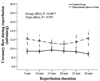
Compared to normal osmotic perfusion, hyperosmotic glucose perfusion signiicantly increased pre-ischemia basal coronary low in isolated rat heart after 5 and 10 minutes of perfusion (Figure 1). After ischemia and reperfusion treatment, coronary low was also signiicantly higher with hyperosmotic glucose than normal osmotic perfusion over the 30 minutes of perfusion (Figure 2).
Ventricular function during ischemia and reperfusion
Rat hearts perfused with hyperosmotic solution took a longer time (10.6±1.5 vs. 5.6±0.7 min) to stop beating during ischemia and a shorter time (13.0±2.5 vs. 19.2±1.5 sec) to resume beating during reperfusion than control hearts. In other words, hyperosmotic hearts were asystolic about 32 minutes as opposed to 43 minutes for control hearts. The reperfusion arrhythmia score (3.3±1.7) and ventricular tachycardia-ibrillation period (15.0±1.2 min) were signiicantly reduced in hyperosmotic hearts compared to control hearts (5.3±1.2, 24.3±2 min respectively) (Table 1). There were no signiicant differences in pre-ischemia HR, LVP, LVEDP, dP/dtmax and
dP/dtmin between the 2 groups. However, HR, LVP, dP/dtmax
and dP/dtmin in both groups signiicantly decreased during
reperfusion (P<0.05, Table 2).
Creatine phosphokinase (CPK) leakage from myocardial tissue and myocardial anti-oxidant enzymes
CPK leakage into the coronary artery, an indication of myocardial cell damage, was signiicantly reduced after reperfusion in hyperosmotic compared to control hearts, as shown by biochemical assays (360±115 vs. 860±124 U/g). Of the two anti-oxidant enzymes measured, hyperosmotic hearts showed a signiicant increase in myocardial catalase in both biochemical assay and Western blotting (Table 1 and Figure 3A, C). However, myocardial contents of SOD in hyperosmotic hearts were similar to those of normal control hearts in both biochemical assay and Western blotting (Table 1 and Figure 3B, D).
Fig. 1 – The effects of normal and hyperosmotic glucose perfusion
on pre-ischemia coronary low in isolated rat heart . (mean±SE,
n=20 in each time point)
Table 1. Ischemic arrest time, heartbeat recovery time, arrhythmia score and VT-VF time after ischemia/reperfusion with hyperosmotic glucose.
Ischemic arrest time (min) Heartbeat recovery time (sec) Arrhythmia score
VT-VF duration (min)
Creatine phosphokinase leakage (U/g ventricular weight) SOD (U/g wet weight)
Catalase (U/g wet weight)
Control 5.62±0.73 19.19±1.99
5.30±0.27 24.46±0.45 861.90±26.36
343±4.7 917±18.9
Hyperosmotic glucose 10.60±1.50 11.24±2.40 3.20±0.33 14.91±0.28 340.10±24.88
354±10.0 1435±17.5
P value
0.005 0.056 <0.001 <0.001 <0.001 0.324 <0.001
I/R ischemia/reperfusion; SOD, superoxide dismutase; VF, ventricular ibrillation; VT, ventricular tachycardia. Data are displayed as mean ± standard error. n=20
Table 2. Heart rate, contractility and relaxation before and after ischemia/reperfusion.
HR (bpm) Pre 30 min LVP (mmHg) Pre
30 min
LVEDP (mmHg) Pre
30 min
dP/dtmax (mmHg/sec)
Pre 30 min
dP/dtmin (mmHg/sec) Pre
30 min
Control
241.00±11.84 100.20±27.81a
88.12±4.77 63.89±5.34a
5.32±1.88 53.28±3.94a
2476.5±218.6 345.0±105.8a
1657.5±137.5 303.0±61.1a
Hyperosmotic glucose
249.70±15.82 198.50±17.23a
86.08±3.50 69.92±3.42a
5.87±1.21 37.16±3.74a
2538.0±129.3 1101.8±97.7a
1881.3±121.6 834.0±46.55a
P value
0.665 0.008
0.734 0.354
0.807 0.008
0.811 <0.001
0.287 <0.001
dP/dtmax, maximal rate of left ventricular rise; dP/dtmin, maximal rate of left ventricular pressure fall; HR, heart
rate; LVEDP, left ventricular end diastolic pressure; LVP, left ventricular pressure. a Signiicant difference
between ischemia and reperfusion in each group using paired –t test. Data are displayed as mean ± standard error
DISCUSSION
In the current study, hyperosmotic perfusion pretreatment with glucose of rat hearts subjected to global ischemia resulted in better ventricular contractile and electrophysiological function, increased coronary low, and less myocardial damage than was seen in normal rat hearts. Hyperosmotic perfusion pretreatment also increased levels of catalase, although levels of a second anti-oxidant enzyme, SOD, were similar to those seen in control hearts.
Endothelial nitric oxide (NO) production has previously been shown to be responsible for the increase in coronary blood low seen after bolus injection of hypertonic saline in anaesthetized pigs [7]. This is a possible mechanism for the increased coronary low seen with hypertonic perfusion pretreatment of glucose in our study, but we did not measure NO production.
Previous research by others has shown that an important change responsible for ischemia/reperfusion damage is the increase in intracellular calcium that occurs during ischemia [8]. Anoxia decreases pH, and the increased Na/H exchange used by the cell to bring the pH back to normal affects Na/Ca exchange in a way that increases intracellular calcium [8]. Myocardial perfusion with luid made hypertonic with either NaCl or sucrose has been shown to decrease intracellular calcium by decreasing the anoxia-induced increase in Na/H exchange [9].
Reactive oxygen species (ROS) have also been implicated in the pathogenesis of ischemia/reperfusion injury [8]. In our study, hyperosmotic hearts showed an increase in the activity of the anti-oxidant enzyme, catalase, but no increase in SOD, the second enzyme studied. A study of diabetic hearts from rats with severe hyperglycaemia also showed an increase in catalase, but not SOD, compared to normal hearts [4]. And a study of hyperosmotic sodium chloride perfusion of normal and stroke-prone spontaneously hypertensive rat hearts also showed increased catalase but not increased SOD, although in this study raising the osmolarity to 400 mOsm/L was able to increase SOD slightly [10]. In our study, levels of catalase were increased 20%, but levels of the marker of myocardial damage, CPK, however, were decreased 60%. It seems doubtful, therefore, that this modest increase in catalase activity could, by itself, cause such a profound lessening of myocardial damage.
An unanswered question is whether beneicial effects for heart by solution made hypertonic by the addition of glucose differs in any way from that provided by solution made hypertonic by the addition of NaCl. Hyperosmotic glucose differs from hypertonic saline in that glucose is a substrate for energy metabolism in the myocardium. In normal conditions, myocardium utilizes fatty acids for aerobic metabolism but in anoxic conditions, it uses glucose
for anaerobic glycolysis. Anaerobic metabolism plays a crucial role in protecting myocardium from death [11]. Hyperosmotic glucose perfusate provides more glucose for the myocardium to produce ATP through glycolysis, an action beneicial for maintaining the activity of Na+-K+ATPase and the ATP-dependent K channel. This action may attenuate myocardium injury. However, because the manner in which the myocardium utilizes metabolic substrates is complex, the possibility that increases in extracellular glucose availability can be beneicial to heart needs further investigation.
Chu et al. [12] also that ischemic myocardium in alloxan-induced-diabetic (DM) group displayed higher expression of cell survival proteins including phospho eNOS, heat shock protein 27, NFkB, and mTOR as compared with ischemic myocardium in normoglycemic (ND), whereas in non-ischemic tissue, expression of these proteins was similar or lower in the DM group. The expression of total Erk 1/2 was similar between ND and DM groups, whereas the expression of phospho-Erk 1/2(Thr202/Tyr204) was lower in the DM group as compared to the ND group [12]. The expression of SAPK/JNK was lower in the DM group [12]. The current study shows that normal rat hearts prefused with hyperosmotic glucose can provide beneicial effects to heart, but these beneicial effects could very well be due to the hyperosmolarity itself rather than glucose.
In our study, rat hearts prefused with hyperosmotic glucose before ischemia/reperfusion increased coronary artery low and decreased coronary artery resistance, increased left ventricular function, and increased levels of catalase but not SOD. In Shen’s et al. [10] study, prefused with hyperosmolar NaCl gave similar results: increased coronary artery low and decreased coronary artery resistance, increased left ventricular function, and increased levels of catalase but not SOD. Shen et al. [10] also compared the effects of a 2 hour perfusion of rats hearts with solution made hyperosmolar with 4 different osmolytes (NaCl, glucose, mannitol, and rafinose) and found no signiicant differences in levels of the cellular constituents measured. They did not, however look at left ventricular function. Our indings and Shen’s et al. [10], therefore, may be due entirely to the hyperosmolarity.
However, one must also consider the use for which a solution is intended. Studies comparing the success of solutions used to protect hearts used for transplantation have focused on protecting cellular energy metabolism [13,14]. Despite the crystalloid solutions studied are not fully able to suppress the deleterious effects of ischemia and reperfusion in the rat heart, Lima et al. [14] had also found that a solution may contain the mannitol, instead of glucose, and the concentrations of K+ (15 mmol/L) and
Ca2
+ (0.25 mmol/L) of solution can contribute to a better
REFERENCES
1. Gurinkel V, Poggetti RS, Fontes B, Costa Ferreira Novo F, Birolini D. Hypertonic saline improves tissue oxygenation and reduces systemic and pulmonary inlammatory response caused by hemorrhagic shock. J Trauma. 2003;54(6):1137-45. the ischemic period. We were investigating hyperosmotic solutions might be beneicial to normal rat heart ex vivo to against ischemia/reperfusion injury during cardiac bypass surgery. In this situation, the physiologic effect of the hyperosmotic luid used on non-cardiac tissue must be considered, and the use of glucose instead of NaCl as the osmolyte might have advantages.
A limitation of this study is that we should compare the use of glucose to mannitol, an osmotically active but metabolically inactive compound, to see whether hyperosmolarity alone was responsible for the beneicial effects of glucose to heart or not. Perfusion with hypertonic mannitol has been shown to increase catalase levels in normal rat hearts [11] but there are no reports to date about its actions on ventricular function and enzymes during ischemia/reperfusion. Other limitations are that NO production, intracellular calcium, and ROS should be measured to elucidate the mechanism behind of these beneicial effects of hyperosmotic perfusion pretreatment. We also like to study the protein proiles of cell survival signaling and mitogen-activated protein kinase signaling to see whether changes in any of these parameters might be involved in beneicial effects of hyperosmotic perfusion pretreatment with glucose to normal rat heart.
In summary, the present study had shown that hyperosmotic perfusion pretreatment may provide beneicial effects to heart from ischemia and reperfusion injury, increase coronary low, and decrease reperfusion arrhythmia. The beneicial effects induced by hyperosmotic perfusion pretreatment with glucose may be attributed partly to the increased antioxidative activity. It is possible that hyperosmotic perfusion pretreatment may be able in the future to be applied during cardiac bypass surgery for its beneicial effects against ischemia/reperfusion injury.
ACKNOWLEDGEMENTS
This study was supported by grants from the National Natural Science Foundation of China (No. 31171099, 30971154 and 30770848).
CONFLICT OF INTEREST
No conlict of interest declared.
2. Oliveira RP, Velasco I, Soriano FG, Friedman G. Clinical review: hypertonic saline resuscitation in sepsis. Crit Care. 2002;6(5):418-23.
3. Chen LB, Liu T, Wu JX, Chen XF, Wang L, Fan CL, et al. Hypertonic perfusion reduced myocardial injury during subsequent ischemia and reperfusion in normal and hypertensive rats. Acta Pharmacol Sin. 2003;24(11):1077-82.
4. Chen H, Shen WL, Wang XH, Chen HZ, Gu JZ, Fu J, et al. Paradoxically enhanced heart tolerance to ischaemia in type 1 diabetes and role of increased osmolarity. Clin Exp Pharmacol Physiol. 2006;33(10):910-6.
5. Skrzypiec-Spring M, Grotthus B, Szelag A, Schulz R. Isolated heart perfusion according to Langendorff: still viable in the new millennium. J Pharmacol Toxicol Methods. 2007;55(2):113-26.
6. Curtis MJ, Walker MJ. Quantiication of arrhythmias using scoring systems: an examination of seven scores in an in vivo model of regional myocardial ischaemia. Cardiovasc Res. 1998;22(9):656-65.
7. Vacca G, Papillo B, Battaglia A, Grossini E, Mary DA, Pelosi G. The effects of hypertonic saline solution on coronary blood low in anaesthetized pigs. J Physiol. 1996;491( Pt 3):843-51.
8. Murphy E, Steenbergen C. Mechanisms underlying acute protection from cardiac ischemia-reperfusion injury. Physiol Rev. 2008;88(2):581-609.
9. Ho HS, Liu H, Cala PM, Anderson SE. Hypertonic perfusion inhibits intracellular Na and Ca accumulation in hypoxic myocardium. Am J Physiol Cell Physiol. 2000;278(5):C953-64.
10. Shen WL, Chen LB, Zhao JX, Guo SJ, Chen YC, Wang LP, et al. Effects of hyperosmotic sodium chloride perfusion on ischemia/reperfusion injury in isolated hearts of normal and stroke-prone spontaneously hypertensive rats. J Clin Exper Cardiol. 2011;2(7):146.
11. Gong J, Li LH, Pei WD, Wang HY, Zheng YL, Zhou GY, et al. Glycolytic and fatty acid metabolic enzyme changes early after acute myocardial ischemia. Zhonghua Xin Xue Guan Bing Za Zhi. 2006;34(6):546-50.
12. Chu LM, Osipov RM, Robich MP, Feng J, Oyamada S, Bianchi C, et al. Is hyperglycemia bad for the heart during acute ischemia? J Thorac Cardiovasc Surg. 2010;140(6):1345-52.
13. Silveira Filho LM, Petrucci O Jr, Carmo MR, Oliveira PP, Vilarinho KA, Vieira RW, et al. Trimetazidine as cardioplegia addictive without pre-treatment does not improve myocardial protection: study in a swine working heat model. Rev Bras Cir Cardiovasc. 2008,23(2):224-34.