RBCCV 44205-1444 DOI: 10.5935/1678-9741.20130011
Analysis of left ventricular function in
patients with heart failure undergoing cardiac
resynchronization
Análise da função ventricular esquerda de pacientes com insuiciência cardíaca submetidos à
ressincronização cardíaca
Ricardo Adala Benfatti
1, Felipe Matsushita Manzano
2, José Carlos Dorsa Vieira Pontes
3, Amaury
Edgardo Mont’Serrat Ávila Souza Dias
4, João Jackson Duarte
5, Guilherme Viotto Rodrigues da
Silva
6, Jandir Ferreira Gomes Junior
7, Neimar Gardenal
81. Master of Science, Assistant Professor of Federal University of Mato Grosso do Sul (UFMS), Campo Grande, MS, Brazil – Main author. 2. Medical Doctor of UFMS, Campo Grande, MS, Brazil – Data
collection.
3. Philosophy Doctor, Full Professor of UFMS, Campo Grande, MS, Brazil – Final review.
4. Assistant Professor of UFMS, Campo Grande, MS, Brazil – Performed surgeries and bibliographic research.
5. Master of Science of UFMS, Campo Grande, MS, Brazil – Performed surgeries and bibliographic research.
6. Resident Doctor of UFMS, Campo Grande, MS, Brazil – Bibliographic research, assistance of surgeries.
7. Master of Science, Campo Grande, MS, Brazil – Performed Surgeries. 8. Master of Science, Campo Grande, MS, Brazil – Co-author in the
writing of the scientiic text.
Work carried out at Federal University of Mato Grosso do Sul, Campo Grande, MS, Brazil.
Correspondence address Ricardo Adala Benfatti
Av. Senador Filinto Muller, S/N – Hospital Universitário – UTI Cardiológica – Campo Grande, MS, Brazil.
E-mail: [email protected]
Article received on September 9th, 2012 Article accepted on December 18th, 2012
Resumo
Fundamentos: O tratamento cirúrgico da insuiciência
cardíaca padrão-ouro é o transplante cardíaco, porém,
em decorrência das diiculdades desse tratamento, outras propostas cirúrgicas têm sido relatadas, entre elas o implante
de ressincronizador cardíaco.
Objetivo: Analisar a função ventricular esquerda, por meio
da ecocardiograia, de pacientes portadores de insuiciência
cardíaca avançada com dissincronia interventricular submetidos a implante de ressincronizador cardíaco.
Métodos: Entre junho de 2006 a junho de 2012, foram avaliados 24 pacientes com idade média de 61,5 ± 11 anos,
portadores de insuiciência cardíaca congestiva avançada em
classe funcional III e IV (NYHA), dissincronia interventricular e tratamento medicamentoso otimizado. Esses pacientes foram submetidos ao implante de ressincronizador cardíaco e avaliados
ecocardiograicamente no pós-operatório de seis meses.
Resultados: Houve melhora signiicativa dos parâmetros ecocardiográicos analisados. A média dos diâmetros diastólicos ventriculares esquerdos reduziu de 69,6 ± 9,8 mm para 66,8 ± 8,8 mm, diâmetros sistólicos de 58,6 ± 8,8 mm para 52,7 ± 8,8 mm e a fração de ejeção, média de 31 ± 8% para 40 ± 7% com nível de signiicância, respectivamente, de 0,019, 0,0004 e 0,0002, estatisticamente signiicativos com nível de signiicância de 0,05.
Conclusão: Houve melhora da função ventricular
esquerda analisada por meio da ecocardiograia, em seis meses, de pacientes portadores de insuiciência cardíaca
avançada submetidos a implante de ressincronizador cardíaco.
Abstract
Background: The gold standard surgical treatment
for heart failure is cardiac transplantation, however, due
to dificulties of this treatment, other surgical proposals
have been reported, including the implantation of cardiac resynchronizer.
Objective: To analyze the left ventricular function by
echocardiography in patients with advanced heart failure with interventricular dyssynchrony undergone implantation of cardiac resynchronizer.
Methods: Between June 2006 and June 2012, 24 patients with average age of 61.5 ± 11 years were evaluated, carriers of advanced congestive heart failure functional class III and IV (NYHA), interventricular dyssynchrony and optimal drug therapy, and submitted implantation of cardiac resynchronizer and postoperative echocardiographically evaluated in six months.
Results: There was signiicant improvement of the analyzed echocardiography parameters. The average left ventricular diastolic diameter decreased from 69.6 ± 9.8 mm to 66.8 ± 8.8 mm, systolic diameters from 58.6 ± 8.8 mm to 52.7 ± 8.8 mm, and ejection fraction, average of 31 ± 8% to 40 ± 7% with level of signiicance, respectively, of 0.019, 0.0004 and 0.0002, statistically signiicant with a signiicance level
of 0.05.
Conclusion: There was a signiicant improvement of left
ventricular function analyzed by echocardiography at six months, in patients with advanced heart failure undergone implantation of cardiac resynchronizer.
Descriptors: Heart failure. Cardiac resynchronization therapy. Echocardiography.
Abbreviations, acronyms & symbols
INTRODUCTION
Heart failure (HF) one of the greatest clinical challenges in the current public health, and it is considered an epidemic on progress, diagnosed in 1% to 2% of developed countries population [1,2].
The HF is one of the most prevalent causes of hospital admissions in Brazil, on the Uniied Health System (SUS). In 2007, there were 1.156.136 hospital admissions for cardiovascular diseases, representing the third leading cause of hospital admissions on the SUS, and HF is the main one [3].
The HF treatment has the purpose of symptoms improvement, reduction of mortality, hospital costs decreasing and prevention of readmissions. The treatment of heart failure consists of: nonpharmacological steps (diet, exercising, stress management, and others), drug therapy and surgery [3].
Studies show that medical treatment should always include a beta blocker and an inhibitor of the angiotensin converting enzyme (ACE), composing the optimized medical treatment. For symptomatic patients, diuretics and digitalis are added. But if there is a disabling symptoms persistence, it is necessary to introduce aldosterone
antagonists (functional class III heart failure by Criteria Committee of the New York Heart Association - NYHA) with strict control of serum potassium and combination of hydralazine + nitrate [3-6].
The search for alternative or complementary methods to drug treatment, which could change the course of the disease, is a major challenge of the researchers [7].
Surgical treatment should be considered for patients resistant to optimized medical treatment. Cardiac transplantation is the main treatment for patients with severe clinical and hemodynamic conditions, which represent signiicant changes in the prognosis of HF, but there are major obstacles to its achievement, as the low amount of donors, adverse responses to immunosuppressants, unfavorable clinical conditions and surgical risk, and other factors that are against the surgery [8].
Therefore, other alternative surgical techniques to heart transplantation, including surgical intervention of mitral valve in dilated cardiomyopathy [9], left ventricular aneurysmectomy and implanting a pacemaker of cardiac resynchronization [10,11] are given being performed to improve the clinical conditions and reduce morbidity and mortality of HF patients.
The cardiac interventricular dyssynchrony is presented AAS
ACE CRT DCM ECG EF HF LBBB LVDD LVEF LVSD NYHA RV SD SUS UFMS
Acetylsalicylic acid
Angiotensin converting enzyme Cardiac resynchronization therapy Dilated cardiomyopathy Electrocardiogram Ejection fraction Heart failure
Left bundle branch block Left ventricle diastolic diameter Left ventricular ejection fraction Left ventricle systolic diameter New York Heart Association Right ventricle
Standard deviation Uniied Health System
as electromechanical regional change, leading to delays in both contraction and relaxation in cardiac muscle. Among the possible mechanisms of biventricular function worsening, the dyssynchronous contraction of the right ventricle stands out, leading to a septal bulging towards the left ventricle. This can unleash a delay in the papillary muscles activation with consequent mitral valve insuficiency [11].
The interventricular dyssynchrony can be analyzed by echocardiography, using the pulsed Doppler, calculating the electromechanical delay between the ventricles by measuring the time interval between the R wave of the electrocardiogram and the beginning of the curve of low speed and aortic pulmonary low. In case the difference between the two intervals is greater than 40 milliseconds, it is an indicative of interventricular dyssynchrony [12,13].
A cardiac resynchronization therapy plays an important role in treating patients with severe heart failure with conduction abnormalities and prolongation of the QRS complex, having as the main representative the left bundle branch block (LBBB), found in about 25-50% of patients [14].
Among the indications for implantation of cardiac resynchronizer with or without internal deibrillator, patients with dilated cardiomyopathy with depressed left ventricular ejection fraction (LVEF) less than 35% are included, patients with ischemic heart disease without surgical or interventional treatment conditions, irreversible structural changes, functional class III or IV with electrocardiogram (ECG) showing QRS>150 milliseconds or QRS 120 to 150 milliseconds with dyssynchrony [15].
Studies show that biventricular resynchronization therapy provides a signiicant medical improvement with a reduction in the number of hospital admissions, improvement in functional class and quality of life. The assessment of ventricular function by echocardiography demonstrates improvement in systolic and diastolic function and ejection fraction [16-20].
The echocardiographic analysis, a cost-effective favorable, reproducible and affordable examination reveals itself as a method not only for indication, but also for postoperative evaluation of patients who underwent cardiac resynchronization therapy, evaluating parameters such as ejection fraction, cardiac synchrony, ventricular remodeling, reducing the degree of mitral valve incompetence and reversal of interventricular electromechanical delay [21-24].
Considering the severity of patients with advanced heart failure with optimized medical treatment in need of surgical treatment and wtih electrocardiographic criteria, medical and echocardiographic for implantation of cardiac resynchronizer. The goal of this research is to analyze left ventricular function through of echocardiography
in patients with advanced heart failure who underwent implantation of cardiac resynchronizer.
METHODS
Casuistry
The echocardiographic data of 24 patients with severe congestive heart failure were analyzed, classiied as NYHA functional class III and IV according to the Criteria Committee of the NYHA, underwent implantation of transvenous cardiac resynchronizer, by Cardiovascular Surgery Service of the discipline of Cardiothoracic Surgery of the Helio Mandetta Faculty of Medicine- Federal University of Mato Grosso do Sul (UFMS), between June 2006 to June 2012, with the approval of the UFMS Ethics Committee in humans in the approval letter - protocol number 2235-CAAE 0354.0.049.000 11, on November 9, 2011.
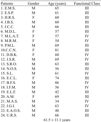
The patients were between 34 and 86 years old, average 61.5 ± 11.1 years old, eight (33%) were female and sixteen (67%) males, according to Table 1. All patients were on maximum allowable drug therapy.
Concerning etiology, idiopathic dilated cardiomyopathy, valvular diseases from mild to severe functional impairment and ischemic cardiomyopathy have been demonstrated.
Table 1. General data of patients.
Patients 1. E.M.S. 2. E.S.P. 3. H.R.S. 4. J.B.S. 5. J.C.C. 6. M.D.L. 7. M.L.A.T. 8. M.R.M. 9. P.M.L. 10.C.C.N. 11. D.B.K. 12. J.S.R. 13. S.R.O. 14. N.O.S. 15. S.L. 16. E.C.L. 17. B.F.S. 18. I.F.M. 19. E.L.F. 20. A.M. 21. M.A.S. 22. J.G.I. 23. E.A.O.S. 24. U.R.S.
Gender M M F M M F F F M F F M M M M F M M M F M M M M
Age (years) 65 61 60 60 66 57 57 54 69 81 60 69 63 56 61 74 86 56 42 69 34 63 48 66 61.5 ± 11.1 years
Functional Class III III III III III III IV IV III III IV IV III III IV III IV IV III III IV IV III III
Inclusion Criteria
The inclusion criteria were: 1) patients with dilated cardiomyopathy (DCM) and congestive heart failure functional class (NYHA) III or IV refractory to optimized drug therapy; 2) patients with DCM without possibility of surgery (coronary artery bypass grafting, valve replacement, left ventricular aneurysm resection or correction of congenital heart disease); 3) left and right bundle branch block standard interventricular conduction disturbance, associated with anterosuperior or left branch induced right ventricular cardiac pacing exclusive block; 4) interventricular dyssynchrony documented on echocardiography; 5) QRS complex duration higher or equal to 120 ms.
Echocardiographic assessment
Echocardiographic evaluation was performed by the same examiner preoperatively and postoperatively with Medson EX 8008 Apparatus, transducers of frequency 2 and 3 MHz in conventional evaluation plans.
The study used the echocardiographic data with, a maximum of one month before the pacemaker implantation multisite to evaluation before, and after six months for postoperative evaluation, shown respectively on Tables 2 and 3. The following variables were used: Left Ventricular Diastolic Diameter, Left Ventricular Systolic Diameter and Ejection Fraction, in the standardized methods in the literature.
Operative technique
The operative technique for implantation of cardiac resynchronizer starts with cardiac monitoring and degermation and antisepsis of the anterior chest wall and neck. Local anesthesia is performed with 2% lidocaine in an area located in the middle third of the right infraclavicular fossa, of approximately 4 cm2. An incision
of approximately 4 cm at the place above 1 cm below the right clavicle is made, with careful hemostasis.
The irst electrode was placed in the right ventricle (RV), followed by the right atrium lead, both by puncture of the right subclavian vein in which the guide wires were inserted, and on them, inserting of the sheaths through which the endocavitary electrodes were passed (active ixation). They were placed in traditional sites (atrium into the right auricle and right ventricle at the end of it) or where the initiation of stimulation was obtained, with acceptable sensing and impedance. The last electrode was placed in the coronary sinus for left ventricular pacing. The electrodes used were: Corox OTW 75 DP (Biotronik®).
The position obtained by the use of radiological anatomy with luoroscopy in left anterior oblique, at an angle of 35 degrees in order to position the electrode of passive ixation on the lateral or posterior vein of the left ventricle. Afterwards, electrophysiological tests took place to verify basic viability and stability. The inal stimulus generator is connected, and it immediately starts its activity veriied by electrocardiogram tracing. Then the closing of plans was held.
Postoperative follow-up
The days of observation and monitoring varied from November 2006 until June 2012. All patients were medicated in order to maintain the optimization of drug therapy for heart failure.
The postoperative evaluation was performed by the Doppler echocardiogram in six months postoperatively.
Statistical analysis
The analysis of quantitative variables was performed by comparing average (previously checked the normality of distributions), using the Student t test. The level of signiicance was P <0.05.
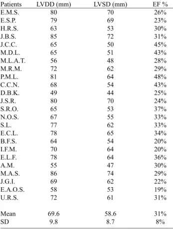
Table 2. Preoperative echocardiographic assessment of patients who underwent implantation of cardiac resynchronizer.
Patients E.M.S. E.S.P. H.R.S. J.B.S. J.C.C. M.D.L. M.L.A.T. M.R.M. P.M.L. C.C.N. D.B.K. J.S.R. S.R.O. N.O.S. S.L. E.C.L. B.F.S. I.F.M. E.L.F. A.M. M.A.S. J.G.I. E.A.O.S. U.R.S.
Mean SD
LVDD (mm) 80 79 63 85 65 65 56 72 81 68 49 80 65 67 77 78 64 70 78 55 86 69 58 72
69.6 9.8
LVSD (mm) 70 69 53 72 50 51 48 62 64 54 44 70 53 55 62 65 54 64 64 47 74 62 53 61
58.6 8.7
EF % 26% 23% 30% 31% 45% 43% 28% 29% 48% 43% 25% 24% 37% 33% 33% 34% 20% 20% 36% 30% 29% 22% 19% 31%
31% 8%
RESULTS
The patients were discharged without complications during the perioperative and no death on the early postoperative.
According to the needs of each patient, there was drug use in their highest tolerated doses, with frequent use of digoxin, furosemide, carvedilol, acetylsalicylic acid (AAS), amiodarone, enalapril, losartan, warfarin and espironalactona.
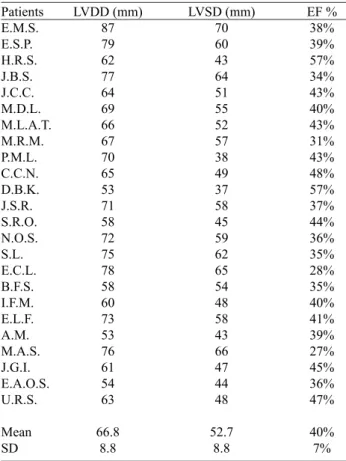
Regarding postoperative data, according to Table 3, the mean left ventricular diastolic diameters were 66.8 ± 8.8 mm, systolic diameters, 52.7 ± 8.8 mm and ejection fraction 40 ± 7%.
Comparing the pre-and postoperatively data, according to Table 4, there was signiicant reduction in echocardiographic parameters and increase in the analyzed ventricular function. The average left ventricular diastolic diameter decreased from 69.6 ± 9.8 mm to 66.8 ± 8.8 mm, systolic diameters of 58.6 mm ± 8.8 to 52.7 ± 8.8 mm and there was an increase
Table 4. Preoperative and postoperative echocardiographic assessment of patients who underwent implantation of cardiac resynchronizer.
Parameters LVDD LVSD EF%
Preoperative 69.6± 9.8 MM 58,6 ± 8,8 MM
31 ± 8%
Postoperative 66.8 ± 8.8 MM 52,7 ± 8,8 MM
40 ± 7%
SD 0.019 0.0004 0.0002
LVDD: left ventricle diastolic diameter; LVSD: left ventricle systolic diameter; EF: ejection fraction; mm: millimeters; SD: standard deviation
Table 3. Postoperative echocardiographic assessment of patients who underwent implantation of cardiac resynchronizer
Patients E.M.S. E.S.P. H.R.S. J.B.S. J.C.C. M.D.L. M.L.A.T. M.R.M. P.M.L. C.C.N. D.B.K. J.S.R. S.R.O. N.O.S. S.L. E.C.L. B.F.S. I.F.M. E.L.F. A.M. M.A.S. J.G.I. E.A.O.S. U.R.S.
Mean SD
LVDD (mm) 87 79 62 77 64 69 66 67 70 65 53 71 58 72 75 78 58 60 73 53 76 61 54 63
66.8 8.8
LVSD (mm) 70 60 43 64 51 55 52 57 38 49 37 58 45 59 62 65 54 48 58 43 66 47 44 48
52.7 8.8
EF % 38% 39% 57% 34% 43% 40% 43% 31% 43% 48% 57% 37% 44% 36% 35% 28% 35% 40% 41% 39% 27% 45% 36% 47%
40% 7%
LVDD: left ventricle diastolic diameter; LVSD: left ventricle systolic diameter; EF: ejection fraction; mm: millimeters; SD: standard deviation
in ejection fraction, an average of 31% ± 8 to 40 ± 7% signiicance level, respectively, 0.019, 0.0004 and 0.0002, statistically signiicant with a signiicance level of 0.05.
DISCUSSION
Heart failure is a serious public health problem, with high annual mortality [25]. In order to decrease the symptoms, complications and increase life expectancy, several forms of treatment are considered, such as pharmacological and nonpharmacological steps with major therapeutic advances in recent decades. However, many patients remain with signiicant symptoms and a high number of hospital admissions, determining reserved prognosis and expensive treatments [26,27]. Where surgical treatment should be considered.
The cardiac resynchronization therapy (CRT) has an important role in the treatment of patients with advanced heart failure with conduction abnormalities and prolongation of the QRS complex, as main representative the left bundle branch block (LBBB), present in approximately 25-50% patients [14,15,28,29].
Studies have shown the medical beneits of CRT. The tests MIRACLE [30] and MUSTIC [31], CONTAK CD [32] showed that CRT determines functional class improvement of HF patients, exercise tolerance (6 minutes walking test peak VO2), reduction in the rate of HF hospitalization and improved quality of life through the Minnesota questionnaire. However, decrease in mortality with CRT and echocardiographic parameters improving were not demonstrated.
A meta-analysis of the CARE-HF [33], MUSTIC [31] and MIRACLE [30] studies, comparing biventricular pacing with optimized medical treatment alone showed a signiicant reduction in hospitalization rates, reducing the risk of death.
increase in ejection fraction was observed, improving quality of life for the Minnesota questionnaire and improvement in functional class.
However, some studies have shown that CRT was able to signiicantly improve as from the third month of follow-up echocardiographic parameters, such as reduced stroke volume and left ventricular end-diastolic and increasing of ejection fraction [26-28].
In contrast with the controversy in the literature regarding the improvement in echocardiographic parameters, this research has demonstrated an increase in ejection fraction of 31% ± 8 to 40 ± 7%, reducing of diastolic and left ventricular systolic diameter from 69.6 ± 9.8 mm to 66.8 ± 8.8 mm and 58.6 mm ± 8.9 to 52.7 ± 8.8 mm, respectively, all parameters statistically signiicant.
This research may infer that transvenous CRT improves beneits in the biventricular systolic function with signiicant improvement in echocardiographic parameters such as ejection fraction and reduction of systolic and diastolic diameters.
CONCLUSION
In the present study, there was improvement in left ventricular function assessed by echocardiography in patients with advanced heart failure who underwent implantation of cardiac resynchronizer.
REFERENCES
1. McAlister FA, Teo KK, Taher M, Montague TJ, Humen D, Cheung L. Insights into the contemporary epidemiology and outpatient management of congestive heart failure. Am Heart J. 1999;138(1 Pt 1):87-94.
2. Abraham WT, Fisher WG, Smith AL, Delurgio DB, Leon AR, Loh E, et al; MIRACLE Study Group. Multicenter InSync Randomized Clinical Evaluation. Cardiac resynchronization in chronic heart failure. N Engl J Med. 2002;346(24):1845-53.
3. Bocchi EA, Braga FGM, Ferreira SMA, Rohde LEP, Oliveira WA, Almeida DR, et al. Sociedade Brasileira de Cardiologia.
III Diretriz Brasileira de Insuiciência Cardíaca Crônica. Arq
Bras Cardiol. 2009;93(1 supl.1):1-71.
4. Dickstein K, Cohen-Solal A, Filippatos G, McMurray JJ, Ponikowski P, Poole-Wilson PA, et al; ESC Committee for Practice Guidelines (CPG). ESC guidelines for the diagnosis and treatment of acute and chronic heart failure 2008: the Task Force for the diagnosis and treatment of acute and chronic heart failure 2008 of the European Society of Cardiology. Developed in collaboration with the Heart Failure Association of the ESC (HFA) and endorsed by the European Society of Intensive Care Medicine (ESICM). Eur J Heart Fail. 2008;10(10):933-89.
5. Remme WJ, Swedberg K; European Society of Cardiology. Comprehensive guidelines for the diagnosis and treatment of chronic heart failure. Task force for the diagnosis and treatment of chronic heart failure of the European Society of Cardiology. Eur J Heart Fail. 2002;4(1):11-22.
6. Hunt SA, Baker DW, Chin MH, Cinquegrani MP, Feldman AM, Francis GS, et al; American College of Cardiology/ American Heart Association. ACC/AHA guidelines for the evaluation and management of chronic heart failure in the
adult: executive summary. A report of the American College
of Cardiology/American Heart Association Task Force on Practice Guidelines (Committee to revise the 1995 Guidelines for the Evaluation and Management of Heart Failure). J Am Coll Cardiol. 2001;38(7):2101-13.
7. Stolf NAG, Jatene AD. História do transplante cardíaco. Rev
Soc Cardiol Estado de São Paulo. 1995;5(6):609-13.
8. Taylor DO, Edwards LB, Boucek MM, Trulock EP, Deng MC, Keck BM, et al. Registry of the International Society
for Heart and Lung Transplantation: twenty-second oficial
adult heart transplant report-2005. J Heart Lung Transplant. 2005;24(8):945-55.
9. Benfatti RA, Pontes JC, Gomes OM, Dias AE, Gomes Júnior JF, Gardenal N, et al. Mitral valve replacement with crossed
papillopexy and annular constriction in heart failure patients.
Rev Bras Cir Cardiovasc. 2008;23(3): 372-7.
10. Horwich T, Foster E, De Marco T, Tseng Z, Saxon L. Effects
of resynchronization therapy on cardiac function in pacemaker patients “upgraded” to biventricular devices. J Cardiovasc Electrophysiol. 2004;15(11):1284-9.
11. Leclercq C, Hare JM. Ventricular resynchronization: current state of the art. Circulation. 2004;109(3):296-9.
12. Silva CES, Barretto ACP. Avaliação ecocardiográfica da
terapia de ressincronização cardíaca. Arq Bras Cardiol.
2005;84(6):503-7.
13. Veiga VC, Abensur H, Rojas SS. Echocardiography in cardiac resynchronization therapy. Arq Bras Cardiol. 2009;93(3):441-5.
14. Aaronson KD, Schwartz JS, Chen TM, Wong KL, Goin JE, Mancini DM. Development and prospective validation of
a clinical index to predict survival in ambulatory patients
referred for cardiac transplant evaluation. Circulation. 1997;95(12):2660-7.
15. Filho MM. Terapia de ressincronização cardíaca (TRC). Jornal
Diagnósticos em Cardiologia. 38a Ed. Nov/Dez 2008.
16. Linde C, Leclercq C, Rex S, Garrigue S, Lavergne T, Cazeau S, et al. Long-term beneits of biventricular pacing in congestive
17. Reuter S, Garrigue S, Bordachar P, Hocini M, Jaïs P, Haïssagueree M, et al. Intermediate-term results of biventricular pacing in heart failure: correlation between clinical and hemodynamic data. Pacing Clin Electrophysiol. 2000;23(11 Pt 2):1713-7.
18. Cazeau S, Leclercq C, Lavergne T, Walker S, Varma C, Linde C, et al. Effects of multisite biventricular pacing in patients with heart failure and intraventricular conduction delay. N Engl J Med. 2001;344(12):873-80.
19. Bakker PF, Meijburg HW, de Vries JW, Mower MM, Thomas AC, Hull ML, et al. Biventricular pacing in end-stage heart failure improves functional capacity and left ventricular function. J Interv Card Electrophysiol. 2000;4(2):395-404.
20. Soares MJF, Oliveira MAB, Braile DM. Marcapasso multisítio no tratamento da insuiciência cardíaca: evolução e resultados.
Reblampa. 2007;20(1):31-5.
21. Kawaguchi M, Murabayashi T, Fetics BJ, Nelson GS, Samejima H, Nevo E, et al. Quantitation of basal dyssynchrony and acute resynchronization from left or biventricular pacing by novel echo-contrast variability imaging. J Am Coll Cardiol. 2002;39(12):2052-8.
22. Breithardt OA, Stellbrink C, Herbots L, Claus P, Sinha AM, Bijnens B, et al. Cardiac resynchronization therapy can reverse abnormal myocardial strain distribution in patients with heart failure and left bundle branch block. J Am Coll Cardiol. 2003;42(3):486-94.
23. Breithardt OA, Stellbrink C, Kramer AP, Sinha AM, Franke A,
Salo R, et al. Echocardiographic quantiication of left ventricular asynchrony predicts an acute hemodynamic beneit of cardiac
resynchronization therapy. J Am Coll Cardiol. 2002;40(3):536-45.
24. Yu CM, Fung WH, Lin H, Zhang Q, Sanderson JE, Lau CP. Predictors of left ventricular reverse remodeling after cardiac resynchronization therapy for heart failure secondary to idiopathic dilated or ischemic cardiomyopathy. Am J Cardiol. 2003;91(6):684-8.
25. McAlister FA, Teo KK, Taher M, Montague TJ, Humen D, Cheung L. Insights into the contemporary epidemiology and outpatient management of congestive heart failure. Am Heart J. 1999;138(1 Pt 1):87-94.
26. Auricchio A, Abraham WT. Cardiac resynchronization therapy: current state of the art: cost versus benefit. Circulation. 2004;109(3):300-7.
27. Kadish A, Mehra M. Heart failure devices: implantable
cardioverter-deibrillators and biventricular pacing therapy.
Circulation. 2005;111(24):3327-35.
28. Souza FSO, Braile DM, Vieira RW, Rojas SO, Mortati NL, Rabelo AC, et al. Aspectos técnicos do implante de eletrodo para estimulação ventricular esquerda através do seio coronariano, com a utilização de anatomia radiológica e eletrograma intracavitário, na terapia de ressincronização
cardíaca. Rev Bras Cir Cardiovasc. 2005;20(3):301-9.
29. Kalil C, Nery PB, Bartholomay E, Albuquerque LC.
Tratamento com cardioversor-desibrilador implantável e ressincronização cardíaca: isolados ou associados? Rev Bras
Cir Cardiovasc. 2006;21(1):85-91.
30. Abraham WT, Fisher WG, Smith AL, Delurgio DB, Leon AR, Loh E, et al. Cardiac resynchronization in chronic heart failure. N Engl J Med. 2002;346(24):1845-53.
31. Cazeau S, Leclercq C, Lavergne T, Walker S, Varma C, Linde C, et al; Multisite Stimulation in Cardiomyopathies (MUSTIC) Study Investigators. Effects of multisite biventricular pacing in patients with heart failure and intraventricular conduction delay. N Engl J Med. 2001;344(12):873-80.
32. Auricchio A, Stellbrink C, Sack S, Block M, Vogt J, Bakker P, et al. Long-term clinical effect of hemodynamically optimized cardiac resynchronization therapy in patients with heart failure and ventricular conduction delay. J Am Coll Cardiol. 2002;39(12):2026-33.
33. Bristow MR, Saxon LA, Boehmer J, Krueger S, Kass DA,
De Marco T, et al. Cardiac-resynchronization therapy with or
without an implantable deibrillator in advanced chronic heart
failure. N Engl J Med. 2004;350(21):2140-50.