Drug-Resistant Genotypes and Multi-Clonality in
Plasmodium falciparum
Analysed by Direct Genome
Sequencing from Peripheral Blood of Malaria Patients
Timothy Robinson1., Susana G. Campino2., Sarah Auburn2,3
, Samuel A. Assefa2, Spencer D. Polley4, Magnus Manske2, Bronwyn MacInnis1,2, Kirk A. Rockett1,2, Gareth L. Maslen2, Mandy Sanders2,
Michael A. Quail2, Peter L. Chiodini4,5, Dominic P. Kwiatkowski1,2, Taane G. Clark5, Colin J. Sutherland4,5* 1Wellcome Trust Centre for Human Genetics, University of Oxford, Oxford, United Kingdom,2Wellcome Trust Sanger Institute, Hinxton, United Kingdom,3Global Health Division, Menzies School of Health Research, Charles Darwin University, Darwin, Australia,4Department of Clinical Parasitology, Hospital for Tropical Diseases, London, United Kingdom,5Faculties of Infectious and Tropical Diseases and Epidemiology and Population Health, London School of Hygiene & Tropical Medicine, London, United Kingdom
Abstract
Naturally acquired blood-stage infections of the malaria parasitePlasmodium falciparumtypically harbour multiple haploid clones. The apparent number of clones observed in any single infection depends on the diversity of the polymorphic markers used for the analysis, and the relative abundance of rare clones, which frequently fail to be detected among PCR products derived from numerically dominant clones. However, minority clones are of clinical interest as they may harbour genes conferring drug resistance, leading to enhanced survival after treatment and the possibility of subsequent therapeutic failure. We deployed new generation sequencing to derive genome data for five non-propagated parasite isolates taken directly from 4 different patients treated for clinical malaria in a UK hospital. Analysis of depth of coverage and length of sequence intervals between paired reads identified both previously described and novel gene deletions and amplifications. Full-length sequence data was extracted for 6 loci considered to be under selection by antimalarial drugs, and both known and previously unknown amino acid substitutions were identified. Full mitochondrial genomes were extracted from the sequencing data for each isolate, and these are compared against a panel of polymorphic sites derived from published or unpublished but publicly available data. Finally, genome-wide analysis of clone multiplicity was performed, and the number of infecting parasite clones estimated for each isolate. Each patient harboured at least 3 clones
ofP. falciparumby this analysis, consistent with results obtained with conventional PCR analysis of polymorphic merozoite
antigen loci. We conclude that genome sequencing of peripheral bloodP. falciparumtaken directly from malaria patients provides high quality data useful for drug resistance studies, genomic structural analyses and population genetics, and also robustly represents clonal multiplicity.
Citation:Robinson T, Campino SG, Auburn S, Assefa SA, Polley SD, et al. (2011) Drug-Resistant Genotypes and Multi-Clonality inPlasmodium falciparumAnalysed by Direct Genome Sequencing from Peripheral Blood of Malaria Patients. PLoS ONE 6(8): e23204. doi:10.1371/journal.pone.0023204
Editor:George Dimopoulos, Johns Hopkins School of Public Health, United States of America
ReceivedApril 1, 2011;AcceptedJuly 8, 2011;PublishedAugust 11, 2011
Copyright:ß2011 Robinson et al. This is an open-access article distributed under the terms of the Creative Commons Attribution License, which permits unrestricted use, distribution, and reproduction in any medium, provided the original author and source are credited.
Funding:SDP is supported by the Foundation for Innovative New Diagnostics; PLC is supported by the UCL Hospitals Comprehensive Biomedical Research Centre for Infection; CJS is supported by the United Kingdom Health Protection Agency. Genome sequencing and the development of software for sequence analysis was funded by the Wellcome Trust core funding to the Sanger Institute (Wellcome 077012/Z/05/Z) and the MRC Centre for Genomics and Global Health (MRC G0600718). None of these funding agencies had any direct role in the design or conduct of the study. The funders had no role in study design, data collection and analysis, decision to publish, or preparation of the manuscript.
Competing Interests:The authors have declared that no competing interests exist.
* E-mail: [email protected]
.These authors contributed equally to this work.
Introduction
Naturally acquired blood-stage infections of the malaria parasite
Plasmodium falciparum typically harbour multiple haploid clones. Different parasite clones may vary significantly in immunogenicity, immune-avoidance mechanisms, susceptibility to drugs, and
transmissibility by different Anophelesmosquito vector species [1–
3]. The polyclonality and diversity of malarial infections together present a major barrier to vaccine development [4,5]. The dif-ferent parasite genotypes present in a single infection can be identified by analysis of polymorphic genetic loci, such as the
merozoite surface protein genes msp1 and msp2, amplified from
peripheral blood samples of infected individuals [3,6]. The
apparent number of clones observed in any single infection thus depends on the diversity [in that individual) of the polymorphic marker used for the analysis, and the relative abundance of rare clones, which often fail to be detected among PCR products derived from numerically dominant clones. However, minority clones are of clinical interest as they may harbour genes conferring drug resistance and thus be selected by treatment, causing therapeutic failure [7,8], or express antigenic variants unaffected by vaccine-elicited immunity [9].
application of clone analysis in malaria infections is in so-called ‘‘PCR correction’’ of recurrent infections in clinical trials of anti-malarial therapy. This can be confounded by the selective emergence after antimalarial treatment of minority clones in the pre-treatment parasite population [20]. Commonly used PCR methods share the limitation that such minority clones are under-represented or may be absent among assay products. This leads to mis-classification of PCR data in clinical trials, and underestima-tion of the extent of allelic polymorphism in any given infecunderestima-tion [8,21–24].
An alternative approach to investigating polyclonality in P.
falciparuminfections is adaptation of patient isolates to culture, and use of cloning and molecular genotyping techniques to analyse
multiplicityin vitro. However, a recent study by Nsobyaet al.[25],
using isolates from Ugandan malaria patients propagatedin vitro,
demonstrated that there are significant inter-genotype differences in the ability to survive in even short-term cultures, leading to a
rapid skewing of the parasite populationin vitro, and an overall loss
of complexity. Thus techniques which avoid expansion of parasite
material, whether by polymerase chain reaction or by in vitro
expansion, are more likely to preserve the complexity and relative abundance of different genotypes in patient isolates of interest.
The recent development of new generation direct sequencing technologies, capable of elucidating whole-genome data from relatively small biological samples, provides a potential new approach to investigate polyclonality in malaria infections. These technologies fractionate DNA samples into random end-tagged
fragments of a uniform size, which are amplifiedin situon a solid
matrix, and then record the sequence of base addition to each growing amplicon. This produces a large number of short, but ‘‘massively’’ parallel sequence (MPS) reads which permit assembly of a partial or full genome provided an established reference genome sequence is available, and sufficient depth (number of reads at each nucleotide position) and breadth (proportion of the genome amplified and sequenced) of coverage are achieved. The single molecule sequencing approach of MPS technologies ensures that each sequence read (or pair of sequence reads if both ends of the molecule are sequenced) is essentially a haplotype, providing great scope for the characterisation of polyclonal infections. Using MPS, it is now possible to derive genome sequence data from a small volume of material from any organism of interest. Although assembly of extended genomic sequences is facilitated by the use of
existing reference sequence, de novoor reference-free approaches
are becoming more widely used [26]. MPS is now being assessed
as a method to examine genome-wide polymorphism in P.
falciparum, using both parasites culturedin vitroand material taken directly from the peripheral blood of people infected with the parasite [27]. However, the AT-rich genome of malaria parasites poses particular challenges for this approach to genome assembly [28], and thus it is unclear how well MPS will perform in analysis ofP. falciparumtaken directly from patients, particularly as natural infections commonly carry multiple clones with distinct genotypes at polymorphic loci.
In this study we use MPS to derive genome-level sequence data
for five P. falciparum parasite isolates prepared directly from
peripheral blood of four malaria patients, after minimal or no amplification of the parasite genome [28]. To evaluate the fidelity of the sequence data generated, and its utility for studies of genomic variation, we first examined structural differences among our isolates by global scanning for copy number variants (CNV). We then assessed sequence diversity at known polymorphic sites among six genes considered to be under strong selective pressure from antimalarial therapy, and among full-length mitochondrial sequences derived from each isolate. Finally, a genome-wide
analysis of multiplicity was performed using other loci, selected empirically, which provided robust estimates of genotype multi-plicity in each patient. These results were compared to con-ventional assessments of polyclonality using polymorphic loci encoding merozoite surface antigens.
Methods
Sample collection
Samples (OX001, OX003, OX005A, OX005B, OX006) were collected from returning travellers attending the Hospital for Tropical Diseases (HTD), or a referring hospital, with malaria symptoms, who were diagnosed positive by malaria films examined in the Department of Clinical Parasitology, and who gave written consent to have their blood samples analysed under a protocol approved by the University College London Hospitals Research Ethics Committee (project ref no. 07/0055). A minimum parasite density of 1% of infected erythrocytes was set as a cut-off to ensure DNA preparations included a high parasite to human white blood cell ratio.
Processing of blood samples
EDTA blood remaining after diagnosis (2–4 mL) was prepared for genomic DNA extraction by removal of human white blood cells within four hours of venesection. Briefly, blood was diluted two to three-fold in phosphate-buffered saline, pH 7.2 (PBS),
layered on to a 2 ml cushion of LymphoprepH(Axis-Shield, UK),
and centrifuged for 30 minutes at 4,000 g in a bench-top centrifuge at room temperature. Upper layers, including leuko-cytes, were removed, and the erythrocyte pellet was washed once in PBS and re-suspended to a total volume of 5 ml with PBS. The
erythrocyte suspension was passed once through a PlasmodipuTM
filter (Euro-Diagnostica), resuspended in 5 ml PBS and pelleted at 4000 g for 10 minutes. The erythrocyte pellet was stored frozen at
220uC, pending DNA extraction using the Qiagen DNA Blood
Maxi (Qiagen, Crawley, UK) extraction kit.
Purified DNA samples were taken to the Wellcome Trust Sanger Institute (WTSI) malaria labs and prepared for MPS sequencing as described elsewhere [27,28]. In parallel, each DNA sample was analysed for multiplicity using a standard nested PCR
method to amplify polymorphic regions ofpfmsp1andpfmsp2[6].
Amplicons were fractionated on agarose gels.
Sequence Analysis
All samples underwent whole genome sequencing on the Illumina Genome Analyzer II platform, as previously described [27]. Briefly, paired 54- or 76-base pair sequence reads were generated on DNA fragments of 200–300bp. The fastq files for each lane of sequence data for each isolate were mapped onto the
P. falciparum3D7 reference genome V2.1.5 (ftp://ftp.sanger.ac.uk/ pub/pathogens/Plasmodium/falciparum/3D7/3D7.version2.1.5/) using the Burrows-Wheeler Aligner (http://bio-bwa.sourceforge.
net), resulting inbamfiles that can be analysed using SAMtools/
nucleotides. All raw sequence data (fastq format) used here are publicly available as fastq files, and as ‘‘Open Access’’ samples (prefix PL) on the WTSI Malaria Programme web-site [29].
Identification of copy number variation
CNV, including deletions and sequence amplifications, may be identified using sequence coverage data. In particular, regions with little or no coverage may be deleted in some or all genomes in the infection, whilst those with coverage in excess of the average may be amplified. A list of small indels from the paired end mapping process with evidence from at least two isolates was constructed using SAM tools with the same Q30 threshold. In particular, small indels are detected by comparing the distance between mapped read pairs to the average insert size of the genomic library [30,31]. For each isolate, we then calculated total coverage at positions in the sequence that were in coding regions and were classified as unique by sliding 50-mers across the reference genome. To further identify regions with CNVs we adopted a similar approach to Yoonet al.[32]. In particular, coverage data were normalised for GC content, and a Z-score statistic calculated using 100-bp windows by subtracting the mean of all windows and dividing by the standard deviation. These Z-scores were converted to p-values
and we report any regions lower than a 1026
threshold. Regions of interest were examined graphically using LookSeq software [33] (available at http://lookseq.sanger.ac.uk/lookseq2/index.html).
Analysis of drug resistance-associated loci
Illumina nucleotide calls were extracted from genome read data at each position for the following six loci, implicated in resistance
to artemisinins or other antimalarials: (i)pfatpase6(PFA0310c, chr
1: 265 447-269 412); (ii)pfmrp1(PFA0590w, chr 1: 465 875–471
344); (iii) pfmdr1 (PFE1150w, chr 5: 957 885-962 144); (iv) pfcrt
(MAL7P1.27, chr 7: 458 990–461 216); (v)pfmrp2(PFL1410c, chr
12: 1 192 877–1 199 204);pfnhe1(P13_0019, chr 13: 170 076–175
991). Sequence from each locus was converted to fasta format after manual removal of introns, and compared to sequence from the other isolates, and the 3D7 reference genome, by alignment in Clustal W (http://www.ebi.ac.uk/Tools). At these loci, coverage depth was typically 40 to 100 reads; multiplicity at a specific site was often observed, indicated by more than one nucleotide being called; a low threshold (2 calls) was set for retaining minor variants in the analysis, irrespective of the total read coverage at that position. Coverage breadth was excellent, being close to 100% for each resistance-associated locus.
Genome-wide screen for minimal multiplicity
For each of our test isolates we inferred the minimal multiplicity by considering the number of distinct haplotypes formed by combinations of three SNP on single or paired reads. We removed any haplotypes that had a frequency of one, and ignored those sets of haplotypes with total frequency (coverage) less than 10, or were located in genomic regions that were not unique (defined using 50-mers, see above).
Results
Patients
Between December 2008 and December 2009, subjects gave consent for genetic studies on parasite isolates derived from the diagnostic blood samples taken as part of their routine care. Five DNA samples from four of these volunteers were deemed suitable for taking into the Illumina DNA sequencing pipeline. For one patient (OX005), two sequential samples, taken at 0 hours (i.e. pre-treatment) and 9 hours after treatment, were analysed in
parallel. All patients were successfully treated and recovered from malaria.
OX001. This patient was a 53-year old Ghanaian who had lived in the UK all their adult life and presented with symptoms of acute malaria after a visit to Accra and the surrounding countryside for a period of six weeks. Having started to feel unwell in Ghana, the patient reported taking one (unidentified) tablet which was bought from a street trader, but reported no other treatment, and did not use chemoprophylaxis.
OX003. This 26-year old Zimbabwean-born Caucasian patient had lived in the UK for four years and reported having malaria 3 times before moving to the UK. The patient presented
with circulating P. falciparum trophozoites and schizonts at a
parasitaemia of 1.3% following a visit to Mozambique (Mapute/ Tete) for a period of three weeks, during which no chemo-prophylaxis or treatment was taken. A single pre-treatment blood sample was analysed.
OX005. Following a visit to family in Ghana, this 49-year-old Ghanaian-born UK resident (of 22 years) presented with severe
malaria and a peripheralP. falciparumparasitaemia of 3.0% (details
presented in reference 34). Two sequential samples, F54840 and F55564, taken prior to treatment and 8 hours later (4 to 5 hours after commencement of quinine treatment), respectively, were analysed.
OX006. This case is also presented in detail in reference 34. Briefly, a 39-year-old European, resident in the UK and with no
previous malaria episodes, presented with severe P. falciparum
hyperparasitaemia (39%) after returning from 10 days in Kenya. One sample, taken approximately 2 hours after commencing intra-venous artesunate therapy, was analysed.
Data for all isolates described are publicly available via the MapSeq portal at the WTSI (http://www.sanger.ac.uk/MapSeq/) and have the following Sanger identifiers within the PF14 study folder: OX001 = PL0001, OX003 = PL0002, OX005 = PL0004, OX006 = PL0005.
Evidence of variation in gene copy number in patient isolates
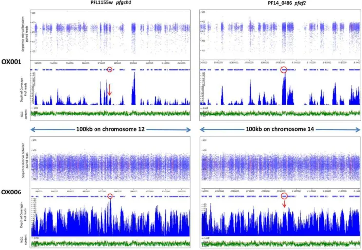
Read coverage, sequence intervals between read pairs and GC content were examined around loci of interest, identified as described in Methods. Two examples of gene amplification are presented in Figure 1, in isolates OX001 and OX006. The richness of data clearly differs between these two isolates, as the former was analysed using an earlier format of the MPS procedure which utilised 54nt reads, whereas 76nt reads were used in analysis
of OX006 (Table 1; Fig. 1). Thepfgch1locus, which has previously
been identified as undergoing amplification in Southeast Asian
Figure 1. Identifying amplifications as areas of high MPS coverage.Length of sequence intervals between paired reads (nt) and coverage (read frequency) are plotted against chromosome position and %GC content for two loci in isolates OX001 and OX006.pfgh1displays high coverage consistent with amplification in OX006, but not OX001.pfef2displays high coverage consistent with amplification in OX001, but not OX006. Loci of interest (red circles) are shown within100 km of genomic context. Red colouring within the read pile-ups signify polymorphic sites within a read in which a non-reference allele is present (i.e. SNP).
parasite isolates [35], showed evidence of excess coverage in isolate OX006, but not in isolate OX001 (Fig. 1), nor in the other 4
isolates (data not shown). The PF14_0486 locus (pfef2) encodes an
EF2 translation elongation factor related to Ef2b isoforms of metazoan eukaryotes, with 65% amino acid sequence identity to
Drosophila melanogaster Ef2b (accession number AY075481). pfef2
displayed high coverage consistent with amplification in isolate OX001, but not in OX006 (Fig. 1).
Evidence of deletion of genomic sequences, defined with respect to the 3D7 reference genome sequence, was found at numerous loci across the genome in each isolate. Figure 2 presents pile-up
graphics [33] for two isoforms ofpfrbp2encoded on chromosome
13 (MAL13P1.176; PF13_0198). The data clearly distinguish between two previously described deletions that are common towards the carboxy terminus of MAL13P1.176, encoding the
b-homologue ofpfrbp2: OX001 and OX005 are to seen to have
Figure 2. Deletions inpfrbp2homologues a and b appear as areas with inflated sequence intervals in four isolates.Two isoforms of RBP2 are encoded by adjacent genes on chromosome 13, arranged head to head and transcribed in opposite directions. 60 kb around these genes are depicted, for four isolates. Loci of interest (red arrows) have either a,600 bp deletion in the carboxy-terminal serine-rich domain of homologue b (red elipses), or a,2–300 bp deletion in the low-complexity protein domain immediately upstream in both genes (blue elipses). Y-axis depicts sequence interval between paired reads. X-axis depicts nucleotide coordinates along the chromosome, as in Figure 1.
apparent read intervals of ,950nt over this domain, which is approximately 600 bp longer than the average read length in that chromosome region (Fig. 2). This fits well with the expected 587 bp deletion in the serine-rich domain, which was recently shown to be common across Africa [35]. In contrast, OX003 and OX004 show evidence of a smaller deletion in the low complexity domain immediately upstream of the serine-rich region. Interest-ingly, this is also seen at the C-terminus of the a-homologue (PF13_0198), which lacks the serine-rich domain and does not exhibit the deletion-polymorphism characteristic of the adjacent b-homologue. Among other loci displaying similar evidence of intra-genic deletion in one or more of our clinical isolates was the
conserved Plasmodium protein (unknown function) PF14_0226 in
which the 12 amino acid repeat motif S(or K or W)TLKEK-KNEMNV occurs in 7 tandem repeats in 3D7, but typically
occurred as 2 to 5 repeats in MPS-derived genome sequences from our isolates. Verification that this observation is not due to ambiguous assembly of repeats could be accomplished simply by amplification and sequencing of this region of the gene.
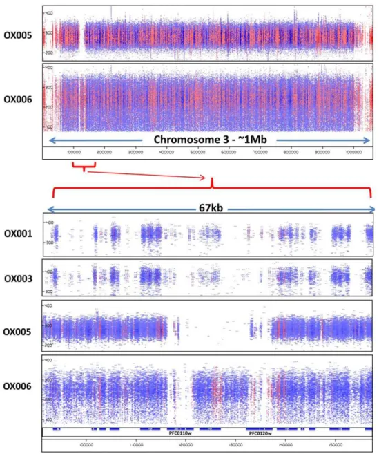
Of particular note was occasional evidence of larger chromo-somal deletions, such as that encompassing one or both of the loci encoding Rh1/Clag3.1 and Rh1/Clag3.2 (PF0110w and PF0120w) at the left end of chromosome 3. In isolates OX005 and OX005A, both genes and the chromosomal region between them appeared to be deleted, totalling over 20 kb of missing chromo-some sequence compared to the reference. This is consistent with reports of variation in the size of this chromosome estimated using
pulse-field gel electrophoresis, with 3D7 carrying a larger 3rd
chromosome than other isolates tested in one study [36]. Thus the
duplication of theclag3locus described in laboratory lines [37] was
Figure 3. Evidence of a major deletion at the right end of chromosome 3 in isolate OX005.Paired reads across the whole of chromosome 3 are presented in pile-up view for two isolates, OX005 and OX006 (upper panel). Y-axis depicts sequence interval between paired reads, and X-axis gives chromosome coordinates as in Figures 1 and 2. A detailed view of,70 kb around theclag3.2andclag3.1loci is also shown for 4 isolates (lower panel). The locus between PFC0110w and PFC0120w is a degeneratevargene lacking a full-length ORF in 3D7 and other parasite sequences in the available databases.
not readily identified in MPS sequence data of our wild isolates.
Interestingly, the PfEMP1 pseudogene between the twoclag3loci
in 3D7 was identified by MPS in some, but not all of our isolates (Fig. 3).
Sequence polymorphism in known loci under drug selection
In order to evaluate the potential of MPS analysis using para-site material taken directly from treated malaria patients for phenotype-genotype association studies, we derived full length data for 6 loci known or considered likely to be under selective pressure from antimalarial drugs (see Methods). Data for all 5 isolates and all 6 loci are presented in Table 2. In each of these genes, known polymorphisms were identified. Both the reference and variant alleles were present in at least one gene from each of the isolates OX001, OX003 and OX006. This indicates that these were polyclonal infections, and that multiple alleles of these genes were present. Both isolates from patient OX005 appeared monoclonal (and identical) at these loci, except for apparent
polyclonality in the repetitive ‘‘hinge’’ region ofpfmdr1and in a
three amino-acid repeat region ofpfmrp2. In both loci, this may
conceivably be caused by ambiguous assembly of highly-repetitive sequences, but we cannot rule out true polyclonality in both these isolates (see further analysis below).
Recent data suggest that the two amino acid changes in theP.
falciparumABC transporter MRP1 observed in isolate OX006 are associated with artemisinin and anti-folate resistance in African studies [38,39], and it is possible that the response to artesunate-treatment of the parasites in this individual may have been delayed
[34]. Examination of the pfmrp1sequence at codon 876 in later
follow-up blood samples from this patient may be instructive, as this locus exhibited mixed wild-type and mutant alleles in the immediately post-treatment sample analysed here. The data
presented onpfmrp2represents the first full-length analyses of this
gene in wild parasites; 8 non-synonymous codons were found in addition to the amino-acid repeat polymorphism at codons 235,
240 and 242. In pfmdr1, which encodes the best characterised
transporter protein in P. falciparum, PgH1, we encountered the
expected polymorphisms at codons 86, 184, 649–654 and 1246. Unexpectedly, our Mozambique isolate also harboured a novel non-synonymous substitution, T496I. This region of the locus is not usually analysed in sequencing studies, and there are no data to suggest whether or not this substitution is relevant to parasite responses to antimalarial treatment.
The less stringent call threshold for SNP used in our analysis of drug-resistance associated loci permitted identification of low frequency amino acid variants at expected positions: for example
in thepfmdr1locus of isolate OX001 at codon 86 there were 46
MPS ‘‘calls’’ for T at nucleotide 256, encoding the resistance-associated tyrosine at this position, and 6 calls for the wild-type nucleotide A, encoding asparagine. Similarly, at codon 1246 there were 42 calls for T at nucleotide 3756, encoding a resistance-associated tyrosine, and 4 calls for G, encoding aspartic acid. Thus, as these low frequency calls generated sequences consistent
with previous studies of the locus, we conclude that they arebona
fideresults, and so caution should be exercised in setting high call
thresholds in MPS studies of potentially polyclonal P. falciparum
isolates. This approach also readily identified substitutions of
interest in the pfdhfr (PFD0830w) and pfdhps (PF08_0095) loci
which are known to modulate parasite sensitivity to the anti-folate drugs (Table 3). All isolates harboured the ‘‘triple mutant’’
haplotype IRNI at codons 51, 59, 108 and 164 of pfdhfr, but
three different haplotypes ofpfdhpswere seen. Only the Kenyan
isolate OX006 harboured the pfdhps 540E mutation associated
with high rates of treatment failure with sulphadoxine-pyrametha-mine, and known to be much more common among travellers returning with malaria from east Africa [40].
In summary, specific analysis from our MPS data of a series of loci implicated in drug response identified expected polymor-phisms in each gene, strongly suggested polyclonality in most if not all isolates, and suggested setting of low call thresholds can assist in identifying low abundance sequences. The analysis identified
previously unknown substitutions in pfmdr1and pfmrp2that now
require evaluation as possible markers of parasite response to antimalarial drugs.
Polymorphism in mitochondrial genome sequences Polymorphic sites in the 5.97 kb mitochondrial genome were derived for each sample by the identification of nucleotides that differed to the reference sequence. As expected the mitochondria had at least ten-fold more coverage than the nuclear genome (Table 1). The full mitochondrial sequence was generated for each patient isolate, and compared against the panel of 32 polymorphic
sites by Joyet al.[41] (Table 3). The parasite isolates were in each
case almost identical to the reference sequence. Three polymor-phisms were found, and two corresponded to sites previously identified by Joy and colleagues. These results indicate that MPS sequencing data derived from uncloned and uncultured parasite material is of a sufficiently high quality for use in phylogenetic studies based on variation in the parasite mitochondrial genome.
Clonal multiplicity estimated from conventional pfmsp2 genotyping
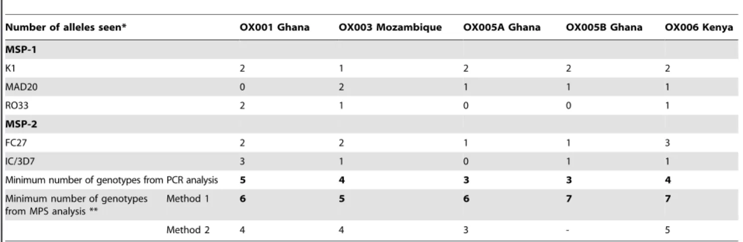
Nested PCR analysis of the pfmsp1 and pfmsp2 locus was
undertaken to estimate the polyclonality of each isolate using conventional methods. The results are presented in Table 4. All isolates except OX005A harboured polyclonal infections as
evidenced by analysis ofpfmsp1andpfmsp2alleles, with five, four,
one, two and six distinct alleles, respectively. It was observed that isolate OX005B, taken 9 hours after initiation of quinine therapy,
harboured an additional P. falciparum genotype not present in
isolate OX005A, taken prior to the first dose.
Genome-wide multiplicity from MPS data
Multiplicity was estimated only for loci in which 3 single-nucleotide polymorphisms (SNP) occurred in close proximity, using two different approaches. The first analysis identified loci
where three SNP were found on a single read of 54 or 76 bp (Table 1). In the second analysis, tri-SNP loci were deployed in which one of the SNP occurred on an independent sequence read
adjacent to the read with the other two SNP (a "2+1’’ SNP trio).
This latter approach could be seen as potentially the more robust, as false 3-SNP haplotypes are less likely to be generated by errors in two adjacent reads, than in a single poor quality sequence read. The first analysis identified 139 polymorphic tri-SNP loci that indicated a multiplicity of 5 genotypes or more across the whole genome in at least one isolate. The majority of these loci did not map to annotated genes in the reference genome, and very few loci displayed high apparent multiplicity in more than one isolate. However, there were clusters of adjacent loci in the same isolate giving high multiplicity estimates, and these suggest the methods deployed were correctly identifying highly polymorphic genomic
regions. In the second more stringent analysis, using ‘‘2+1’’ SNP
trios across paired reads, 52 tri-SNP haplotypes were identified. Multiplicity estimates derived from both methods are compared to those derived from PCR data in Table 5.
Estimates of clonal multiplicity derived from MPS analysis were similar, but slightly higher than those obtained from analysis of
msp1andmsp2alleles by PCR. Therefore, we examined the data
for evidence that the same loci were generating high multiplicity estimates in more than one isolate, and thus might be generally useful tools for clonal multiplicity studies, and also looked for overlap between the two methods used. Two highly repetitive loci contributed to elevated estimates of multiplicity in more than one isolate, but these were only seen when using the first (less stringent) method. The first of these, on chromosome 7, lies in the distal sub-telomeric repeat region, and thus is unlikely to provide an accurate estimate of clone multiplicity. The second locus, within the gene
P10_0265, which encodes a conserved Plasmodium protein of
unknown function, is within a region of open reading frame that is highly repetitive. A tri-nucleotide repeat (CAA/CAG) occurs 67 times in the 3D7 reference sequence, encoding an extensive poly-glutamine tract. Analysis of read-length at this locus [33] con-firmed apparent high multiplicity around these repeat sequences in each isolate, but it is likely this is the result of mapping inconsistencies due to repeat overlaps. (Confirmation of the utility of this locus for estimating multiplicity would require empirical size comparison of appropriate PCR amplicons from different isolates.)
These findings support the use of the more stringent ‘‘2+1’’ SNP
trio approach for identification of multiplicity at polymorphic loci.
Table 3.SNP and inferred haplotypes inpfdhfrandpfdhpsloci.
Isolate ID
Chromosome coordinate Gene ID OX001 OX003 OX005A OX005B OX006
MAL4:755220 PFD0830w T T T T T
MAL4:755243 PFD0830w C C C C C
MAL4:755391 PFD0830w A A A A A
MAL4:755558 PFD0830w A A A A A
DHFR haplotype codons 51_59_108_164 IRNI IRNI IRNI IRNI IRNI
MAL8:550802 PF08_0095 T T G G T
MAL8:550806 PF08_0095 G G G G G
MAL8:551114 PF08_0095 A G A A G
MAL8:551238 PF08_0095 C C C C C
DHPS haplotype codons 436_437_540_581 SGKA SGKA AGKA AGKA SGEA
Discussion
In this study, we have demonstrated that MPS analysis of the
genomes of P. falciparumparasites isolated directly from infected
malaria patients generates high quality data that can be used to identify known and unknown polymorphisms in loci of interest. In particular we demonstrated the utility of this approach to identify genomic sites where both previously described and novel CNV occurred. We were also able to elucidate, directly from MPS data, drug-resistance associated haplotypes of candidate loci, and suc-ceeded in generating high quality mitochondrial genome sequence data suitable for phylogenetic studies. Some of the polymorphisms identified in drug resistance-associated loci and mitochondrial genomes have been previously described, but others were identified here for the first time and can now be investigated in more detail.
The main objective of the study was to investigate the utility of MPS analysis for estimating polyclonality in natural parasite isolates, as accurate estimation of clone multiplicity is important for studies of drug efficacy and parasite population diversity [42,43]. Using a genome-wide approach, reproducible estimates of clonal multiplicity in our clinical isolates were obtained, and these were similar to estimates of clonal multiplicity estimated by standard methods based on well-characterised size and sequence
polymor-phisms in thepfmsp1andpfmsp2genes. Thus we have confirmed by
two completely independent methods a high multiplicity in all of our patients. Multiplicity was also seen in our analysis of drug resistance-associated loci, but as expected of loci under strong directional selection, in each case only two alleles at each position were observed, reflecting lower overall diversity in these genes.
How does a traveller, in many cases only briefly exposed to malaria infection risk, come to harbour multiple parasite clones?
Previous studies have quantified the risk to be one P. falciparum
infection per 1.366105 UK traveller weeks in an endemic area.
This suggests the probability of a single individual receiving more than one infective bite per week is approximately one in one hundred thousand [44,45]. However, as these studies were based on Thai data, it is likely multiple inoculations are more likely in the
African locations where our patients acquired theirP. falciparum
infections: the precise chance that an individual received multiple inoculations will differ depending on the endemicity of infection in the area visited, microheterogeneity in mosquito biting and infection rates, use of and adherence to bite prevention and chemoprophylaxis and the fact that multiple areas of differing risk may have been visited by the same traveller [13]. Nevertheless, the
likelihood is that in most imported cases ofP. falciparuminfection,
the entire clonal repertoire entered the host in a single bite. Thus our patients’ genetically complex parasite infections are expected to reflect the complexity circulating in endemic area human populations; further, as mixed gametocyte genotypes have been shown to circulate in African studies [46,47], recombinant zygotes, heterozygous at polymorphic loci, are expected to be common. Thus an infection with multiplicity 6 must be the product of at least 3 genetically unrelated oocysts, each of which was heterozygous. This oocyst diversity could have built up in the mosquito over a few days as, once infected, mosquitoes are known to continue feeding on subsequent nights [48]. A single female
Anopheles may thus have several developing oocysts at slightly different stages of maturity. This would imply that each sporozoite inoculum may be presenting in a single event the progeny of several blood meals in which the mosquito was infected with a variety of parasite genotypes.
MPS data are well suited to long-range investigation of copy number variations in both coding and non-coding genomic DNA.
Using tools freely available on-line at the WTSI, we were able to identify in our MPS data, almost to the exact base-pair, a
previously described 587 bp deletion in the b-homologue ofpfrbp2
[49]. The evidence for genomic amplification ofpfef2(PF14_0486)
in OX001 is of interest. Although this nuclear locus encodes a eukaryotic-type cytoplasmic translation elongation factor similar to
those of Drosophila, prokaryotic-type EF genes in the Plasmodium
apicoplast genome encode a group of proteins known to be the parasite targets of antibacterial drugs such as clindamycin and doxycycline [50]. It is unknown whether amplification of
cyto-plasmic elongation factor genes such aspfef2might play a role in
parasite drug responses.
An unexpected finding was the evidence suggesting two rhopH1/clag3 genes on chromosome 3, and the var pseudo gene located between them, were deleted in one of our isolates. Paired read data from other isolates was also suggestive of partial or full deletion of the clag sequences. A possible explanation for this is that mistakes in the assembly of MPS end-reads were made due to the fact that these genes belong to a multi-gene family dispersed across the genome at several loci. However, large areas of null read coverage were not common in our dataset, and other members of the clag gene family on chromosomes 2, 7 and 9 were examined and displayed similar or better read coverage than their immediate genomic context. Further alignment of GENBANK sequences for clag3.1 and clag3.2 loci from 3D7, FVO and 7G8 laboratory
strains demonstrated remarkable conservation (.90% sequence
identity at nucleotide level) among these loci. Therefore, extreme polymorphism at these loci is an unlikely explanation for poor read coverage. Given the observations of Kemp et al. that chromosome 3 can vary significantly in size on pulse-field electrophoresis [36] and the evidence of mutually exclusive expression of clag3.1 and clag3.2 proteins in different cultured lines derived from 3D7, suggesting functional redundancy [37], our data may be
inter-preted as a demonstration thatP. falciparum can propagate
per-fectly well in vivo without one or both of the clag loci on
chromosome 3. Unfortunately, genomic material from our
patients was not prepared in such a way as to permit whole chromosome preparations for analysis on pulse-field electropho-resis, in order to verify this interpretation.
Concluding remarks
This study provides proof-of-principle that MPS data from
material obtained directly from malaria-infected Homo sapiens is
suitable for a variety of genomic, phylogenetic, parasitological and clinical analyses. A great strength of MPS in general is the prospect of high-throughput analysis of a large number of genomes in parallel [27]; we have only examined a handful of genomes, and thus have not taken advantage of this aspect of the technology. However, an important feature of our analysis is that we have used well characterised clinical material with follow-up post-treatment DNA samples available, permitting further studies of any locus of interest identified in the pre-treatment isolate. Thus, we have been able to compare the pre-treatment genome sequence, for one of our patients, with the sequence of an isolate taken after 9 hours of antimalarial treatment. It follows that, in some settings, genomic-level follow-up studies of drug selection signals are eminently feasible.
Acknowledgments
We thank the malaria patients at HTD who gave consent for genomic analyses to be carried out on their parasite isolates, and Dr Tom Doherty for helpful discussions.
Author Contributions
Conceived and designed the experiments: TR SGC SA DPK TGC CJS. Performed the experiments: TR SGC SA SAA SDP TGC CJS. Analyzed the data: SGC SA TGC CJS. Contributed reagents/materials/analysis tools: SGC SA MM BM KAR GLM MS MAQ DPK SDP PLC TGC CJS. Wrote the paper: TR TGC CJS. Revised and commented on the manuscript: SGC SA PLC.
References
1. Meyer CG, May J, Arez AP, Gil JP, Do Rosario V (2002) Genetic diversity of Plasmodium falciparum: asexual stages. Trop Med Int Health 7: 395–408.
2. Nicastri E, Paglia MG, Severini C, Ghirga P, Bevilacqua N, et al. (2008) Plasmodium falciparum multiple infections, disease severity and host character-Table 5.Clonal multiplicity estimated from polymorphic amplicon sizes inpfmsp1andpfmsp2PCR assays compared to estimates from MPS analysis.
Number of alleles seen* OX001 Ghana OX003 Mozambique OX005A Ghana OX005B Ghana OX006 Kenya
MSP-1
K1 2 1 2 2 2
MAD20 0 2 1 1 1
RO33 2 1 0 0 1
MSP-2
FC27 2 2 1 1 3
IC/3D7 3 1 0 1 1
Minimum number of genotypes from PCR analysis 5 4 3 3 4
Minimum number of genotypes from MPS analysis **
Method 1 6 5 6 7 7
Method 2 4 4 3 - 5
*Each of the three allelic families occur in a mutually exclusive manner in a singlemsp1gene; similarly for the two allelic families ofmsp1. Thus the minimum number of genotypes is taken as the larger of the allele totals for the two genes.
istics in malaria affected travellers returning from Africa. Travel Med Infect Dis 6: 205–209.
3. Fa¨rnert A, Tengstam K, Palme IB, Bronner U, Lebbad M, et al. (2002) Polyclonal Plasmodium falciparum malaria in travellers and selection of antifolate mutations after proguanil prophylaxis. Am J Trop Med Hyg 66: 487–491.
4. Kilama W, Ntoumi F (2009) Malaria: a research agenda for the eradication era. Lancet 374: 1480–1482.
5. Takala SL, Plowe CV (2009) Genetic diversity and malaria vaccine design, testing and efficacy: preventing and overcoming ‘vaccine resistant malaria’. Parasite Immunol 31: 560–573.
6. Snounou G (2002) Genotyping of Plasmodium spp. Nested PCR Methods Mol Med 72: 103–116.
7. Gadalla NB, Elzaki SE, Mukhtar E, Warhurst DC, El-Sayed B, et al. (2010) Dynamics of pfcrt alleles CVMNK and CVIET in chloroquine-treated Sudanese patients infected with Plasmodium falciparum. Malaria J 9: 74.
8. Sutherland CJ (2008) Comparing highly efficacious antimalarial drugs. PLoS Med 5: e228.
9. Flu¨ck C, Scho¨pflin S, Smith T, Genton B, Alpers MP, et al. (2007) Effect of the malaria vaccine Combination B on merozoite surface antigen 2 diversity. Infect Genet Evol 7: 44–51.
10. Ariey F, Hommel D, Le Scanf C, Duchemin JB, Peneau C, et al. (2001) Association of severe malaria with a specific Plasmodium falciparum genotype in French Guiana. J Infect Dis 184: 237–241.
11. Kimura E, Mattei D, di Santi SM, Scherf A (1990) Genetic diversity in the major merozoite surface antigen of Plasmodium falciparum: high prevalence of a third polymorphic form detected in strains derived from malaria patients. Gene 91: 57–62.
12. Kun JF, Schmidt-Ott RJ, Lehman LG, Lell B, Luckner D, et al. (1998) Merozoite surface antigen 1 and 2 genotypes and rosetting of Plasmodium falciparum in severe and mild malaria in Lambare´ne´, Gabon. Trans R Soc Trop Med Hyg 92: 110–114.
13. Jelinek T, Peyerl-Hoffmann G, Mu¨hlberger N, Wichmann O, Wilhelm M, et al. (2002) Molecular surveillance of drug resistance through imported isolates of Plasmodium falciparum in Europe. Malar J 1: 11.
14. Scherf A, Mattei D, Sarthou JL (1991) Multiple infections and unusual distribution of block 2 of the MSA1 gene of Plasmodium falciparum detected in west African clinical isolates by polymerase chain reaction analysis. Mol Biochem Parasitol 44: 297–299.
15. Kun JFJ, Missinou MA, Lell B, Sovric M, Knoop H, et al. (2002) New emerging Plasmodium falciparum genotypes in children during the transition phase from asymptomatic parasitemia to malaria. Am J Trop Med Hyg 66: 653–658. 16. Ofosu-Okyere A, Mackinnon MJ, Sowa MP, Koram KA, Nkrumah F, et al.
(2001) Novel Plasmodium falciparum clones and rising clone multiplicities are associated with the increase in malaria morbidity in Ghanaian children during the transition into the high transmission season. Parasitology 123: 113–123. 17. Ranjit MR, Das A, Chhotray GP, Das BP, Das BN, et al. (2004) The PfCRT
(K76T) point mutation favours clone multiplicity and disease severity in Plasmodium falciparum infection. Trop Med Int Health 9: 857–861. 18. Hallett RL, Dunyo S, Ord R, Jawara M, Pinder M, et al (2006) Treatment of
malaria in Gambian children with chloroquine plus sulphadoxine-pyrimeth-amine favours survival and transmission to mosquitoes of multi-drug-resistant Plasmodium falciparum. PLoS Clin Trials 1: e15.
19. Sutherland CJ, Alloueche A, McRobert A, Ord A, Leggat J, et al. (2002) Genetic complexity of Plasmodium falciparum gametocytes isolated from the peripheral blood of treated Gambian children. Am J Trop Med Hyg 66: 700–705. 20. Juliano JJ, Gadalla N, Sutherland CJ, Meshnick SR (2010) The perils of PCR:
can we accurately ‘correct’ antimalarial trials? Trends Parasitol 26: 119 - 124. 21. Basco LK, Tahar R, Escalante A (2004) Molecular epidemiology of malaria in
Cameroon. XVIII. Polymorphisms of the Plasmodium falciparum merozoite surface antigen-2 gene in isolates from symptomatic patients. Am J Trop Med Hyg 70: 238–244.
22. Takala SL, Escalante AA, Branch OH, Kariuki S, Biswas S, et al. (2006) Genetic diversity in the Block 2 region of the merozoite surface protein 1 (MSP-1) of Plasmodium falciparum: additional complexity and selection and convergence in fragment size polymorphism. Infect Genet Evol 6: 417–424.
23. Farnert A, Snounou G, Rooth I, Bjorkman A (1997) Daily dynamics of Plasmodium falciparum subpopulations in asymptomatic children in a holoendemic area. Am J Trop Med Hyg 56: 538–547.
24. Kiwanuka GN (2009) Genetic diversity in Plasmodium falciparum merozoite surface protein 1 and 2 coding genes and its implications in malaria epidemiology: a review of published studies from 1997-2007. J Vector Borne Dis 46: 1–12.
25. Nsobya SL, Kiggundu M, Joloba M, Dorsey G, Rosenthal PJ (2008) Complexity of Plasmodium falciparum clinical samples from Uganda during short-term culture. J Infect Dis 198: 1554–1557.
26. Zerbino DR, Birney E (2008) Velvet: algorithms for de novo short read assembly using de Bruijn graphs. Genome Res 18: 821–9.
27. Manske M, Miotto O, Campino S, Auburn S, Almagro-Garcia J, et al. (2010) Next generation sequencing analysis of Plasmodium falciparum diversity within the host and across populations. (Revised version under review).
28. Kozarewa I, Ning Z, Quail MA, Sanders MJ, Berriman M, et al. (2009) Amplification-free Illumina sequencing-library preparation facilitates improved mapping and assembly of (G+C)-biased genomes. Nat Methods 6: 291–295. 29. http://www.sanger.ac.uk/research/projects/malariaprogramme-kwiatkowski/
sequencing.html.
30. Tuzun E, Sharp AJ, Bailey JA, Kaul R, Morrison VA, et al (2005) Fine-scale structural variation of the human genome. Nat Genet 37: 727–732. 31. Korbel JO, Urban AE, Affourtit JP, Godwin B, Grubert F, et al. (2007)
Paired-end mapping reveals extensive structural variation in the human genome. Science 318: 420–426.
32. Yoon S, Xuan Z, Makarov V, Ye K, Sebat J (2009) Sensitive and accurate detection of copy number variants using read depth of coverage. Genome Res 19: 1586–1592.
33. Manske HM, Kwiatkowski DP (2009) LookSeq: a browser-based viewer for deep sequencing data. Genome Res 19: 2125–2132.
34. Beshir K, Hallett RL, Eziefula AC, Bailey R, Watson J, et al (2010) Measuring the efficacy of anti-malarial drugs in vivo: quantitative PCR measurement of parasite clearance. Malaria J 9: 312.
35. Nair S, Miller B, Barends M, Jaidee A, Patel J, et al. (2011) Adaptive copy number evolution in malaria parasites. PLoS Genet 4: e1000243.
36. Kemp DJ, Thompson JK, Walliker D, Corcoran LM (1987) Molecular karyotype of Plasmodium falciparum: Conserved linkage groups and expendable histidine-rich protein genes. Proc Natl Acd Sci USA 84: 7672–7676. 37. Cortes A, Carret C, Kaneko O, Yim Lim BYS, Ivens A, et al (2007) Epigenetic
silencing of Plasmodium falciparum genes linked to erythrocyte invasion. PloS Pathogens 3: 1023–1035.
38. Dahlstro¨m S, Ferreira PE, Veiga MI, Sedighi N, Wiklund L, et al. (2009) Plasmodium falciparum multidrug resistance protein 1 and artemisinin-based combination therapy in Africa. J Infect Dis 200: 1456–1464.
39. Dahlstro¨m S, Veiga MI, Ma˚rtensson A, Bjo¨rkman A, Gil JP (2009) Polymorphism in PfMRP1 (Plasmodium falciparum multidrug resistance protein 1) amino acid 1466 associated with resistance to sulfadoxine-pyrimethamine treatment. Antimicrob Agents Chemother 53: 2553–2556.
40. Sutherland CJ, Fifer H, Pearce RJ, bin Reza F, Nicholas M, et al. (2009) Novel pfdhps haplotypes among imported cases ofPlasmodium falciparummalaria in the UK. Antimicr Agents Chemother 53: 3405–3410.
41. Joy DA, Feng X, Mu J, Furuya T, Chotivanich K, et al (2003) Early origin and recent expansion of Plasmodium falciparum. Science 300: 318–321. 42. Juliano JJ, Porter K, Mwapasa V, Sem R, Rogers WO, et al. (2010) Exposing
malaria in-host diversity and estimating population diversity by capture-recapture using massively parallel pyrosequencing. Proc Natl Acad Sci U S A 107: 20138–20143.
43. Juliano JJ, Kwiek JJ, Cappell K, Mwapasa V, Meshnick SR (2007) Minority-variant pfcrt K76T mutations and chloroquine resistance, Malawi. Emerg Infect Dis 13: 872–877.
44. Druilhe P, Daubersies P, Patarapotikul J, Gentil C, Chene L, et al. (1998) A primary malarial infection is composed of a very wide range of genetically diverse but related parasites. J Clin Invest 101: 2008–2016.
45. Hill DR, Behrens RH, Bradley DJ (1996) The risk of malaria in travellers to Thailand. Trans R Soc Trop Med Hyg 90: 680–681.
46. Babiker HA, Abdel-Wahab A, Ahmed S, Suleiman S, Ranford-Cartwright LC (1999) Detection of low level Plasmodium falciparum gametocytes using reverse transcriptase polymerase chain reaction. Mol Biochem Parasitol 99: 143–148. 47. Sutherland CJ, Alloueche A, McRobert A, Ord A, Leggat J, et al. (2002) Genetic
complexity of Plasmodium falciparum gametocytes isolated from the peripheral blood of treated Gambian children. Am J Trop Med Hyg 66: 700–705. 48. Gouagna LC, Bancone G, Yao F, Yameogo B, Dabire´ KR, et al. (2010) Genetic
variation in human HBB is associated with Plasmodium falciparum transmis-sion. Nat Genet 42: 328–331.
49. Ahouidi AD, Bei AK, Neafsey DE, Sarr O, Volkman S, et al. (2010) Population genetic analysis of large sequence polymorphisms in Plasmodium falciparum blood-stage antigens. Infect Genet Evol 10: 200–206.