wide.(1) Therefore, the number of lung transplant candidates has grown significantly in the last 10 years, whereas the number of organs accepted for transplant is very small. Only 15-20% of the donor lungs available for transplant (donors with brain death) are actually usable. As a result of this disparity, the waiting time for transplan-tation increases, as does mortality among lung transplant candidates on waiting lists. Some European centers have reported that up to 50% of lung transplant candidates die while on waiting lists.(2,3)
In 1983, the first successful lung trans-plant was performed by the Toronto Lung Transplant group. Since then, improvements in the techniques for lung preservation, as well as improvements in postoperative management (intensive care, antibiotic therapy and immuno-suppression), have made lung transplantation a well-established treatment for patients with end-stage lung disease. According to data from the International Society for Heart and Lung Transplantation registries, approximately 2,000 transplants are performed annually
world-Ex vivo lung perfusion: initial Brazilian experience*
Perfusão pulmonar ex vivo: experiência nacional inicial
Paulo Manuel Pêgo-Fernandes, Israel Lopes de Medeiros, Alessandro Wasum Mariani, Flávio Guimarães Fernandes, Fernando do Valle Unterpertinger, Marcos Naoyuki Samano,
Eduardo de Campos Werebe, Fábio Biscegli Jatene
Abstract
In the last 20 years, lung transplantation has become the standard treatment for patients with end-stage lung disease. However, less than 20% of the donor lungs available for transplant are actually usable. This disparity between the growing number of recipients and the small number of donors has resulted in increased mortality among lung transplant candidates on waiting lists. Strategies such as the utilization of organs from marginal donors have proven ineffective in increasing the number of transplants. In 2000, a new method for reconditioning human lungs that had been previously rejected for transplantation was developed in Sweden. We describe our initial experience with ex vivo lung perfusion.
Keywords: Lung transplantation; Organ preservation; Organ preservation solutions; Thoracic surgery.
Resumo
Nos últimos 20 anos, o transplante pulmonar tornou-se o tratamento padrão para algumas pneumopatias graves em estágio terminal. Menos de 20% dos pulmões doados para transplante são realmente utilizados. Essa despro-porção entre o crescente número de candidatos ao transplante pulmonar e o reduzido número de doadores resulta em aumento da mortalidade nas filas de espera. Estratégias, como o uso de órgãos de doadores marginais, não se mostraram efetivas em aumentar o número de transplantes. Em 2000, na Suécia, foi desenvolvido um método novo para recondicionar pulmões humanos rejeitados para transplante. Descrevemos nossa experiência inicial com a perfusão pulmonar ex vivo.
Descritores: Transplante de pulmão; Preservação de órgãos; Soluções para preservação de órgãos; Cirurgia
torácica.
* Study carried out at the Heart Institute, Faculdade de Medicina da Universidade de São Paulo – FMUSP, University of São Paulo School of Medicine – Hospital das Clínicas and at the FMUSP Laboratory for Medical Research 61, São Paulo, Brazil.
Correspondence to: Paulo Manuel Pêgo Fernandes. Avenida Dr. Enéas de Carvalho Aguiar, 44, 2º andar, Bloco 2, Sala 9, Cerqueira César, CEP 05403-900, São Paulo, SP, Brasil.
Tel 55 11 3069-5248. E-mail: [email protected]
Financial support: This study received financial support from the Fundação de Amparo à Pesquisa do Estado de São Paulo (FAPESP, Foundation for the Support of Research in the State of São Paulo), as well as from the companies Farmoterápica and Braile Biomédica.
published a report on six patients who received initially rejected lungs after they were recondi-tioned through AVLP.(9)
Most of the lungs that are not used for trans-plantation are rejected due to poor blood gas analysis results, that is, PaO2 < 300 mmHg, with FiO2 of 100% and positive end-expiratory pres-sure (PEEP) of 5 cmH2O. The solution used in EVLP, marketed under the name Steen Solution® (Vitrolife, Göteborg, Sweden), is a solution of extracellular electrolyte composition that contains albumin and dextran and therefore has high oncotic pressure.(10) Dextran, in addition to increasing oncotic pressure, has an antithrom-botic effect, since it covers the surface of the endothelial cells and platelets. This covering has a beneficial effect on the pulmonary microcir-culation and preserves the alveolar-capillary barrier, reducing the extravasation of water and protein during perfusion.(11) Since it is important that EVLP be performed without edema forma-tion, the temperature is gradually increased until normothermia (37°C) is achieved and the flow is controlled so that pulmonary artery pressure (PAP) does not exceed 20 mmHg.(10) Alveolar recruitment maneuvers (manual ventilation, increased PEEP) can be performed to expand areas of atelectasis.
The success of EVLP in reconditioning rejected lungs, increasing their oxygenation capacity, is due to the displacement and reduc-tion of the alveolar and interstitial edema by the high oncotic pressure of the perfusion solution, Brazilian national data show an even worse
situation. In the state of São Paulo in 2006, only 4% of the donor lungs for transplant were usable. In the same period, the rate at which livers and kidneys available for transplantation were utilized was higher than 90%, according to data from the São Paulo State Department of Heath.(4) This can be explained, in part, by the greater susceptibility of the lungs to the deleterious effects of brain death (hemodynamic instability, endocrine insufficiency, inflammatory response, hypothermia and arrhythmias) and to ICU complications (prolonged intubation, pneu-monia, barotrauma and excessive crystalloid injection).
In recent years, various strategies have been proposed in an attempt to increase the number of transplanted lungs, including living donor transplantation, xenotransplantation and non-heart-beating donor transplantation. However, having been limited to a small number of patients due to technical and ethical limita-tions, these strategies did not have the expected impact on the number of transplants.(5)
Another attempt made by some centers in order to increase the number of usable lungs was the expansion of the criteria used to select the so-called ideal donors. A greater number of marginal donors, that is, donors over 55 years of age, smokers (more than 20 pack-years) and donors with a localized infiltrate on chest X-ray, came to be used. Although many studies have shown similar short-term survival rates, the use of marginal lungs in high-risk recipients, such as those with severe pulmonary hypertension, is associated with higher thirty-day mortality rates.(6) Therefore, this strategy also did not significantly reduce the number of patients on waiting lists.
It became evident that a method that would allow a more accurate assessment of donor lungs and that, at the same time, could improve the quality of such organs was necessary. A new method for ex vivo lung assessment was devel-oped and used for the first time in humans when a lung from a non-hear-beating donor was trans-planted by Steen et al. in Sweden in 2000.(7) Ex vivo lung perfusion (EVLP) can also be used for the reconditioning of marginal lungs and lungs rejected for transplantation, as demonstrated in an experimental study, through the increase in PaO2 after EVLP.(8) In early 2009, the same group
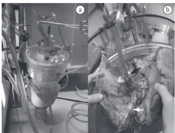
Figure 1 - In a), venous reservoir and membrane
of the left atrium, simplifying the technique and eliminating the need for the special atrial cannula used by those authors. The system is filled with 1500 mL of Steen Solution®. We chose to use the acellular solution, since obtaining human blood for study is not simple. In addition, we were able to reduce the necessary volume of perfusate using oxygenators, reservoirs and pediatric tubes (Braile Biomédica). Through the addition of trometamol (Addex-THAM®; Fresenius-Kabi AB, Uppsala, Sweden), pH is adjusted between 7.35 and 7.45. A flow of 40% of the estimated cardiac output (Qt) is used for the perfusion of both lungs. This value is calculated using the following formula: Qt = 3 × body surface area.
EVLP is initiated with the solution at 20°C and an initial flow of 10% of the calculated value. The temperature is gradually increased up to 37°C within the first 30 min. The flow also increases slowly, reaching 30% of the calculated value within 30 min and reaching the maximum flow (40% × Qt) within 60 min. If PAP is as high as 20 mmHg before the maximum flow is reached, the latter is kept at a lower value. It is imperative that PAP be kept below 20 mmHg in order to minimize edema formation. When the temperature of the perfusate reaches 32°C, ventilation is initiated using a size 8 endotra-cheal tube introduced into the trachea. At this point, a gas (7% CO2 and 93% N2, at a flow rate of approximately 1 L/min) is released into the membrane oxygenator, which will actually deoxygenate the perfusate coming from the pulmonary veins so that the solution that enters the pulmonary artery has the same gas concen-trations as those of the venous blood.
Mechanical ventilation is set at the following parameters: tidal volume between 6 and 8 mL/kg; RR of 7 breaths/min; PEEP of 5 cmH2O; and FiO2 of 100%. When the perfusion flow reaches the maximum calculated value, perfusate samples as well as to the expansion of prior areas of
atel-ectasis.(8,9)
Our research protocol began in January of 2009, after having been approved by the Ethics Committee of the Hospital das Clínicas da Faculdade de Medicina da Universidade de São Paulo (HC-FMUSP, University of São Paulo School of Medicine Hospital das Clínicas) and after an extensive discussion with the HC-FMUSP Organização de Procura de Órgãos (OPO, Organ Procurement Organization), the Santa Casa de São Paulo Hospital OPO and the São Paulo State Department of Health Transplant Center. When a donor is reported, the transplant teams are informed. If the lungs are rejected for transplan-tation (usually due to poor blood gas analysis results), the research consent form is presented to the family members. If authorized, we collect the lungs in the usual manner, together with the other teams. Cold (4°C)Perfadex® (Vitrolife), infused through the pulmonary artery trunk (50 mL/kg), is used as a preservation solution, whereas cold (4°C) saline is poured over the lungs for topical cooling. After removal, the set formed by the two lungs is stored in a cooler and kept at 4°C for 10 h.
After this period, EVLP is initiated (Figure 1). The system is formed by a containment box (Vitrolife), a centrifugal pump (Braile Biomédica, São José do Rio Preto, Brazil), a heat exchanger (Fisics Biofísica, São Paulo, Brazil), a membrane oxygenator (Braile Biomédica) and a venous reservoir. Initially, a cannula (Vitrolife), which has a small tube to be connected to the pressure transducer, is introduced into the pulmonary artery so that PAP is continuously monitored. The solution returning through the pulmonary veins flows directly into the containment box, being drained into the reservoir by the action of gravity. In contrast to what has been reported in one study,(12) we do not use closed cannulation
Table 1 - PaO2 measured in donors (in situ) prior to collection and after (ex vivo) lung perfusion. Donor Age, years Duration of
intubation, days
PaO2,a mmHg PaO
2, mmHg Duration of EVLP,c
min
In situ Ex vivo
1 70 6 92 457 60
2 48 3 365b 463 40
3 60 8 179 480 60
EVLP: ex vivo lung perfusion. aMeasured with a FiO
2 of 100%. bAlthough PaO2 was greater than 300 mmHg, the lungs were
rejected due to the presence of purulent secretion on bronchoscopy. cDuration of ex vivo lung perfusion until blood gases
2. Hornby K, Ross H, Keshavjee S, Rao V, Shemie SD. Non-utilization of hearts and lungs after consent for donation: a Canadian multicentre study. Can J Anaesth. 2006;53(8):831-7.
3. Punch JD, Hayes DH, LaPorte FB, McBride V, Seely MS. Organ donation and utilization in the United States, 1996-2005. Am J Transplant. 2007;7(5 Pt 2):1327-38. 4. Fernandes PM, Samano MN, Junqueira JJ, Waisberg
DR, Noleto GS, Jatene FB. Lung donor profile in the State of São Paulo, Brazil, in 2006. J Bras Pneumol. 2008;34(7):497-505.
5. de Perrot M, Snell GI, Babcock WD, Meyers BF, Patterson G, Hodges TN, et al. Strategies to optimize the use of currently available lung donors. J Heart Lung Transplant. 2004;23(10):1127-34.
6. Pierre AF, Sekine Y, Hutcheon MA, Waddell TK, Keshavjee SH. Marginal donor lungs: a reassessment. J Thorac Cardiovasc Surg. 2002;123(3):421-7; discussion, 427-8.
7. Steen S, Sjöberg T, Pierre L, Liao Q, Eriksson L, Algotsson L. Transplantation of lungs from a non-heart-beating donor. Lancet. 2001;357(9259):825-9.
8. Wierup P, Haraldsson A, Nilsson F, Pierre L, Scherstén H, Silverborn M, et al. Ex vivo evaluation of nonacceptable donor lungs. Ann Thorac Surg. 2006;81(2):460-6. 9. Ingemansson R, Eyjolfsson A, Mared L, Pierre L,
Algotsson L, Ekmehag B, et al. Clinical transplantation of initially rejected donor lungs after reconditioning ex vivo. Ann Thorac Surg. 2009;87(1):255-60.
10. Steen S, Liao Q, Wierup PN, Bolys R, Pierre L, Sjöberg T. Transplantation of lungs from non-heart-beating donors after functional assessment ex vivo. Ann Thorac Surg. 2003;76(1):244-52; discussion 252.
11. de Perrot M, Liu M, Waddell TK, Keshavjee S. Ischemia-reperfusion-induced lung injury. Am J Respir Crit Care Med. 2003;167(4):490-511.
12. Cypel M, Yeung JC, Hirayama S, Rubacha M, Fischer S, Anraku M, et al. Technique for prolonged normothermic ex vivo lung perfusion. J Heart Lung Transplant. 2008;27(12):1319-25.
are collected from the pulmonary veins for blood gas analysis.
In the three cases performed to date, we observed a significant improvement in PaO2 (Table 1). The second case already had a satis-factory PaO2, having been rejected due to the extensive pneumonia.
These three cases represent our initial experi-ence, which was encouraging. After appropriate training, it is not difficult to perform EVLP, and the difference found in the blood gas analysis results indicates a significant improvement in the gas exchange capacity of the organs assessed. We will continue our studies of EVLP in order to gain a better understanding, through assess-ment of pathological anatomy, of how EVLP affects the lung tissue, so that it can soon be applied in clinical practice.
Acknowledgments
We would like to thank Vitrolife, the HC-FMUSP/OPO, the Santa Casa de Misericórdia de São Paulo Hospital OPO and the São Paulo State Department of Health Transplant Center.
References
About the authors
Paulo Manuel Pêgo-Fernandes
Associate Professor, Department of Thoracic Surgery, Faculdade de Medicina da Universidade de São Paulo – FMUSP, University of São Paulo School of Medicine – São Paulo, Brazil.
Israel Lopes de Medeiros
Doctoral Student. Postgraduate Program in Thoracic and Cardiovascular Surgery, Faculdade de Medicina da Universidade de São Paulo – FMUSP, University of São Paulo School of Medicine – São Paulo, Brazil.
Alessandro Wasum Mariani
Intern. Lung Transplant Team of the Heart Institute, Faculdade de Medicina da Universidade de São Paulo – FMUSP, University of São Paulo School of Medicine – Hospital das Clínicas, São Paulo, Brazil.
Flávio Guimarães Fernandes
Medical Student. Faculdade de Medicina da Universidade de São Paulo – FMUSP, University of São Paulo School of Medicine – São Paulo, Brazil.
Fernando do Valle Unterpertinger
Medical Student. Faculdade de Medicina da Universidade de São Paulo – FMUSP, University of São Paulo School of Medicine – São Paulo, Brazil.
Marcos Naoyuki Samano
Attending Physician. Department of Thoracic Surgery, Faculdade de Medicina da Universidade de São Paulo – FMUSP, University of São Paulo School of Medicine – São Paulo, Brazil.
Eduardo de Campos Werebe
Attending Physician. Department of Thoracic Surgery, Faculdade de Medicina da Universidade de São Paulo – FMUSP, University of São Paulo School of Medicine – São Paulo, Brazil.
Fábio Biscegli Jatene