J Bras Pneumol. 2008;34(8):631-634
631
Case Report
slowly progressing anterior mediastinal teratoma. This is an extremely rare case, not only in terms of the presence of this type of tumor in an elderly patient but also in terms of its evolution to malignancy. This, together with the fact that, in the literature, we have found only three studies of carcinoid tumors arising within mediastinal teratomas, has motivated us to write this article.
Case report
A 66-year-old male patient, born in the city of São Luiz (in the state of Maranhão) and a resident of the city of São Paulo (in the state of São Paulo), was admitted to
Introduction
Teratomas are germ cell tumors originating from the three embryonic germ layers. These tumors can be found at multiple sites, and most are benign. They are the second most common tumors of the anterior mediastinum, after thymomas, and are most often seen in young adults.(1) They
account for 8 to 13% of all tumors occurring in this region. If they are treated surgically, the prognosis is quite favo-rable. Although there is little data in the literature regarding the evolution of a teratoma to malignancy, it is known to be an extremely rare occurrence.(2,3)
We report the case of a 66-year-old patient submitted to resection of a carcinoid tumor (revealed by anato-mopathological examination) that had arisen within a
Mediastinal teratoma with malignant degeneration*
Teratoma de mediastino com degeneração maligna
Fabiano Alves Squeff1, Eduardo Salvador Gerace2, Roberto Saad Júnior3,
Márcio Botter4, Roberto Gonçalves5, Juliana Fracalossi Paes6
Abstract
Here, we report the case of a patient with a slowly-progressing anterior mediastinal teratoma submitted to surgical resection. The anatomopathological examination of the sample revealed malignant degeneration to carcinoid tumor. Such evolution is very rare, and we have found only three related studies in the literature. We describe the clinicopathological features of the tumor and discuss the treatment administered. The evolution was satisfactory, and the patient was submitted to oncological treatment.
Keywords: Mediastinal neoplasms; Teratoma; Carcinoid tumor; Mediastinum/surgery.
Resumo
No presente artigo, relatamos o caso de um paciente portador de teratoma de mediastino anterior, de evolução lenta, o qual foi submetido à ressecção cirúrgica. O exame anatomopatológico da peça revelou degeneração maligna para tumor carcinóide. Tal evolução é extremamente rara, sendo encontrados na literatura apenas três artigos correlatos. Apresentamos uma descrição clínico-patológica do tumor e, por fim, discutimos a conduta terapêutica. Houve evolução satisfatória, e o paciente foi submetido a tratamento oncológico.
Descritores: Neoplasias do mediastino; Teratoma; Tumor carcinóide; Mediastino/cirurgia.
* Study carried out in the Department of Thoracic Surgery of the Faculdade de Ciências Médicas da Santa Casa de São Paulo – FCMSCSP, Santa Casa School of Medical Sciences in São Paulo – São Paulo, Brazil.
1. Former Resident in the Department of Thoracic Surgery. Faculdade de Ciências Médicas da Santa Casa de São Paulo – FCMSCSP, Santa Casa School of Medical Sciences in São Paulo – São Paulo, Brazil.
2. Fourth-year Medical Resident in the Department of Thoracic Surgery. Faculdade de Ciências Médicas da Santa Casa de São Paulo – FCMSCSP, Santa Casa School of Medical Sciences in São Paulo – São Paulo, Brazil.
3. Full Professor in the Department of Thoracic Surgery. Faculdade de Ciências Médicas da Santa Casa de São Paulo – FCMSCSP, Santa Casa School of Medical Sciences in São Paulo – São Paulo, Brazil.
4. Instructor in the Department of Thoracic Surgery. Faculdade de Ciências Médicas da Santa Casa de São Paulo – FCMSCSP, Santa Casa School of Medical Sciences in São Paulo – São Paulo, Brazil.
5. Graduate Student in the Department of Surgery. Faculdade de Ciências Médicas da Santa Casa de São Paulo – FCMSCSP, Santa Casa School of Medical Sciences in São Paulo – São Paulo, Brazil.
6. Resident Physician in the Department of Pathology. Faculdade de Ciências Médicas da Santa Casa de São Paulo – FCMSCSP, Santa Casa School of Medical Sciences in São Paulo – São Paulo, Brazil.
Correspondence to: Fabiano Alves Squeff. Rua Senador Olegário Pinto, Quadra 16/L12, Jundiaí, CEP 75110-630, Anápolis, GO, Brasil. Tel 55 62 3324-7614. E-mail: [email protected]
632 Squeff FA, Gerace ES, Saad Júnior R, Botter M, Gonçalves R, Paes JF
J Bras Pneumol. 2008;34(8):631-634
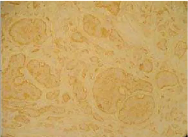
cystic structures, sometimes covered with epidermis with sebaceous glands and hair follicles, sometimes covered with ciliated cylindrical epithelium with a respiratory pattern. Smooth muscle bundles, adipose tissue organized in lobes, lymphoid aggregates, and cartilage were found. In certain areas, there was proliferation of cells with regular nuclei, with salt-and-pepper chromatin. Those cells formed irregular clusters and were positive for chromogranin in the immunohistochemical analysis, which resulted in a diagnosis of a carcinoid tumor arising within a mediastinal cystic teratoma (Figure 3).
The patient underwent adjuvant chemotherapy with cisplatin and bleomycin and, up to postopera-tive month 4, was in good health. There were no signs of disease recurrence, and the levels of tumor markers were normal.
Discussion
In the case reported, the patient had been asymptomatic for ten years, since the first tomog-raphy was performed. When he sought treatment at our facility, we discussed whether it would be neces-sary to perform surgery, since he was asymptomatic, and the radiological imaging results had remained unchanged for ten years. A review of the literature, however, showed that, despite being rare, malignant the outpatient clinic presenting good general
health status. The patient, who had no complaints, also had no history of morbidity or smoking. Radiological imaging, performed at another facility ten years prior, had revealed an anterior mediastinal tumor measuring approximately 4 cm in diameter. Therefore, we performed another round of chest X-ray and computed tomography scans of the chest. The new images revealed a tumor (3.0 x 4.0 cm) with spiculated margins, located in the right ante-rior mediastinum, with the same characteristics found on the scans performed previously (Figure 1). Laboratory test results and pulmonary function test results were normal. Tumors markers were normal. The syndromic diagnosis of anterior mediastinal tumor was confirmed, and surgical treatment was indicated.
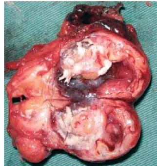
The patient was submitted to a right postero-lateral thoracotomy. During surgery, we observed a yellowish tumor presenting a cystic consistency, measuring 5.0 x 3.5 x 3.0 cm, and weighing 35 g. The tumor had areas of calcification and was located in the anterior mediastinum, on the ante-rior wall of the supeante-rior vena cava, infiltrating the pericardium and the anterior segment of the left upper lobe. Dissection was performed through the opening of the pleura. In addition, partial resection of the pericardium and pulmonary segmentectomy were necessary. The mediastinum was free of lymph node enlargement. The postoperative evolution was favorable and uneventful. Sample sectioning revealed hair and sebaceous secretion within the tumor (Figure 2). Microscopy revealed an embry-onal neoplasm characterized by proliferation of
Figure 1 - Tomography scan of the chest (mediastinal window).
Mediastinal teratoma with malignant degeneration
J Bras Pneumol. 2008;34(8):631-634
633
Histologically, cystic teratomas are character-ized by cystic and solid areas. They can contain teeth, skin, and hair (ectoderm layer); cartilage and bone (mesoderm layer); and bronchial, intestinal, or pancreatic tissue (endoderm layer).(5,8) In the case
reported here, we found cartilage, epidermal tissue, and bone tissue, as well as hair.
The anatomopathological examination of the surgical sample revealed a teratoma that had evolved to malignancy, defined as growth of malignant non-germ cells in a preexisting teratoma. This evolution is rare in gonads, as well as in extragonadal sites, and remains indeterminate. There are reports of its occurrence in patients submitted to chemotherapy or radiotherapy. In dermoid cysts of the ovary, evolu-tion to malignancy is described in approximately 1-2% of cases.(3) In 1987, a case of carcinoid tumor
arising within a mediastinal germ cell neoplasm was described in a patient who had been treated with chemotherapy and radiotherapy.(9)
In the literature, there are few cases docu-menting the evolution of mediastinal teratomas to malignancy. In 1985, three such cases were described: one case of adenocarcinoma, one case of sarcoma, and one case of malignant neuroepithe-lioma.(10) In 1989, there was a report of squamous
cell carcinoma arising from an anterior mediastinal teratoma.(11) In 1994, there was a case of
well-dif-ferentiated adenocarcinoma arising from a mature cystic teratoma of the mediastinum.(12)
In the case reported here, a primary carcinoid tumor was found to have arisen within the teratoma. Such evolution is extremely rare, and there have only three such cases have been published in journals indexed for the Latin American and Caribbean Health Sciences Literature database or for Medline.(1,4,13)
The anatomopathological examination of the sample revealed cells with regular nuclei and a salt-and-pepper pattern within the teratoma. This region presented immunohistochemical positivity for chromogranin, which is characteristic of a carci-noid tumor.(14)
Mature teratomas are usually treated with surgical resection. The most common approach is median sternotomy,(14) although, in the present
case, we opted for right thoracotomy due to the right juxtamediastinal location of the tumor. We had access to the entire tumor. Tumor dissection was performed near the superior vena cava, and the pericardial “tamponade” was removed so that the tumors can arise within teratomas, and we therefore
decided to perform the operation. To our surprise, we encountered a carcinoid tumor, as confirmed through anatomopathological examination.
Mature teratomas are tumors that originate from at least two of the three primary germ cell layers, being most commonly found in children and young adults, with no gender-related differences. They are the most common germ cell tumors, accounting for 75% of all cases, and, after the gonads, the medi-astinum is the site most frequently affected. In the anterior mediastinum, their incidence ranges from 8 to 20%.(2,4) The majority are
anatomopathologi-cally well-differentiated, benign, and slow-growing Therefore, they are usually diagnosed based on an incidental radiological finding, as in the case reported. In adults, approximately two thirds of all cases are asymptomatic.(2,5)
Cystic teratomas, such as that seen in the case reported here, rarely cause symptoms, principally due to their cystic component. When there are symptoms, the most common is chest pain. When there is infec-tion, cyst rupture can occur, causing hemoptysis, pleural empyema, acute respiratory insufficiency, and cardiac tamponade.(1) Expectoration of hair is a
rare but pathognomonic symptom.(6)
The syndromic diagnosis is made through chest X-ray and computed tomography scans of the chest. It presents as an encapsulated tumor with well-de-fined borders and size, containing cystic areas and bone elements. Nuclear magnetic resonance imaging is useful to evaluate the relationship between the mediastinum and the hilar structures.(6,7)
634 Squeff FA, Gerace ES, Saad Júnior R, Botter M, Gonçalves R, Paes JF
J Bras Pneumol. 2008;34(8):631-634
operation could be oncologically curative. One group of authors(15) described a series of 95 resections of
mediastinal teratomas over 50 years. In that long-term study, three patients underwent lobectomy, five underwent partial pulmonary resection, and seven underwent pericardiectomy.
Video-assisted thoracoscopy has been used in the resection of mediastinal teratomas with prom-ising results.(16) In the case reported here, however,
evolution to malignancy, with infiltration of tissues and adjacent organs, would make it difficult to use such access.
Due to the carcinoid tumor, adjuvant treatment was required. The therapeutic effects of irradiation on carcinoid tumors are unknown. Radiotherapy has a palliative effect on symptomatic bone metastases and local disease control. The use of long-acting analogs of somatostatin should be considered when the patient presents carcinoid syndrome and high levels of biochemical markers. One group of authors(17) used routine therapy with intravenous
octreotide preoperatively, intraoperatively, and post-operatively in 82 patients with advanced carcinoid tumors submitted to surgery. None of the patients presented carcinoid syndrome. Carcinoid tumor with lymph node metastasis presents a higher recurrence rate. The efficacy of chemotherapy is controversial, although there are reports that chemotherapy, used in cases of locoregional and distant disease, affects survival.(16,17) The most common cause of death in
such patients is progression to advanced meta-static disease. The overall survival rate in patients receiving aggressive surgical treatment is 50 to 80%. However, where there is metastasis, the five-year survival ranges from only 18 to 30%.(17,18)
References
1. Yetkin U, Orgencalli A, Yuncu G, Gurbuz A. Large mediastinal teratoma originating from the aortic adventitia. Tex Heart Inst J. 2004;31(3):309-12.
2. Castro Jr MA, Rosemberg NP, Castro MA, Castro AP, Wietzycoscki C, Mespaque C. Mediastinal teratoma
mimicking pleural effusion on chest X-rays. J Bras Pneumol. 2007;33(1):113-5.
3. Lancaster KJ, Liang CY, Myers JC, McCabe KM. Goblet cell carcinoid arising in a mature teratoma of the mediastinum. Am J Surg Pathol. 1997;21(1):109-13.
4. Lancaster KJ, Liang CY, Myers JC, McCabe KM. Goblet cell carcinoid arising in a mature teratoma of the mediastinum. Am J Surg Pathol. 1997;21(1):109-13.
5. Zisis C, Rontogianni D, Stratakos G, Voutetakis K, Skevis K, Argiriou M, et al. Teratoma occupying the left hemithorax. World J Surg Oncol. 2005;3:76.
6. Strollo DC, Rosado de Christenson ML, Jett JR. Primary mediastinal tumors. Part 1: tumors of the anterior mediastinum. Chest. 1997;112(2):511-22.
7. Saini ML, Krishnamurthy S, Kumar RV. Intrapulmonary mature teratoma. Diagn Pathol. 2006;1:38.
8. Thomson JC.Tumores de Células Germinativas. In: Saad Jr R, Carvalho WR, Neto MX, Forte V, editors. Cirurgia Torácica Geral. São Paulo: Atheneu; 2006. p. 893-8.
9. Warren JS, Yum MN. Carcinoid tumor arising in a treated primary germ cell tumor of the mediastinum. South Med J. 1987;80(2):259-61.
10. Knapp RH, Hurt RD, Payne WS, Farrow GM, Lewis BD, Hahn RG, et al. Malignant germ cell tumors of the mediastinum. J Thorac Cardiovasc Surg. 1985;89(1):82-9.
11. Wirtanen GW, Stephenson JA, Wiley AL Jr. Primary anterior mediastinal malignant teratoma. A case report with long-term survival. Cancer. 1989;63(9):1823-5.
12. Morinaga S, Nomori H, Kobayashi R, Atsumi Y. Well-differentiated adenocarcinoma arising from mature cystic teratoma of the mediastinum (teratoma with malignant transformation). Report of a surgical case. Am J Clin Pathol. 1994;101(4):531-4.
13. Rudnicka L, Papla B, Malinowski E. Mature cystic teratoma of the mediastinum containing a carcinoid. A case report. Pol J Pathol. 1998;49(4):309-12.
14. Biasi P. Tumores Malignos Menos Freqüentes. In: Saad Jr R, Carvalho WR, Neto MX, Forte V, editors. Cirurgia Torácica Geral. São Paulo: Atheneu; 2006. p. 481-5.
15. Takeda S, Miyoshi S, Ohta M, Minami M, Masaoka A, Matsuda H. Primary germ cell tumors in the mediastinum: a 50-year experience at a single Japanese institution. Cancer. 2003 ;97(2):367-76.
16. Cheng YJ, Huang MF, Tsai KB. Video-assisted thoracoscopic management of an anterior mediastinal teratoma: report of a case. Surg Today. 2000;30(11):1019-21.
17. Boudreaux JP, Putty B, Frey DJ, Woltering E, Anthony L, Daly I, et al. Surgical treatment of advanced-stage carcinoid tumors: lessons learned. Ann Surg. 2005;241(6):839-45; discussion 845-6.