Acute mediastinitis. Restropective analysis of 21 cases*
MARCELO CUNHA FATURETO1, MILTON ALVES DAS NEVES-JUNIOR2, THASSIO CUNHA DE SANTANA2
ABSTRACT
Objective: To evaluate the epidemiological and clinical aspects of acute mediastinitis and to characterize its treatment.
Methods: A retrospective study conducted through review of the medical charts of patients diagnosed with acute mediastinitis at the Hospital Escola da Faculdade de Medicina do Triângulo Mineiro (Triângulo Mineiro Medical School Hospital) between 1987 and 2004. Results: A total of 21 patients were studied. Most (76.2%) were male, and the mean age was 52.5 years. Six patients (28.6%) died. The most common cause (in 38.1%) was median sternotomy, followed by esophageal perforation (in 33.3%) and cervical infection (in 14.3%). Staphylococcus aureus and Staphylococcus epidermidis were the causative agents most frequently isolated. In most cases, the treatment of choice was antibiotic therapy accompanied by surgery. The most frequent complications of the acute mediastinitis were pleural effusions (in 23.8%) and osteomyelitis (in 19.0%). The average hospital stay was 26.6 days. Conclusion: Acute mediastinitis is a serious complication of some diseases and procedures. Despite its low incidence, the mortality rate is high. Staphylococcus aureus and Staphylococcus epidermidis are the most common causative agents. The treatment used was antibiotic therapy accompanied by surgery.
Keywords: Mediastinitis/diagnosis; Mediastinitis/drug therapy; Mediastinitis/epidemiology; Mediastinitis/surgery; Mediastinitis/etiology. Drainage/methods; Anti-bacterial agents/therapeutic use; Empyema, pleural/etiology; Osteomyelitis/ etiology; Respiratory insufficiency/etiology; Retrospective studies.
* Study conducted at the Faculdade de Medicina do Triângulo Mineiro (FMTM, Triângulo Mineiro School of Medicine), Uberaba (MG) Brazil.
1. Masters in Thoracic Surgery from the Faculdade de Medicina do Triângulo Mineiro (FMTM, Triângulo Mineiro School of Medicine), Uberaba (MG) Brazil
2. Medical Student at the Faculdade de Medicina do Triângulo Mineiro (FMTM, Triângulo Mineiro School of Medicine), Uberaba (MG) Brazil
Correspondence to: Marcelo Cunha Fatureto. Av. Leopoldino de Oliveira 4458, apto. 401, Centro, Uberaba, MG. CEP: 38060-000. Tel: 55 34 3332-2155. E-mail: [email protected]
INTRODUCTION
Acute mediastinitis is defined as an inflammatory process of the connective tissue of the mediastinum.(1-2) It is an entity whose incidence is
low, ranging from 0.2% to 5% among patients submitted to median sternotomy,(3) but whose
mortality rate is high. In more recent studies, acute mediastinitis mortality has been estimated to range from 15.4% to 50%.(1,4) In some studies, mediastinitis
was found to be more prevalent among males.(5-6)
There are several the factors that increase the risk for developing acute mediastinitis. These include smoking, diabetes mellitus, chronic obstructive pulmonary disease, long-term corticosteroid use and long stays in intensive care units.(3)
Inflammation of the mediastinum may have several causes, chief among which are median sternotomy(3,5) and esophageal perforation
(Boerhaave's syndrome, dilatations, foreign bodies, etc.),(7-10) as well as head and neck suppurations such
as peritonsillar abscesses, deep cervical abscesses,
etc.(4,11-12) Other less common causes include pleural
empyema, osteomyelitis of the vertebrae and ribs, retroperitoneal abscesses and subphrenic abscesses.(3)
Notable among the infectious agents found in cultures obtained from inflamed mediastinal tissue are Staphylococcus aureus, Staphylococcus epidermidis, Pseudomonas sp and Escherichia coli, the last having been correlated with a high mortality rate.(3,5) However,
in some cases, the exudate culture may be negative, which is usually due to previous use of antibiotics.(3)
There are several treatment options for acute mediastinitis. Drainage is necessary in order to remove the exudate contained within the mediastinal space. Antibiotic therapy, in combination with drainage, is introduced immediately after culture collection, albeit empirically.
In addition to mediastinal drainage and antibiotic therapy, specific treatment of the cause of mediastinitis is required whenever possible.(13)
In cases of suppuration in the neck area, removal of the necrotic tissue and drainage of the area are performed.(11-12) In cases of esophageal perforation,
two methods of treatment have been proposed: cervical-access esophagectomy followed by cervical esophagostomy in combination with gastrostomy, or primary suture of the injured wall.(9)
Various complications have been described in cases of acute mediastinitis, including renal
insufficiency, respiratory insufficiency, sepsis and pleural empyema.(5,8)
Within this context, our objective was to study the epidemiological and clinical aspects of acute mediastinitis in order to better understand the disease. In addition, we evaluated the efficacy of the treatments proposed.
METHODS
We carried out a retrospective study through review of the medical charts of patients diagnosed with acute mediastinitis at the Hospital Escola da Faculdade de Medicina do Triângulo Mineiro (Triângulo Mineiro School of Medicine Teaching Hospital) between 1987 and 2004. This review was carried out in the Department of Information and Methods of the hospital. The following data were registered: gender, age, risk factors for acute mediastinitis, etiologies of this pathology, infectious agents that cause mediastinitis, treatment employed, complications, length of hospital stay and patient condition at discharge.
The present study was appraised and approved by the Ethics in Research Committee of the Triângulo Mineiro School of Medicine.
In order to analyze the data obtained, we used the methods of frequency counting and percentage, in addition to calculation of means and standard deviations. Comparison between the variables was carried out using the chi-square test - or Fisher's exact test when one of chi-square cells was smaller than five.(14)
RESULTS
A total of 21 patients diagnosed with acute mediastinitis were studied. Of those, 16 (76.2%) were male and 5 (23.8%) were female. The mean age of the patients was 52.5 years (± 17.096), ranging from 23 to 79 years of age. Of the 21 patients studied, 6 died, resulting in a general mortality rate of 28.6%. Most of the patients (52.4%) presented no risk factors. However, 38.1% were smokers, and smoking has been identified in the literature as a risk factor for the condition.
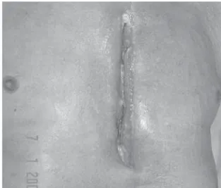
cases), spontaneous perforation (Boerhaave's syndrome; 2 cases), traumatic perforation (1 case) and perforation by a foreign body (1 case). Head and neck infections accounted for 3 (14.3%) of the cases. Of those 3, 2 were cases of deep cervical abscess and 1 was a case of Ludwig's angina (Figures 1, 2 and 3). Mediastinoscopy, pyopericarditis and pleural empyema were each responsible for 1 case (4.8%).
Culture of the exudates revealed that the acute mediastinitis was caused by a single microorganism in 42.9% of the cases and by multiple microbes in 23.8%. In 33.3% of the cases, the disease-causing agent was not isolated. There were no statistically significant differences between the number of microorganisms present in the cultures and the mortality rate (p = 0.51). The agents S. aureus and S. epidermidis were found in 7 cases each, whereas Pseudomonas sp., E. coli, Klebsiella pneumoniae, Enterococcus cloacae, Proteus sp. and Acinetobacter sp were found in 1 case each.
Antibiotic therapy was used exclusively in only 1 case (4.8%). Antibiotic therapy in combination with mediastinal drainage was used in 9 cases (42.9%) and antibiotic therapy in combination with specific treatment of the cause of mediastinitis was used in 4 cases (19.0%). A combination of antibiotic therapy, mediastinal drainage and specific treatment of the cause was used in 7 cases (33.3%).
In our facility, antibiotic therapy is administered (empirically) immediately after collection of the material for culture. We typically use a broad-spectrum antimicrobial combination in order to eliminate gram-positive, gram-negative and anaerobic microorganisms. After the result of the culture is known, the antibiotic therapy is adjusted according to the result obtained in the exam. For gram-positive microorganisms, we use either oxacillin (especially in patients whose culture was positive for S. aureus, which is sensitive to this antimicrobial agent) or vancomycin. For gram-negative microorganisms, we use a combination of cefepime, ceftriaxone (or ceftazidime) and amikacin (or gentamicin), the first three also attacking gram-positive microorganisms. For anaerobic microorganisms, we use clindamycin or metronidazole.
When the cause of the mediastinitis was median sternotomy, less invasive treatments were typically adopted. Of the 8 cases with this etiology, 5 were submitted only to antibiotic therapy (using one of the regimens described above) and extensive
mediastinal drainage. Drainage of the incision site, total sternum removal and partial sternum removal (the last two both in combination with a chest muscle flap) were used in 1 case each. In this group (patients with median sternotomy-related mediastinitis), the mortality rate was 25%, which is very similar to the overall mortality rate.
When the etiology of mediastinitis was esophageal perforation, either primary closure or proximal esophagostomy, involving gastrostomy and esophagectomy, was used. In 3 of the 7 cases of esophageal perforation, the second option was used, and 1 death occurred. In the 4 remaining cases, primary closure was the procedure chosen, and 2 of these patients died. Among the patients with mediastinitis resulting from esophageal perforation, the mortality rate was 42.9%, which is high in comparison to the overall mortality rate.
For cases in which mediastinitis was caused by cervical abscess, the procedure used was always surgical drainage, provided that collection did not extend beyond the brachiocephalic veins. Among such cases, the mortality rate was 33.3%.
In the 3 cases of mediastinitis caused by other
Figure 1 Mediastinitis caused by Boerhaave's syndrome
etiologies, no deaths occurred (in these cases, there was no specific treatment of the cause).
Complications of acute mediastinitis included pleural empyema (23.8%), osteomyelitis (19%), respiratory insufficiency (9.5%) and others (19.1%). In 28.6% of the cases, there were no complications. The mean length of hospital stay among patients diagnosed with acute mediastinitis was 26.6 days (± 19.), ranging from 6 to 77 days.
DISCUSSION
Acute mediastinitis continues to be a much feared complication of various diseases and procedures. In our study, as well as in the literature, the incidence of the disease was low (1.17 cases per year in our hospital), although the mortality rate was quite high.(1,4)
Notable among the causes of acute mediastinitis are the cases resulting from median sternotomy. Since the number of cardiac surgical procedures using this approach is on the rise, this etiology has become the most important and also the most widely studied.(3,5-6) However, esophageal perforation should
not be forgotten since it is responsible for the highest mortality rates.(7-10,15)
Among the infectious agents involved, S. aureus and S. epidermidis are the most frequently found. Similar results have been reported in the literature, and some studies have also shown that the isolation of E. coli is correlated with a high mortality rate.(3,5)
It is important to bear in mind that the culture of the exudates may be negative, which is typically
attributable to prior use of antibiotics.(3)
The treatment of acute mediastinitis is based on three basic points: antibiotic therapy, extensive drainage of the mediastinal cavity and treatment of the specific cause.(13) In cases of mediastinitis
occurring as a complication of median sternotomy, the literature indicates that the prognosis is better when the patient is submitted to a specific treatment, with drainage of the incision site and omental flap rotation.(3,5) However, despite the small number of
cases in our study, this type of treatment did not reduce the mortality rate among these patients.
Another controversial point is the treatment of choice for esophageal perforation, especially in cases of Boerhaave's syndrome. Some authors prefer primary resection of the esophagus, whereas others believe superior esophagectomy to be the ideal.(8-9) In our study,
both types of treatment presented similar prognoses. Considering the data found in our study and that in the literature,(1,6) we believe that it is important to consider
a diagnosis of acute mediastinitis for all patients submitted to median sternotomy or to any esophageal manipulation and presenting subsequent fever or other signs of significant infection, especially (in those submitted to esophageal procedures) around the head and neck. The great diagnostic challenge continues to be spontaneous esophageal perforation (Boerhaave's syndrome), which manifests only as a nonspecific history of vomiting and signs of toxemia. It is important to emphasize that, in these cases, a better prognosis is closely correlated with early diagnosis.(8-9)
As soon as there is clinical suspicion, a chest X-ray
Figure 2 - Mediastinitis caused by deep cervical infection - Tomography of the neck and chest showing cervical-mediastinal abscess after suppurative thyroiditis
should be requested. This may reveal signs of mediastinitis or its complications, such as mediastinal emphysema and pleural effusion. Tomography of the chest using a window for the mediastinum complements the chest X-ray, revealing mediastinal abscesses, emphysemas, sternal involvement, etc., in more detail.(1,6)
In suspected cases of Boerhaave's syndrome, a contrast esophagogram is extremely important since it confirms the diagnosis by showing the extravasation of the contrast medium into the mediastinum.
The most important laboratory exams are culture and antibiogram of the mediastinal secretion. Control exams should be requested in order to detect complications such as renal insufficiency as early as possible.
In our facility, after collection of material for culture, empirical antibiotic therapy is initiated and continues until the result of the antibiogram is known. Generally, this initial treatment is oriented according to the etiology. In cases of median sternotomy-related acute mediastinitis, the initial antibiotic therapy includes the administration of oxacillin and a third-generation or higher cephalosporin (ceftriaxone, cefepime or ceftazidime), combined or not with an aminoglycoside (amikacin or gentamicin). In cases of cervical abscesses, we administered a third-generation or higher cephalosporin combined with clindamycin. Finally, in cases of esophageal perforation, a combination of crystalline penicillin, clindamycin (or metronidazole) and an aminoglycoside is the treatment used. Boerhaave's syndrome constitutes an exception, in which, due to its severity and usually late diagnosis, we opt for a combination of a third-generation or higher cephalosporin, combined with clindamycin and oxacillin or even vancomycin.
In conclusion, acute mediastinitis is a severe complication of some diseases and procedures. Despite a low incidence, the mortality rate is high. Hospital stays are longer among patients with mediastinitis resulting from infection with S. aureus and S. epidermidis, which are the most common etiologic agents.
REFERENCES
1. Macrí P, Jiménez MF, Novoa N, Varela GA. Descriptive of a series of patients diagnosed with acute mediastinitis. Arch Bronconeumol. 2003;39(9):428-30.
2. Souza VC, Freire ANM, Tavares-Neto, J. Mediastinite pós-esternotomia longitudinal para cirurgia cardíaca: 10 anos de análise. Rev Bras Cir Cardiovasc. 2002;17(3):266-70. 3. Sampaio DT, Alves JCR, Silva AF, Lobo Jr. NC, Simões D, Faria W, et al. Mediastinite em cirurgia cardíaca: tratamento com epíploon. Rev Bras Cir Cardiovasc. 2000;15(1):23-31.
4. Katsetos MC, Tagbo AC, Lindberg MP, Rosson RS. Esophageal perforation and mediastinitis from fish bone ingestion. South Med J. 2003;96(5):516-20. 5. Sancho LM, Minamoto H, Fernandez A, Sennes LU,
Jatene FB. Descending necrotizing mediastinitis: a retrospective surgical experience. Eur J Cardiothorac Surg. 1999;16(2):200-5.
6. Izadi K, Lazow SK, Berger JR. Mediastinitis secondary to an odontogenic infection. A case report. NY State Dent J. 2003;69(10):28-30.
7. Salo JA, Isolauri JO, Heikkila LJ, Markkula HT, Heikkinen LO, Kivilaakso EO et al.: Management of delayed esophageal perforation with mediastinal sepsis. Esophagectomy or primary repair? J Thorac Cardiovasc Surg. 1993;106(6): 1088-91.
8. Brommelstroet M, Rosa JFT, Boscardim PCB, Schmidlin CA, Shibata S. Mediastinite descendente necrosante pós-angina de Ludwig. J Pneumol. 2001;27(5):269-71. 9. Sung SW, Park JJ, Kim YT, Kim JH. Surgery in thoracic
esophageal perforation: primary repair is feasible. Dis Esophagus. 2002;15(3):204-9.
10. Schimin LC, Batista RL, Mendonça FCC. Mediastinite no Hospital de Base do Distrito Federal: incidência em seis anos. Rev Bras Cir Cardiovasc. 2002;17(2):36-9. 11 . J a m p l is R W, M c Fa d d e n P M . I n f e c t i o n o f t h e
mediastinum and the superior vena caval syndrome. In: Shields TW. General thoracic surgery. 3rd ed. London: Lea & Febiger; 1989. p.1085-95.
1 2 . Soldati G, Di Piero A, Bassani L, Di Vito A, Rossi M. Boerhaave syndrome. A case report and review of the literature. Minerva Chir. 2000;55(12):873-9. Italian. 13. Martinez-Ordaz JL, Cornejo-Lopez GB, Blanco-Benavides
R. Boerhaave’s Syndrome. Case report and literature review. Rev Gastroenterol Mex. 2002;67(3):190-4. Spanish. 14. Bernini CO, Curi N. Mediastinite. In.: Corrêa Netto, A. Clínica
cirúrgica. 4ª ed. São Paulo: Sarvier; 1994. p.341-4. 15. Moore DS. Inferência para tabela de dupla entrada. In.: