J Bras Pneumol. 2008;34(5):294-297
Original Article
* Study carried out at the University Hospital, Universidade Estadual de Campinas – Unicamp, State University at Campinas – School of Medicine, Campinas, Brazil. 1. Physiotherapist of the Emergency Department Ward. University Hospital, Universidade Estadual de Campinas – Unicamp, State University at Campinas – School of Medicine, Campinas, Brazil.
2. Chief of the Emergency Department. University Hospital, Universidade Estadual de Campinas – Unicamp, State University at Campinas – School of Medicine, Campinas, Brazil.
3. Chief of the Lung Diseases Division. University Hospital, Universidade Estadual de Campinas – Unicamp, State University at Campinas – School of Medicine, Campinas, Brazil.
Correspondence to: Armando Carlos Franco de Godoy. Rua Hercules Florence, 100, apto 23, Botafogo, CEP 13020-170, Campinas, SP, Brasil. Tel 55 19 3231-4742. Email: [email protected]
Submitted: 1 June 2007. Accepted, after review: 13 August 2007.
**A versão completa em português deste artigo está disponível em www.jornaldepneumologica.com.br
Endotracheal tube cuff pressure alteration after changes
in position in patients under mechanical ventilation*
,**
Alteração da pressão intra-cuff do tubo endotraqueal após mudança da posição em pacientes sob ventilação mecânica
Armando Carlos Franco de Godoy1, Ronan José Vieira2, Eduardo Mello De Capitani3
Abstract
Objective: The purpose of this study was to investigate endotracheal tube cuff pressure (Pcuff) alteration in patients under mechanical ventilation after changes in position. Methods: All selected patients were initially placed in the 35° semi-Fowler position, with Pcuff adjusted to 20 mmHg, and randomly divided into two groups. Group A, in which patients were moved to the lateral decubitus position, facing away from the ventilator (measurement designated Pcuff A1), returned to the initial position (measurement designated Pcuff A2), moved to a lateral decubitus position, facing the ventilator (measurement designated Pcuff A3) and then returned to the initial position (measurement designated Pcuff A4); and Group B, in which patients were moved to the lateral decubitus position, facing the ventilator (measurement designated Pcuff B1), returned to the initial position (measurement designated Pcuff B2), moved to the lateral decubitus position; facing away from the ventilator (measurement designated Pcuff B3) and then returned to the initial position (measurement designated Pcuff B4). Results: The study comprised 70 patients, 31 allocated to group A and 39 allocated to group B. Values >22 mmHg were observed in 142(50.7%) of the 280 Pcuff measurements taken, and values <18 mmHg were observed in 14 (5%). When moved from the 35° semi-Fowler position to the lateral decubitus position, facing away from the ventilator, 58 (82.2%) of the patients presented mean Pcuff values in the higher range (>22 mmHg).
Conclusions: Changes in body position can cause significant Pcuff variations in patients under mechanical ventilation.
Keywords: Pressure; Intubation, intratracheal; Posture; Supine position.
Resumo
Objetivo: O objetivo deste trabalho foi investigar a alteração da pressão intra-cuff (Pcuff) do tubo endotraqueal em pacientes sob ventilação
mecânica, após alteração de sua posição corporal. Métodos: Todos os pacientes selecionados eram inicialmente colocados em posição de
semi-Fowler (35°), Pcuff em 20 mmHg, e divididos aleatoriamente em dois grupos. Grupo A: a Pcuff era medida após mover-se o paciente para decúbito lateral, costas voltadas para o ventilador (denominada Pcuff A1); após retornar o paciente à posição inicial (denominada Pcuff A2); após mover o paciente para decúbito lateral, de frente para o ventilador (denominada Pcuff A3); e após retornar o paciente, novamente, à posição inicial (denominada Pcuff A4). No Grupo B: a Pcuff era medida após mover-se o paciente para decúbito lateral, de frente para o ventilador (denominada Pcuff B1); após retornar o paciente à posição inicial (denominada Pcuff B2); após mover o paciente para decúbito lateral, costas voltadas para o ventilador (denominada Pcuff B3); e após retornar o paciente, novamente, à posição inicial (denominada
Pcuff B4). Resultados: Foram incluídos 70 pacientes no estudo, 31 no grupo A e 39 no grupo B. Valores >22 mmHg foram observados em
142 (50,7%) das 280 medidas de Pcuff realizadas, e valores <18 mmHg, em 14 (5%). Quando movidos da posição de semi-Fowler (35°) para decúbito lateral, costas voltadas para o ventilador, 58 (82,2%) dos pacientes apresentaram valores médios de Pcuff mais altos, >22 mmHg.
Conclusões: Mudanças na posição corporal dos pacientes sob ventilação mecânica podem alterar significativamente a Pcuff.
Descritores: Pressão; Intubação intratraqueal; Postura; Decúbito dorsal.
Introduction
In mechanically ventilated patients, the endotracheal tube cuff should remain inflated in order to prevent gas leakage and aspiration of oropharyngeal contents into
the lungs.(1) It is important that the cuff pressure (Pcuff)
Endotracheal tube cuff pressure alteration after changes in position in patients under mechanical ventilation
J Bras Pneumol. 2008;34(5):294-297
295
The second measurement, designated Pcuff A2, was taken after the patient had been returned to the 35° semi-Fowler position. The third measurement, designated Pcuff A3, was taken after the patient had been moved to the lateral decubitus position facing the ventilator. The fourth measurement, designated Pcuff A4, was taken after the patient had again been returned to the 35° semi-Fowler position. In group B, the maneuvers were the same as those employed in Group A but followed a different sequence: Pcuff B1 was taken after the patient had been moved from the 35° semi-Fowler position to the lateral decubitus position, facing the ventilator; Pcuff B2 was taken after the patient had been returned to the 35° semi-Fowler position; Pcuff B3 was taken after the patient had been moved to the lateral decubitus position, facing away from the ventilator; and Pcuff B4 was taken after the patient had again been returned to the 35° semi-Fowler position. The entire process followed a cross-over design in which all patients were submitted to the same changes in position with the only difference being in the initial maneuver to move the patient into the lateral decu-bitus position: facing away from the ventilator in group A and facing the ventilator in group B.
In the statistical analyses, mean Pcuff values were compared using analysis of variance in the BioEstat program, version 3.0 for Windows (Conselho Nacional de Desenvolvimento Científico e Tecnológico, Brasília, Brazil). Values of p ≤ 0.05 were considered statistically significant.
Results
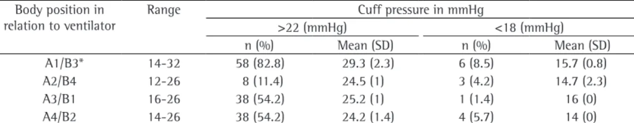
Between January and March of 2006, a total of 70 patients were selected for inclusion in the study: 31 were allocated to group A, and 39 were allocated to group B. Table 1 shows the means, standard devi-ations and Pcuff ranges, according to the changes in patient position, stratified into those in which the mean was above 22 mmHg and those in which it was below 18 mmHg.
Values >22 mmHg were observed in 142 (50.7%) of the 280 Pcuff measurements taken, and values <18 mmHg were observed in 14 (5%).
As can also be seen in Table 1, 58 (82.2%) of the patients presented mean Pcuff values in the higher range (>22 mmHg) when moved from the 35° semi-Fowler position to the lateral decubitus position, facing away from the ventilator. In comparison with wall can be damaged by high Pcuffs,(2,3) and that
low Pcuffs can promote microaspiration of oropha-ryngeal contents, predisposing to the occurrence of nosocomial pneumonia and poor ventilation effi-cacy due to gas leakage.(3-5)
Various factors are thought to promote Pcuff variations: changes in tracheal muscle tone; hypo-thermia; hyperhypo-thermia; diffusion of anesthetic gas into the tube cuff; and changes in endotracheal tube position.(4-6)
The purpose of this study was to investigate Pcuff alterations in adult patients under mechanical ventilation during changes in patient position.
Methods
This study was carried out in the Emergency Room of the State University at Campinas University Hospital, and the protocol was approved by the Hospital Ethics Committee. Patients were recruited and selected sequentially. Inclusion criteria were being at least 18 years of age, being under mechan-ical ventilation for no more than 24 h and being orotracheally intubated with an endotracheal tube that has a high-volume/low-pressure cuff. Patients in which changes from the recumbent position were contraindicated were excluded. Each patient was evaluated only once. The Pcuff was calibrated using a three-way stopcock: the pilot balloon of the endotracheal tube was connected to one of the stopcock ports; an analogical manometer, previously calibrated (in mmHg) using a reference manometer, was connected to one of the two remaining ports; and a 5-mL syringe was connected to the third port, allowing the Pcuff to be regulated by adjusting the plunger of the syringe. The measurement set remained connected to the pilot balloon during the maneuvers for changes in patient position.
296 Godoy ACF, Vieira RJ, De Capitani EM
J Bras Pneumol. 2008;34(5):294-297
the damage to the respiratory system caused either simply by the presence of the endotracheal tube or by Pcuff variation: regular and periodic control of the Pcuff(1); use of endotracheal tubes that are of
an appropriate diameter(6); use of a Pcuff regulating
valve in the pilot balloon; use of high-volume/low-pressure cuffed endotracheal tubes; and continuous aspiration of the oropharyngeal content.(1) Although
the advent of high-volume/low-pressure cuffed endotracheal tubes in the early 1970s decreased the frequency of postintubation tracheal injuries, this type of cuff, when inflated with great volumes, can reach pressures that damage tissues after 2-4 h.(15)
To our knowledge, there have been no studies investigating changes in patient position associated with Pcuff variation during mechanical ventila-tion. In the present study, we found Pcuff values above 22 mmHg and below 18 mmHg in the two groups studied. The variation in Pcuff might have been due to compression or decompression of the endotracheal tube over the cuff caused by move-ment and changes in the position of the mechanical ventilator circuit. However, Pcuff variation can also be attributed to the cuff membrane lying in folds, independent of changes in patient position.(1) The
mean Pcuff values, as well as the number of Pcuff values above or below reference values, observed for Pcuffs A1 and B3 (after the patient had been moved from 35° semi-Fowler position to the lateral decubitus position, facing away from the ventilator) were higher than those observed for Pcuffs A3 and B1 (after patient had been moved from 35° semi-Fowler position to lateral decubitus position, facing the ventilator). This difference, which was signifi-cant (p < 0.05), might be attributable to the greater strain put on the ventilator air circuit by facing patients away from the ventilator.
the other positions, the difference was significant (p < 0.01).
Discussion
Our results suggest that changing patient posi-tion during mechanical ventilaposi-tion can lead to significant alterations in Pcuff.
Various factors can induce lesions in the respira-tory tract of mechanically ventilated patients. Such factors include the following: inadequate airway humidification; a high fraction of inspired oxygen; insufficient heating of administered gases; frequent tracheal suction; prolonged endotracheal intubation; prolonged mechanical ventilation; and inappro-priate Pcuff values.(7-11) Some researchers suggest
that, among these factors, inappropriate Pcuff values (Pcuff variation) is of great importance in the genesis of postintubation injuries diagnosed in the respiratory tract. Variations in Pcuff can damage the tracheal wall and promote oropharyngeal content aspiration, leading to nosocomial pneumonia.(7-9)
When Pcuff remains between 18 and 22 mmHg, damage to the tracheal wall tends to be mini-mized or averted. However, after 2-12 h at a Pcuff of approximately 20 mmHg, a local inflammatory process can begin in areas that are in direct contact with the cuff. The severity of the process is propor-tional to the duration of intubation.(8) Conversely,
if Pcuff is kept below 18 mmHg, microaspirations of oropharyngeal content can occur.(12) The section
immediately above the cuff collects oropharyngeal and upper airway secretions, being the source of material for microaspirations if the air cuff loses pressure.(13,14)
Previous studies have reported several prophy-lactic measures that should be taken in order to avoid
Table 1 - Means, standard deviations and ranges of values above 22 mmHg and below 18 mmHg in mechanically ventilated patients after changes in position.
Body position in relation to ventilator
Range Cuff pressure in mmHg
>22 (mmHg) <18 (mmHg)
n (%) Mean (SD) n (%) Mean (SD)
A1/B3* 14-32 58 (82.8) 29.3 (2.3) 6 (8.5) 15.7 (0.8)
A2/B4 12-26 8 (11.4) 24.5 (1) 3 (4.2) 14.7 (2.3)
A3/B1 16-26 38 (54.2) 25.2 (1) 1 (1.4) 16 (0)
A4/B2 14-26 38 (54.2) 24.2 (1.4) 4 (5.7) 14 (0)
Endotracheal tube cuff pressure alteration after changes in position in patients under mechanical ventilation
J Bras Pneumol. 2008;34(5):294-297
297
with ventilator-associated pneumonia. Eur Respir J. 1996;9(8):1729-35.
6. Mehta S, Mickiewicz M. Pressure in large volume, low pressure cuffs: its significance, measurement and regulation. Intensive Care Med. 1985;11(5):267-72.
7. Berlauk JF. Prolonged endotracheal intubation vs. tracheostomy. Crit Care Med. 1986;14(8):742-5.
8. Sarper A, Ayten A, Eser I, Ozbudak O, Demircan A. Tracheal stenosis aftertracheostomy or intubation: review with special regard to cause and management. Tex Heart Inst J. 2005;32(2):154-8.
9. Fan CM, Ko PC, Tsai KC, Chiang WC, Chang YC, Chen WJ, et al. Tracheal rupture complicating emergent endotracheal intubation. Am J Emerg Med. 2004;22(4):289-93.
10. Badenhorst CH. Changes in tracheal cuff pressure during respiratory support. Crit Care Med. 1987;15(4):300-2. 11. Nseir S, Di Pompeo C, Pronnier P, Beague S, Onimus
T, Saulnier F, et al. Nosocomial tracheobronchitis in mechanically ventilated patients: incidence, aetiology and outcome. Eur Respir J. 2002;20(6):1483-9.
12. Rumbak MJ. The pathogenesis of ventilator-associated pneumonia. Semin Respir Crit Care Med. 2002;23(5):427-34.
13. Bernhard WN, Yost L, Joynes D, Cothalis S, Turndorf H. Intracuff pressures in endotracheal and tracheostomy tubes. Related cuff physical characteristics. Chest. 1985;87(6):720-5.
14. Wunderink RG. Nosocomial pneumonia, including ventilator-associated pneumonia. Proc Am Thorac Soc. 2005;2(5):440-4.
15. Klainer AS, Turndorf H, Wu WH, Maewal H, Allender P. Surface alterations due to endotracheal intubation. Am J Med. 1975;58(5):674-83.
16. Wiswell TE, Turner BS, Bley JA, Fritz DL, Hunt RE. Determinants of tracheobronchial histologic alterations during conventional mechanical ventilation. Pediatrics. 1989;84(2):304-11.
Various authors have recommended that, in order to prevent scarring and nosocomial pneu-monia, mechanically ventilated patients should be submitted to regular and periodic changes in posi-tion.(5,11,16) However, we believe that, during such
changes in position, special care should be taken to monitor and, if necessary, recalibrate the Pcuff.
Moving mechanically ventilated patients from the 35° semi-Fowler position to the lateral decu-bitus position can cause significant variations in Pcuff. In the routine care of such patients, regular Pcuff measurement and adjustment after changes in body position should be encouraged.
References
1. Farré R, Rotger M, Ferre M, Torres A, Navajas D. Automatic regulation of the cuff pressure in endotracheally-intubated patients. Eur Respir J. 2002;20(4):1010-3.
2. Schmidt WA, Schaap RN, Mortensen JD. Immediate mucosal effects of short-term, soft-cuff, endotracheal intubation. A light and scanning electron microscopic study. Arch Pathol Lab Med. 1979;103(10):516-21.
3. Vyas D, Inweregbu K, Pittard A. Measurement of tracheal tube cuff pressure in critical care. Anaesthesia. 2002;57(3):275-7.
4. Mahul P, Auboyer C, Jospe R, Ros A, Guerin C, el Khouri Z, et al. Prevention of nosocomial pneumonia in intubated patients: respective role of mechanical subglottic secretions drainage and stress ulcer prophylaxis. Intensive Care Med. 1992;18(1):20-5.