J Bras Pneumol. 2008;34(3):181-184
181
Case Report
* Study carried out at the Hospital das Forças Armadas – HFA, Armed Forces Hospital – Brasília, Brazil. 1. Radiologist. Hospital das Forças Armadas – HFA, Armed Forces Hospital – Brasília, Brazil.
2. Full Professor of Radiology. Universidade Federal Fluminense – UFF, Fluminense Federal University – Niterói, Brazil.
3. Radiologist. Instituto do Coração do Distrito Federal – Incor/DF, Heart Institute of the Federal District of Brasília – Brasília, Brazil. 4. Resident in Radiology. Hospital das Forças Armadas – HFA, Armed Forces Hospital – Brasília, Brazil.
5. Physician in the Department of Pathology. Faculdade de Medicina da Universidade de São Paulo – FMUSP, University of São Paulo School of Medicine – São Paulo, Brazil.
6. Head of the Department of Pulmonology. Hospital das Forças Armadas – HFA, Armed Forces Hospital – Brasília, Brazil. Correspondence to: Rosane Rodrigues Martins. SQSW 305, Bloco H, apto 105, CEP 70673-506, Brasília, DF, Brasil. Tel 55 61 3233-7220. E-mail: [email protected]
Submitted: 20 December 2006. Accepted, after review: 18 April 2007.
Chronic eosinophilic pneumonia secondary to
long-term use of nitrofurantoin: high-resolution
computed tomography findings*
Pneumonia eosinofílica crônica secundária ao uso prolongado de nitrofurantoína: achados da tomografia computadorizada de alta resolução do tórax
Rosane Rodrigues Martins1, Edson Marchiori2, Sérgio Lopes Viana3,
Luiz Sérgio Pereira Grillo Júnior4, Vera Luiza Capelozzi5, Laércio Moreira Valença6
Abstract
The authors report the case of a female patient who developed chronic eosinophilic pneumonia secondary to long-term use of nitrofurantoin for prophylaxis of recurrent urinary tract infections due to urethral stenosis. On high-resolution computed tomography scans, the pulmonary reaction to nitrofurantoin most commonly manifests as an interstitial-alveolar pattern in both lung bases. However, in this case, the alterations were most pronounced in the periphery of the upper lobes. In itself, this tomographic profile is strongly indicative of chronic eosinophilic pneumonia. The patient had previously been submitted to an open lung biopsy. The diagnosis of chronic eosinophilic pneumonia was confirmed through a review of the biopsy.
Keywords: Pneumonia; Pulmonary eosinophilia; Nitrofurantoin/adverse effects; Tomography, X-ray computed.
Resumo
Os autores relatam o caso de uma paciente com estenose de uretra que desenvolveu pneumonia eosinofílica crônica secundária ao uso prolongado de nitrofurantoína como profilaxia para infecção urinária de repetição. A paciente havia sido submetida a uma biópsia pulmonar a céu aberto. É dada ênfase aos achados da tomografia computadorizada de alta resolução do tórax, já que, embora as alterações pulmonares associadas à toxicidade da nitrofurantoína geralmente sejam basais e bilaterais, no caso aqui descrito, as lesões de natureza interstício-alveolares situaram-se nas regiões subpleurais dos lobos superiores. Esses achados, por si só, são muito sugestivos de pneumonia eosinofílica crônica. O diagnóstico foi confirmado por meio da revisão da biópsia.
Descritores: Pneumonia; Eosinofilia pulmonar; Nitrofurantoína/efeitos adversos; Tomografia computadorizada por raios X.
Introduction
Nitrofurantoin has been widely used as an efficient and relatively safe medication in the treatment of genitouri-nary infections, and complications due to its use are rare. However, pulmonary parenchymal lesions have occasionally been associated with long-term nitrofurantoin therapy. The authors report the case of a patient with chronic eosinophilic pneumonia secondary to long-term nitrofurantoin use, with emphasis on the findings obtained through high-resolution computed tomography (HRCT) scans of the chest.
Case report
corti-182 Martins RR, Marchiori E, Viana SL, Grillo Jr LSP, Capelozzi VL, Valença LM
J Bras Pneumol. 2008;34(3):181-184
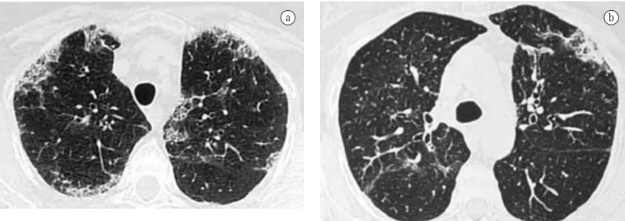
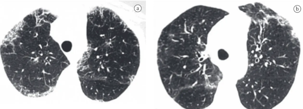
stitium accompanied by faint ground-glass opacities, these alterations being present only in the upper lobes, and alveolar opacity being observed in the anterior segment of the left upper lobe (Figure 2). There were no mediastinal alterations. A control HRCT scan performed four months later showed no significant changes, except for an improvement in the area of alveolar opacity in the left upper lobe (Figure 3).
Dyspnea and rales progressively resolved over the course of the treatment, and the dose of oral corticos-teroid was progressively reduced until the medication was completely discontinued, after fifteen months of treatment. After the corticosteroid therapy had been discontinued, there was no recurrence of symptoms.
Discussion
Nitrofurantoin pulmonary toxicity was first reported in 1957 by Fisk.(1) Since then, there have
been various reports of nitrofurantoin-induced pulmonary reactions, with polymorphic profiles that include pulmonary fibrosis,(2,3) alveolar hemorrhage,(4)
bronchiolitis obliterans organizing pneumonia (BOOP),(5) lupus-like syndrome,(6) desquamative,
usual, and nonspecific interstitial pneumonias,(7)
allergic reactions,(8) diffuse alveolar damage,(9) and
eosinophilic pneumonia.(10)
Nitrofurantoin-induced pulmonary disease has two principal forms of presentation: the acute form, which is more common, develops hours to days after the use of the medication, and is probably related to a hypersensitivity reaction or to an immunolog-costeroid therapy previously performed in another
facility was unsuccessful, and an open-lung biopsy was then performed, resulting in a histopathological diagnosis of diffuse interstitial fibrosis.
In the pathological history, the report of uterine cancer at the age of 35, which was treated with radiotherapy and chemotherapy, was worthy of note. As a complication of that condition, the patient presented urethral stenosis, with recurrent episodes of urinary tract infection. During the preceding three years, the management of the urinary tract infection had included self-catheterization every 4 h and prophylaxis with nitrofurantoin. The physical examination revealed only a scar from a left thora-cotomy, decreased chest expansion, and crackling rales in the lung bases.
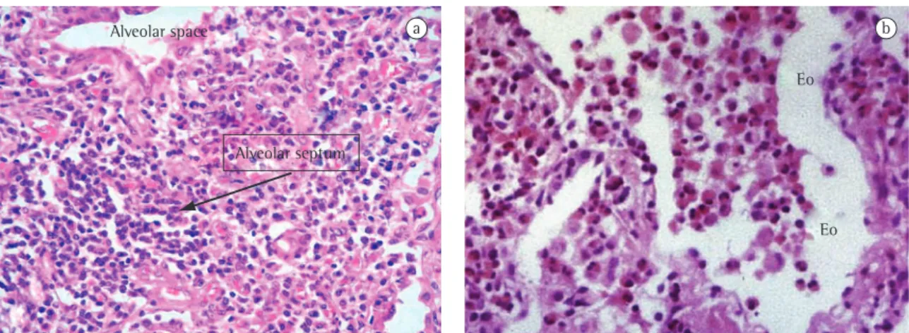
The review of the lung biopsy by a patholo-gist resulted in a histological diagnosis of chronic eosinophilic pneumonia, possibly attenuated by previous use of steroids, accompanied by alterations resulting from bronchial obstruction. There was infiltration by eosinophils and mononuclear cells throughout the alveolar septa and alveolar spaces (Figure 1).
Since the anatomopathological findings might be associated with a reaction to nitrofurantoin, this medication was discontinued, and oral corticos-teroid therapy was initiated.
A chest X-ray performed four months after the initial medical visit showed areas of subpleural inter-stitial thickening in the upper lobes. An HRCT scan, performed concomitantly, revealed focal subpleural thickening of the intralobular and interlobular
inter-a Alveolar space
Alveolar septum
Figure 1 - Chronic eosinophilic pneumonia: a) detail of the septal alveolar thickening (arrow) caused by eosinophil
and mononuclear cell infiltration, H&E ×200; and b) infiltration by eosinophils (Eo) throughout the alveolar septa and within the alveolar spaces, H&E ×200.
b
Eo
Chronic eosinophilic pneumonia secondary to long-term use of nitrofurantoin: high-resolution computed tomography findings
J Bras Pneumol. 2008;34(3):181-184
183
In the case presented here, the histopathological diagnosis was chronic eosinophilic pneumonia, since there have been no specific reports of this disease being diagnosed (based on HRCT findings) as an adverse reaction to nitrofurantoin. The principal chronic alterations associated with nitrofurantoin are usually found in the lung bases,(3,16,17) although
sparse peribronchovascular alterations have been described in patients presenting BOOP as a reaction to nitrofurantoin administration.(5) There can be an
overlap between the clinical, radiological, and path-ological findings of chronic eosinophilic pneumonia and those of BOOP. However, in many cases, HRCT helps to differentiate between these two diseases.
In the case in question, the findings were predom-inantly in the upper lobes and were principally subpleural. If only the HRCT findings are consid-ered, the profile was highly suggestive of chronic eosinophilic pneumonia.(18-20) The favorable clinical
evolution after the discontinuation of the medica-tion, as well as the absence of recurrence after the discontinuation of the corticosteroid therapy, made it possible to establish a causal relationship between the pulmonary condition and the use of nitrofuran-toin. The persistence of the imaging findings in the upper lobes translated to chronic interstitial altera-tions that were irreversible.
Having knowledge regarding the various acute and chronic pulmonary manifestations of nitro-furantoin toxicity is essential for diagnosing the pulmonary reaction to the drug, as is taking a patient history regarding the use of this medica-tion,. Some authors recommend that patients using ical reaction; and the chronic form, which appears
after months or years of continuous use and is probably directly related to oxidant-mediated tissue injury.(11-13)
The acute form is clinically characterized by tachypnea, fever, nonproductive cough, tachy-cardia, chest pain, skin rash, and arthralgia. Chest X-rays show a reticular or alveolar pattern in both lung bases, with septal lines and pleural effusion, mimicking pulmonary edema. Peripheral eosi-nophilia and reduced diffusing capacity of the lung for carbon monoxide are typical, and pulmonary function test results can include hypoxemia and a restrictive pattern.(13-15) The differential diagnosis
should primarily include pulmonary embolism, infection, myocardial infarction, and heart failure. In addition, the temporal relationship between the onset of the symptoms and the initiation of the medication, as well as the fact that there is signifi-cant improvement after discontinuation of the medication, are fundamental.
The chronic form has a more insidious onset, with progressive dyspnea, dry cough, tachypnea, and no fever. The most common finding on chest X-rays is interstitial thickening in the lung bases.(3,16)
Pulmonary function tests reveal a restrictive pattern, with hypoxemia and reduced diffusing capacity of the lung for carbon monoxide. The differential diag-nosis includes idiopathic pulmonary fibrosis and other causes of interstitial thickening in the lung bases. Within a few months after discontinuation of the drug, symptoms typically improve, with or without regression of the interstitial thickening.
a b
Figure 2 - High-resolution computed tomography scan of the chest performed in January 2004. Slices slightly above
184 Martins RR, Marchiori E, Viana SL, Grillo Jr LSP, Capelozzi VL, Valença LM
J Bras Pneumol. 2008;34(3):181-184
10. Rossi SE, Erasmus JJ, McAdams HP, Sporn TA, Goodman PC. Pulmonary drug toxicity: radiologic and pathologic manifestations. Radiographics. 2000;20(5):1245-59. 11. Cooper JA Jr, White DA, Matthay RA. Drug-induced
pulmonary disease. Part 2: Noncytotoxic drugs. Am Rev Respir Dis. 1986;133(3):488-505.
12. Suntres ZE, Shek PN. Nitrofurantoin-induced pulmonary toxicity. In vivo evidence for oxidative stress-mediated mechanisms. Biochem Pharmacol. 1992;43(5):1127-35. 13. Witten CM. Pulmonary toxicity of nitrofurantoin. Arch Phys
Med Rehabil. 1989;70(1):55-7.
14. Holmberg L, Boman G. Pulmonary reactions to nitrofurantoin. 447 cases reported to the Swedish Adverse Drug Reaction Committee 1966-1976. Eur J Respir Dis. 1981;62(3):180-9. 15. Santos MS, Costa JC, Moraes RC, Valença LM. Reação
pulmonar a nitrofurantoína. J Pneumol. 1978;(4):20-3. 16. Brutinel WM, Martin WJ 2nd. Chronic nitrofurantoin
reaction associated with T-lymphocyte alveolitis. Chest. 1986;89(1):150-2.
17. Cleverley JR, Screaton NJ, Hiorns MP, Flint JD, Müller NL. Drug-induced lung disease: high-resolution CT and histological findings. Clin Radiol. 2002;57(4):292-9. 18. Mayo JR, Müller NL, Road J, Sisler J, Lillington G. Chronic
eosinophilic pneumonia: CT findings in six cases. AJR Am J Roentgenol. 1989;153(4):727-30.
19. Webb WR, Müller NL, Naidich DP. High-resolution CT of the lung. 3rd ed. Philadelphia: Lippincott Williams & Wilkins; 2000. p. 367-70.
20. Souza CA, Müller NL, Johkoh T, Akira M. Drug-induced eosinophilic pneumonia: high-resolution CT findings in 14 patients. AJR Am J Roentgenol. 2006;186(2):368-73. nitrofurantoin for prolonged periods be submitted
to X-rays every six months as a tool for the early diagnosis of pulmonary alterations.
References
1. Fisk AA. Brief recording: anaphylactoid reaction to nitrofurantoin. N Engl J Med. 1957;256(22):1054. 2. Jick SS, Jick H, Walker AM, Hunter JR. Hospitalizations for
pulmonary reactions following nitrofurantoin use. Chest. 1989;96(3):512-5.
3. Willcox PA, Maze SS, Sandler M, Benatar SR. Pulmonary fibrosis following long-term nitrofurantoin therapy. S Afr Med J. 1982;61(19):714-7.
4. Averbuch SD, Yungbluth P. Fatal pulmonary hemorrhage due to nitrofurantoin. Arch Intern Med. 1980;140(2):271-3. 5. Cameron RJ, Kolbe J, Wilsher ML, Lambie N. Bronchiolitis
obliterans organising pneumonia associated with the use of nitrofurantoin. Thorax. 2000;55(3):249-51.
6. Selroos O, Edgren J. Lupus-like syndrome associated with pulmonary reaction to nitrofurantoin. Report of three cases. Acta Med Scand. 1975;197(1-2):125-9.
7. Bone RC, Wolfe J, Sobonya RE, Kerby GR, Stechschulte D, Ruth WE, et al. Desquamative interstitial pneumonia following long-term nitrofurantoin therapy. Am J Med. 1976;60(5):697-701.
8. DeMasi CJ. Allergic pulmonary infiltrates probably due to nitrofurantoin. Arch Intern Med. 1967;120(5):631-4. 9. Magee F, Wright JL, Chan N, Currie W, Karr G, Hogg J, et al.
Two unusual pathological reactions to nitrofurantoin: case reports. Histopathology. 1986;10(7):701-6.
Figure 3 - High-resolution computed tomography scans of the chest (a and b), performed in May 2004, showing no
changes, except for an improvement in the alveolar opacity in the left upper lobe.