Crutch-related acute arterial thrombosis in upper limb:
case report
Oclusão arterial aguda de membro superior associada à utilização de muleta:
relato de caso
Maurício dos Reis Basílio1, Alex Aparecido Cantador1, Giovani José Dal Poggetto Molinari1, Fábio Hüsemann Menezes1
Abstract
Case report of an acute arterial obstruction in the upper limb secondary to thrombosis of the axillary artery caused by chronic use of crutches. he authors make a brief review of the literature and discuss it in relation to the present case.
Keywords: axillary artery; thrombosis; crutches.
Resumo
Relato de caso de obstrução arterial aguda do membro superior por trombose da artéria axilar secundária ao uso crônico de muleta. Os autores fazem uma breve revisão da literatura, discutindo o presente caso.
Palavras-chave: artéria axilar; trombose; muletas.
1Universidade Estadual de Campinas – UNICAMP, School of Medical Sciences, Department of Surgery, Discipline of Vascular Diseases, Campinas, SP, Brazil
Financial support: None.
Conlicts of interest: No conlicts of interest declared concerning the publication of this article. Submitted: November 12, 2013. Accepted: May 20, 2014.
INTRODUCTION
Acute thrombosis of the axillary artery resulting from injury caused by chronic use of crutches is a
rare event. Dificulty in walking is the most common
reason for using crutches, usually secondary to orthopedic problems and neurological sequelae. Incorrect use of underarm crutches results in axillary compression for long periods and can cause localized trauma leading to structural changes, formation of thrombus, and/or aneurysmal degeneration at the site,
which in turn can lead to acute arterial obstruction.1-4
This case report is intended to call attention to the type of clinical presentation found in this rare form of upper limb arterial obstruction and the importance of its prevention.
CLINICAL CASE
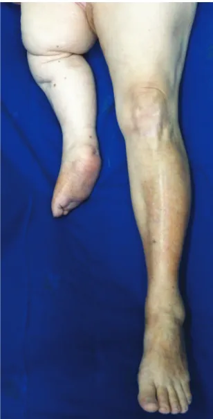
The patient was a 67-year-old white woman who had undergone multiple surgical interventions
during childhood because of osteomyelitis of the right femur, resulting in limb hypoplasia as a sequela (Figure 1). She adapted herself to using a home-made
crutch as an aid in walking, which she used under
the right axilla (Figures 2 and 3). She presented
systemic arterial hypertension, for which she was
on Enalapril, depressive syndrome, being treated
with Fluoxetine and Alprazolam, and had a past
history of an adenocarcinoma of the left breast, that
had been treated with quadrantectomy and left-side axillary clearance 5 years previously and was still taking Tamoxifen. Four days prior to the admission
to our service she began to suffer continuous pain,
low temperature and pallor in the right upper limb.
On the day of admission she sought medical attention
in her home town, where she was given a bolus
infusion of 10,000 i.u. of unfractionated heparin
and was then maintained on continuous infusion of unfractionated heparin at a dosage of 625 i.u. per
hour. She also received analgesia. Some hours later
her clinical status worsened with sudden cyanosis of the right upper limb associated with pain and pallor and she was referred to our service on the basis of
suspected acute arterial obstruction. She stated that she had not suffered pain in the limb during physical exercise previously. On admission to our service she exhibited a good general appearance, although
in continuous pain with reduced temperature and
loss of sensitivity in the upper right limb, but
with motricity preserved and no arrhythmia on
clinical examination or on the electrocardiogram.
Continuous wave Doppler showed an absence of arterial blood low and presence of venous low in
the affected limb, and she was diagnosed with a
Rutherford type II-B acute arterial occlusion. She
was transported to the operating room, where an embolectomy was attempted via the brachial artery. It was possible to clear the distal arterial bed, but restoration of anterograde low was unsuccessful
because it proved impossible to advance the Fogarty
catheter proximally. The decision was taken to construct an axillobrachial graft with a reversed
great saphenous vein. During the operation the patient suffered a coagulation disorder, probably because of the accumulation of the doses of heparin she had already been given plus the dose given intraoperatively (5,000 i.u. of unfractionated heparin
in bolus), which was reversed by administration of protamine sulphate. On the irst day after surgery
she suffered an occlusion of the graft. In view of the previous 4 days’ clinical history, the hypothesis was
that the distal bed could be obstructed. The patient
underwent embolectomy of the graft and
intra-arterial thrombolysis of the distal bed of the forearm and hand, using recombinant tissue plasminogen
activator, achieving restoration of low through the graft, radial pulse and good perfusion to the ingers. Subsequent recovery was uneventful and at 30 days the radial pulse was still present and the limb was
viable.
DISCUSSION
The most common cause of acute arterial obstruction of upper limbs is attributed to emboli from cardiac sources.5-7 Symptomatic arterial
thrombosis of an upper limb is a rare event. The primary hypothesis for the cause of such cases is distal embolization or thrombosis of aneurysms of the subclavian-axillary transition secondary to thoracic outlet syndrome.7 Other causes that can provoke
aneurysmal degeneration of this segment include the
inlamatory diseases, particularly giant-cell arteritis,
and repetitive trauma. Previously published reports of similar cases illustrate that the clinical presentation of arterial thrombosis is different from the presentation of embolism, since it is more gradual and patients
generally seek medical care several days after onset
of the symptoms. It may also be observed that some
patients who use crutches present with pain in the
upper limb during physical activity. Table 1 lists the times from onset of symptoms to admission
of patients with arterial obstruction in some of the
case reports that have been published to date. In the majority of cases, initial clinical presentation is of
Figure 2. Patient using crutch. Observe that the crutch is higher than the axillary pit and is used at an open angle with the body leaning in the contralateral direction. here is no hand grip.
low perfusion for a period varying from hours to days preceding the deinitive occlusion deined as severe ischemia, as was seen in this case, since the patient initially had symptoms of low perfusion and pain,
reduced temperature and pallor in the limb 4 days before her admission. Later the condition deteriorated
over a short period of time with worsening of the
existing symptoms and emergence of cyanosis and paresthesia of the limb.
It should be remembered that the use of crutches is one possible cause of vascular injury leading to acute
arterial obstruction in patients who need the help of
ortheses.4 The trauma caused by the crutch against
the axilla generates chronic repetitive traumatic
compression of the axillary artery, which is capable of provoking the formation of an aneurysm or localized thickening of the artery wall.5,6,8 An arterial dilation
can often be the site of local formation of mural
thrombi, which, if displaced, can cause acute arterial
Table 1. Time from onset of symptoms to admission and prior presence/absence of pain during physical efort in cases described in the literature.
Reference Case Time from onset of ischemic
symptoms to admission and prior presence/absence of the
symptom pain during efort
Brooks AL et al.1 Case 1 5 days
Case 2 3 months + pain during efort
Case 3 3 weeks
Aboot WM et al.2 Case 1 18 hours
Case 2 6 months + pain during efort
Case 3 18 hours + pain during efort
Case 4 Not described
Case 5 2 days
Case 6 1 day
Case 7 Not described
De Luccia N et al.3 Case 1 8 hours
Achramek A et al.4 Case 1 6 hours
Case 2 2 weeks
Ettien J T5 Case 1 3 days
McFall B et al.6 Case 1 Not described
Konishi T et al.7 Case 1 3 days
Moon I S et al.8 Case 1 7 years - 8 hours
Case 2 7 months + pain during efort
Furukawa K et al.9 Case 1 5 years + pain during efort
he present report Case 4 days
obstruction. When present, aneurysms should be treated as soon as they are diagnosed.9 All patients
chronically reliant on crutches should be taught
how to use them correctly, in terms of coniguration and itting adjustments. When indicated, Canadian
crutches should be preferred to axillary support crutches.3,8,9
CONCLUSIONS
The use of crutches may be associated with
thrombosis and formation of aneurysm of the axillary artery through a mechanism of chronic injury, although the condition is rare. Aneurysms of the axillary artery should be repaired to prevent episodes of acute arterial obstruction. Canadian
crutches should be preferred for patients with walking dificulties, whenever indicated, in order to
reduce the occurrence of aneurysms/thromboses of the axillary artery.
REFERENCES
1. Brooks AL, Fowler SB. Axillary artery thrombosis after prolonged use of crutches. J Bone Joint Surg Am. 1964;46:863-4. PMid:14162522.
2. Abbott WM, Darling RC. Axillary artery aneurysms secondary to crutch trauma. Am J Surg. 1973;125(4):515-20. http://dx.doi. org/10.1016/0002-9610(73)90092-5. PMid:4693046
3. Luccia N, Albers MT, Wolosker M. Axillary artery aneurysm due to the use of crutches. Rev Paul Med. 1979;94(3-4):87-9. PMid:549197.
4. Schramek A, Hashmonai M, Abrahamson J. Axillary artery thrombosis due to crutch trauma. Angiology. 1974;25(7):467-9. http://dx.doi.org/10.1177/000331977402500707. PMid:4843182
5. Ettien JT. Crutch-induced aneurysms of the axillary artery. Am Surg. 1980;46(4):267-9. PMid:7386992.
6. McFall B, Arya N, Soong C, Lee B, Hannon R. Crutch induced axillary artery injury. Ulter Med J. 2004;73:50-2
7. Konishi T, Ohki S, Saito T, Misawa Y. Crutch-induced bilateral brachial artery aneurysms. Interact Cardiovasc Thorac Surg. 2009;9(6):1038-9. http://dx.doi.org/10.1510/icvts.2009.219832. PMid:19783546
8. Moon IS, Hwang JK, Kim JI. Recurrent upper extremity embolism due to a crutch-induced arterial injury: a different cause of upper extremity embolism. Ann Vasc Surg. 2010;24(4):554.e7-12. http:// dx.doi.org/10.1016/j.avsg.2009.11.005. PMid:20097518 9. Furukawa K, Hayase T, Yano M. Recurrent upper limb ischaemia
Correspondence
Fábio Hüsemann Menezes Rua Deusdeti Martins Gomes, 122 CEP 13084-723 – Campinas (SP), Brazil Tel.: +55 (19) 3521-9450, Fax: +55 (19) 3288-0202 E-mail: [email protected]
Author information
MRB is a medical student at School of Medical Sciences, Universidade Estadual de Campinas AAC is a resident physician (Discipline of Vascular Diseases), Department of Surgery, School of Medical Sciences, Universidade Estadual de Campinas GJDPM is a primary physician, Discipline of Vascular Diseases, Department of Surgery, School of Medical Sciences, Universidade Estadual de Campinas FHM is a PhD, assistant professor, Discipline of Vascular Diseases, Department of Surgery, School of Medical Sciences, Universidade Estadual de Campinas
Author contributions
Conception and design: MRB, FHM Analysis and interpretation: MRB, FHM Data collection: AAC, GJDPM Writing the article: MRB, AAC, FHM Critical revision of the article: GJDPM, MRB, AAC, FHM Final approval of the article*: GJDPM, MRB, AAC, FHM Statistical analysis: N/A Overall responsibility: MRB, FHM Obtained funding: None.