einstein. 2014;12(2):254-5
LEARNING BY IMAGES
Cystic carcinoid tumor of the pancreas diagnosed by
endoscopic ultrasound-guided fine needle aspiration of the
cystic wall: an unusual presentation and diagnosis
Tumor carcinoide cístico de pâncreas diagnosticado por punção da parede do cisto guiada
por ecoendoscopia: apresentação e diagnóstico incomuns
Rogério Colaiacovo1, Ana Carolina Figueiredo de Castro1, Ricardo Leite Ganc1, Christina Shiang1,
Renée Zon Filippi1, Ângelo Paulo Ferrari Junior1
1 Department of Endoscopy, Hospital Israelita Albert Einstein, São Paulo, SP, Brazil.
Corresponding author: Ana Carolina Figueiredo de Castro − Rua Doutor Cesário Mota Júnior, 112 – Vila Buarque – Zip code: 01221-906 – São Paulo, SP, Brazil – Phone: (55 11) 2176-7000 E-mail: [email protected]
Received on: June 25, 2012 − Accepted on: Dec 1, 2013 DOI: 10.1590/S1679-45082014AI2516
Figure 1. Neuroendocrine cyst
Figure 2. After fine needle aspiration
Figure 3. Endoscopic ultrasound-guided fine needle aspiration of the cyst wall
255 Cystic carcinoid tumor of the pancreas diagnosed by endoscopic ultrasound-guided fine needle aspiration of the cystic wall
einstein. 2014;12(2):254-5
Cystic carcinoid tumors of the pancreas represent a subgroup of malignant potential and difficult diagnosis.(1,2)
Endoscopic ultrasound-guided fine needle aspiration (EUS-FNA) is an effective tool to evaluate these lesions.(1-4)
A 52-year old man was referred to the Hospital Israelita Albert Einstein to investigate a pancreatic cyst. Endoscopic ultrasound revealed a cystic lesion, measuring 2cm in the pancreatic tail, without septations and communication with the pancreatic duct (Figures 1 and 2). The FNA fluid showed normal amylase (67U/L) and low CEA (11,2ng/mL) levels.
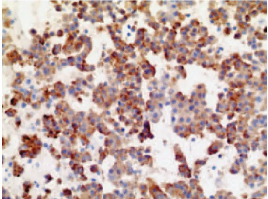
Histological examination of fragments of the cystic wall obtained by FNA (Figure 3) revealed a carcinoid tumor, confirmed by chromogranin and synaptofisin immunohistochemistry analyses (Figure 4).
DISCUSSION
The preoperative diagnosis of cystic pancreatic carcinoid tumor is important due to their malignant potential and possibility of ressection.(5)
Computed tomography (CT) and EUS may not help making diagnosis, since the radiological aspect is often
interpreted as a pancreatic mucinous cystadenoma, such as in this report.
EUS-FNA is a highly accurate method to diagnose pancreatic carcinoid tumors.(2-4) The few studies available
show a high agreement between cytology and pathology.(2-4)
In this report a rare lesion is described, and the diagnosis was possible only after the histological study of the cystic wall fragments obtained by EUS-FNA.
This case report shows the efficacy of EUS-FNA in an unusual diagnosis.
REFERENCES
1. Ballarin R, Masetti M, Losi L, Di Benedetto F, Di Sandro S, De Ruvo N, et al. Cystic pancreatic neuroendocrine neoplasms with uncertain malignant potential: report of two cases. Surg Today. 2009;39(2):162-7.
2. Charfi S, Marcy M, Bories E, Pesanti C, Caillol F, Giovannini M, et al. Cystic pancreatic endocrine tumors: an endoscopic ultrasound-guided fine-needle aspiration biopsy study with histologic correlation. Cancer Cytopathol. 2009;117(3):203-10.
3. Raddaoui E. Clinical utility and diagnostic accuracy of endoscopic ultrasound-guided fine needle aspiration of pancreatic lesions: Saudi Arabian experience. Acta Cytol. 2011;55(1):26-9.
4. Figueiredo FA, Giovannini M, Monges G, Charfi S, Bories E, Pesenti C, et al. Pancreatic endocrine tumors: a large single-center experience. Pancreas. 2009;38(8):936-40.