Approach of an Ultrasound Bronchoscope for
Mediastinal Staging of Lung Cancer
Kyung Jong Lee1, Gee Young Suh1, Man Pyo Chung1, Hojoong Kim1, O Jung Kwon1, Joungho Han2,
Sang-Won Um1*
1Division of Pulmonary and Critical Care Medicine, Department of Medicine, Samsung Medical Center, Sungkyunkwan University School of Medicine, Seoul, Korea, 2Department of Pathology, Samsung Medical Center, Sungkyunkwan University School of Medicine, Seoul, Korea
Abstract
Background: We evaluated the utility of a combined approach using endobronchial ultrasound-guided transbronchial needle aspiration (EBUS-TBNA) and transesophageal bronchoscopic ultrasound-guided fine-needle aspiration (EUS-FNA-B/E) for mediastinal staging of lung cancer.
Methods: An EBUS-TBNA database was analyzed retrospectively. EUS-FNA-B/E was performed after EBUS-TBNA when mediastinal lymph nodes were not accessible using EBUS-TBNA or when tissue sampling using EBUS-TBNA alone was inadequate.
Results:During the study period, 44 patients were enrolled. EBUS-TBNA and EUS-FNA-B/E were performed on 79 and 52 lymph nodes, respectively. The sensitivity, specificity, and accuracy of mediastinal N-staging using EBUS-TBNA alone were 79%, 100%, and 84%, respectively. The sensitivity, specificity, and accuracy of mediastinal N-staging using a combination of EBUS-TBNA and EUS-FNA-B/E were 100%, 100%, and 100%, respectively. Significant differences in sensitivity (P= 0.008) and accuracy (P= 0.004) of mediastinal N-staging were evident when EBUS-TBNA alone and the combined procedure were compared. The nodal stage shifted higher after use of the EUS-FNA-B/E procedure in six cases (13%). No serious complication associated with the procedures was noted.
Conclusions:Use of a combination of EBUS-TBNA and EUS-FNA-B/E can afford better sensitivity and accuracy of mediastinal N-staging compared with use of EBUS-TBNA alone. Such combined procedures should be considered for examination of lesions that are inaccessible or difficult to access by EBUS-TBNA.
Citation:Lee KJ, Suh GY, Chung MP, Kim H, Kwon OJ, et al. (2014) Combined Endobronchial and Transesophageal Approach of an Ultrasound Bronchoscope for Mediastinal Staging of Lung Cancer. PLoS ONE 9(3): e91893. doi:10.1371/journal.pone.0091893
Editor:Min Wu, University of North Dakota, United States of America
ReceivedOctober 18, 2013;AcceptedFebruary 16, 2014;PublishedMarch 14, 2014
Copyright:ß2014 Lee et al. This is an open-access article distributed under the terms of the Creative Commons Attribution License, which permits unrestricted use, distribution, and reproduction in any medium, provided the original author and source are credited.
Funding:The authors have no support or funding to report.
Competing Interests:The authors have declared that no competing interests exist. * E-mail: [email protected]
Introduction
Lung cancer is the leading cause of mortality associated with malignancy despite recent advances in lung cancer management strategies [1]. The possibility of resection is one of the most significant factors influencing the treatment of lung neoplasms if no obvious distant metastasis is evident.
Thus, mediastinal lymph node assessment is important when assessing the resectability of lung cancer in the absence of extra- or intra-thoracic metastasis [2,3]. Mediastinoscopy plays a key role in staging of mediastinal lymph nodes suspicious for metastasis on positron-emission-tomography imaging, although systemic lymph node dissection requires the use of general anesthesia [4].
Endobronchial ultrasound-guided transbronchial needle aspira-tion (EBUS-TBNA) has been developed to diagnose and stage non-small cell lung cancer; the technique is an alternative to mediastinoscopy [5]. EBUS-TBNA cannot access all mediastinal lymph nodes. A combination of endoscopic ultrasound-guided
fine-needle aspiration (EUS-FNA) and EBUS-TBNA can afford a more accurate and systematic assessment of the mediastinum [6]. However, use of this combination is associated with several limitations when mediastinal node staging is performed in clinical practice. The procedure requires expert endoscopists and expen-sive equipment, increasing medical costs and the time required for lung cancer evaluation [7]. It would be better if EBUS-TBNA and EUS-FNA could be performed sequentially, in the same setting, by the same operator.
Materials and Methods
Study Subjects
Data collected between May 2010 and February 2012 at the Samsung Medical Center (Seoul, South Korea) were respectively analyzed. A total of 605 patients underwent EBUS-TBNA for staging and diagnosis of primary lung cancer, extrapulmonary malignancies, lymphoma, tuberculous lymphadenitis, and sarcoid-osis. The study was approved by the Institutional Review Board of the Samsung Medical Center. The requirement for informed consent of the individual patients was waived given the retrospec-tive nature of the study by the Institutional Review Board of the Samsung Medical Center. Patient information was anonymized and de-identified prior to analysis. Patients who underwent EBUS-TBNA followed by EUS-FNA-B/E to diagnose and stage confirmed or suspicious lung cancer were included. Prior to each procedure, PET/CT was routinely used for assessment of mediastinal lymph nodes and other metastatic sites. Patients exhibiting metastasis to extrathoracic sites were excluded. Patients underwent EUS-FNA-B/E after EBUS-TBNA when mediastinal lymph nodes were not accessible by EBUS-TBNA or when adequate tissue samples could not be obtained using EBUS-TBNA alone.
EBUS-TBNA
EBUS-TBNA was performed in a bronchoscopy suite by two bronchoscopists (KJ Lee and SW Um). A routine endobronchial inspection was initially performed. A convex-probe ultrasound bronchoscope fitted with a linear transducer (7.5 MHz) was used in both EBUS-TBNA and EUS-FNA-B/E (CP-EBUS, BF-UC206F-OL8, Olympus, Tokyo, Japan). Lymph node images were also processed using an ultrasonic scanner (EU-C2000; Olympus, Tokyo, Japan). Conscious sedation with midazolam was applied during the EBUS-EBNA procedure, which was performed on an inpatient basis. Nebulized lidocaine (4%) was used to achieve local anesthesia prior to each procedure. Bolus doses of 1.3% lidocaine were administered during the procedure, as needed. The use of EBUS-TBNA to assess mediastinal lymph nodes was indicated when nodes more than 10 mm in diameter (short axis diameter) on chest CT or when increased mediastinal lymph node FDG uptake (compared to that of surrounding tissue) was evident. A 22-gauge needle (NA-201SX-4022; Olympus) was used to obtain TBNA biopsy material. Lymph node station was defined in accordance with the international staging system of the International Association for the Study of Lung Cancer (IASLC) [8]. All aspirate specimens were smeared and immediately fixed on glass slides and sent to a pathologist for cytological and/or histological examination. We did not implement Rapid On-Site Cytopathological Evaluation (ROSE).
Transesophageal bronchoscopic ultrasound-guided fine-needle aspiration (EUS-FNA-B/E) using a EBUS bronchoscope
After completion of EBUS-guided TBNA, the EBUS broncho-scope was re-inserted through the esophagus from the pharynx. To facilitate insertion of the bronchoscope and visualization of the esophagus, oxygen (1–2 l/min) was delivered through a working channel [9]. The scope was inserted gently, and the aortic arch, descending aorta, left main pulmonary artery, and heart were searched for landmarks. A 22-gauge needle was used to sample lymph nodes through the esophageal wall. All aspirate specimens were smeared and fixed immediately on glass slides and sent to a pathologist for cytological and/or histological examination.
Definitions of diagnostic standards
Final nodal stage was defined based on evaluation of the data from all of EBUS-TBNA, EUS-FNA-B/E, and surgery. Metastasis was defined by pathological confirmation via EBUS-TBNA, EUS-FNA-B/E, mediastinoscopy, or mediastinal lymph node dissec-tion. Benign lymph nodes were confirmed by surgery to treat suspected lesions. Patients who had yielded benign results on EBUS-TBNA and EUS-FNA-B/E but whose benign status was not confirmed surgically were excluded from diagnostic perfor-mance analyses.
Statistical analysis
Mediastinal N-staging results from PET-CT, EBUS-TBNA, and the combination of EBUS-TBNA and EUS-FNA-B/E, were analyzed. Comparisons of mediastinal staging afforded by use of EBUS-TBNA alone and the combined procedure were made using McNemar’s test and the Bennett method [10]. Comparisons of mediastinal N-staging afforded by PET/CT and the combina-tion of EBUS-TBNA and EUS-FNA-B/E were also made. The sensitivity, specificity, positive predictive value (PPV), negative predictive value (NPV), and accuracy of mediastinal N staging were analyzed on a per-person basis using standard definitions. All tests were two-tailed and statistical significance was defined when the comparativePvalue was,0.05. The SPSS software (version 20.0; SPSS Inc., Chicago, IL,) was used for statistical analysis.
Results
Study patients
We included 44 patients. Of these, 82% were males and the median age was 66 years (range, 43–86) years. The median procedure time from the start of EBUS-TBNA to the end of EUS-FNA-B/E was 40 min (range, 15–70 min). No serious complica-tion was associated with use of the EBUS-TBNA or EUS-FNA-B/ E procedures. Baseline characteristics of all patients are shown in Table 1.
Lymph nodes examined
Totals of 79 and 52 lymph nodes were examined using EBUS-TBNA and EUS-FNA-B/E, respectively (Table 2). The median number of aspirations per lymph node was 2 (range, 1–4). The
Table 1.Demographic characteristics of the patients (n = 44).
Characteristics Number (%) or median (range)
Age, years 66.0 (43–86)
Male/female (%) 36 (81.8)/ 8 (18.2) Histological type
Adenocarcinoma 19 (43.2)
Squamous cell carcinoma 20 (45.5) Small cell carcinoma 3 (6.8)
NSCLC,NOS 2 (4.5)
Nodal size, mm
Short axis 10.0 (4–58)
Long axis 14.0 (6–72)
Procedure time, min 40 (15–70) Aspiration per lymph node 2 (1–4)
most frequently aspirated nodes by in EBUS-TBNA were the subcarinal lymph nodes (7) while the left paratracheal lymph nodes (in the 4L region) were most often accessed using EUS-FNA-B/E (Table 2). Both EBUS-TBNA and EUS-FNA-B/E were per-formed at 19 nodal stations (2 at station 1R, 11 at station 4L and 6 at station 7). Among the 79 lymph nodes examined by EBUS-TBNA, 24, 53, and 2 yielded malignant, benign, and inadequate results by cytology and 26, 52, and 1 malignant, benign, and inadequate results by histology, respectively. Of the 52 lymph nodes examined by EUS-FNA-B/E, 21, 30, and 1 yielded malignant, benign, and inadequate results by cytology and 24 and 28 had malignant and benign by histology.
Final nodal stage
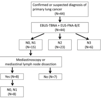
The final stages of study patients are summarized in Figure 1. Six and 23 patients were confirmed as having N3 and N2 disease after combined EBUS-TBNA and EUS-FNA-B/E, respectively. Of 15 patients who yielded benign results after the combined procedure, 8 underwent mediastinoscopy (n = 4) or mediastinal lymph node dissection (n = 4); these patients were confirmed to have N0 or N1 disease. Seven patients who did not undergo surgical confirmation of benign status were excluded from diagnostic performance analyses.
Mediastinal N-staging analyses
The sensitivity, specificity, PPV, NPV, and accuracy of mediastinal N-staging using PET/CT, EBUS-TBNA, and com-bined EBUS-TBNA and EUS-FNA-B/E data are summarized in Table 3. The sensitivity of mediastinal N-staging by PET-CT was 93%, the specificity 37%, the PPV 84%, the NPV 60%, and the accuracy was 81%. Use of EBUS-TBNA alone for mediastinal N-staging yielded a sensitivity of 79%, a specificity of 100%, a PPV of 100%, an NPV of 57%, and an accuracy of 84%. The combined procedure yielded a sensitivity of 100%, a specificity of 100%, a PPV of 100%, an NPV of 100%, and an accuracy of 100%. Significant differences in mediastinal N-staging sensitivity
(p= 0.008) and accuracy (P= 0.004) were evident when EBUS-TBNA alone and the combined procedure were compared. Differences occurred in the diagnostic specificity (P= 0.025) and PPV (P= 0.039) between PET/CT and the combined procedure. Six patients (13.6%) were up-shifted to higher nodal stages after EUS-FNA-B/E data were added to EBUS-TBNA information. Nodal stage changes are summarized in Table 4. EUS-FNA-B/E could detect nodal metastasis in stations 1R, 4L, 5, 7, and 8.
Discussion
In the present study, the sensitivity of mediastinal N-staging afforded by combined EBUS-TBNA and EUS-FNA-B/E was significantly higher than that of EBUS-TBNA alone. Upstaging was noted in six of 44 patients (13.6%) by adding EUS-FNA-B/E. Table 2.Mediastinal lymph nodes examined by
endobronchial ultrasound-guided transbronchial needle aspiration (EBUS-TBNA) and transesophageal bronchoscopic ultrasound-guided fine-needle aspiration (EUS-FNA-B/E).
Nodal stations Number of lymph nodes
EBUS-TBNA (n = 79) EUS-FNA-B/E (n = 52)
1R 2 4
2R 8 1
3P 0 2
4R 21 0
4L 18 24
5 0 7
7 23 6
8 0 6
10L 0 2
11R 2 0
11L 5 0
EBUS-TBNA: endobronchial ultrasound-guided transbronchial needle aspiration; EUS-FNA-B/E: transesophageal bronchoscopic ultrasound-guided fine-needle aspiration.
doi:10.1371/journal.pone.0091893.t002
Figure 1. Diagnostic algorithm of study patients. EBUS-TBNA, endobronchial ultrasound-guided transbronchial needle aspiration; EUS-FNA-B/E, transesophageal bronchoscopic ultrasound-guided fine-needle aspiration.
doi:10.1371/journal.pone.0091893.g001
Table 3.Diagnostic performances of PET/CT, EBUS-TBNA, and the combination of EBUS-TBNA and EUS-FNA-B/E for the detection of mediastinal metastasis on a per person basis.
Sensitivity Specificity PPV NPV Accuracy
PET-CT 93.1(27/29) 37.5 (3/8) 84.3 (27/32) 60.0 (3/5) 81.0 (30/37) EBUS 79.3 (23/29) 100 (8/8) 100 (23/23) 57.1 (8/14) 83.7 (31/37) EBUS+EUS 100 (29/29) 100 (8/8) 100 (29/29) 100 (8/8) 100 (37/37) p-value* 0.083 0.025 0.039 0.205 0.004
p-value** 0.008 NA NA 0.053 0.004
Data are presented as n (%).
* p-values from the comparisons of PET-CT and EBUS+EUS-FNA-B/E. ** p-values from the comparisons of EBUS-TBNA and EBUS+EUS-FNA-B/E. PPV: positive predictive value; NPV: negative predictive value; PET-CT: positron-emission tomography with computed tomography; EBUS-TBNA: endobronchial ultrasound-guided transbronchial needle aspiration; EUS-FNA-B/E:
transesophageal bronchoscopic ultrasound-guided fine-needle aspiration; NA: not applicable.
Five of these six patients were re-staged from N0 to N2 or N3, and one patient was re-staged from N2 to N3.Mediastinal lymph node evaluation permitting exact staging is important in predicting prognosis and planning treatment strategies for lung cancer [11]. FDG-PET assessment of mediastinal lymph nodes yielded a sensitivity of 73% and a specificity of 92% [12]. Despite this excellent specificity of integrated PET-CT, these data suggest that tissue samples should be obtained to confirm mediastinal node staging. Currently, video-assisted mediastinoscopy is the gold standard for evaluation of all accessible mediastinal lymph nodes in lung cancer patients. A review of video-assisted mediastino-scopic lymphadenectomy in patients with NSCLC reported a sensitivity of 90% and a false-negative rate of 7%. Moreover, the procedure-related morbidity and mortality rates of mediastino-scopic lymphadenectomy are 2% and 0.08%, respectively [3].
EBUS-TBNA is a non-invasive procedure used to aspirate mediastinal nodes employing an ultrasonic probe adopted for use in bronchoscopy. The procedure has several advantages, including a lower risk of morbidity and mortality and an increase in accessible regions, including the hilar and interlobar lymph nodes, compared with mediastinoscopy [13]. A meta-analysis performed by Guet al. showed that use of the technique afforded a sensitivity of 93% and a specificity of 100% [14]. EBUS-TBNA does not allow access to the paraaortic or paraesophageal lymph nodes via a transbronchial approach. The technique also cannot be used to access the deep left lower paratracheal station because of the limited angulation afforded by bronchoscopy [15].
To overcome these limitations, a combined procedure including EUS-FNA was developed. The use of EUS-FNA with EBUS-TBNA enabled accurate and systematic assessment of mediastinal lymph nodes [16]. This combined approach was associated with a significantly higher sensitivity and specificity for non-small cell lung cancer staging than were EBUS-TBNA or EUS-FNA alone [17]. EUS-FNA can access additional lymph node stations that are inaccessible using an endobronchial approach. The systematic approach has a sensitivity of 68–96% and an NPV of 75–97% [17–19]. The results suggest that use of the combined procedure enables more accurate mediastinal staging.
A transesophageal approach using a single EBUS bronchoscope was performed in the same bronchoscopy suite for more accurate
evaluation of mediastinal staging in this study. In total, 112 lymph nodes were sampled in 44 patients by a single ultrasound bronchoscope through the bronchus and esophagus. We sampled the same lymph node using EUS-FNA-B/E if EBUS-TBNA was unable to acquire sufficient tissue. This could occur because the lymph node was located in a deep station that was not readily visible using the ultrasound probe or because there was insufficient tissue from aspiration due to poor alignment. Only 71% of all sampled lymph nodes were accessible and sampled by EBUS-TBNA alone, but the proportion of accessible lymph nodes increased with the addition of the transbronchial approach using a single ultrasound bronchoscopy. In this study, 19 nodal stations (2 at station 1R, 11 at station 4L, and 6 at station 7) were examined by both EBUS-TBNA and EUS-FNA-B/E at the bronchoscopist’s discretion, as it seemed that sufficient tissue was not obtained by EBUS-TBNA alone. Material aspirated from three nodal stations using EBUS-TBNA and EUS-FNA-B/E yielded different results. These three nodal stations were diagnosed as metastatic by EUS-FNA-B/E despite being negative on EBUS-TBNA. The diagnostic sensitivities of EBUS-TBNA and EUS-FNA-B/E were 75% (9/12) and 100% (12/12) for these 19 nodal stations, respectively.
The advantages of the combined procedure using a single ultrasonic bronchoscope have been reported in two studies. Boet al. evaluated the use of combined B-FNA and EBUS-TBNA in 150 patients and claimed a 97% diagnostic accuracy for detection of mediastinal metastasis [20]. Herthet al. also used a transbronchial approach followed by transesophageal needle aspiration employing an ultrasonic bronchoscope and reported a 96% sensitivity of cancer detection and a 96% NPV on examination of 619 nodes from 139 patients with suspected lung cancer [21]. We also found that the accuracy of mediastinal staging was improved after addition of EUS-FNA-B/E in clinical practice. The combined procedure yielded more accurate results than those afforded by PET-CT or EBUS-TBNA alone.
Use of EUS-FNA overcomes the limitations of EBUS-TBNA for mediastinal nodal staging of lung cancer. However, the combina-tion of EBUS-TBNA and EUS-FNA increases the medical cost and delays the diagnosis, as EUS-FNA would need to be performed by expert endoscopists during a different session. The combination of EBUS-TBNA and EUS-FNA-B/E using a single Table 4.The six cases of increased mediastinal stage after addition of EUS-FNA-B/E.
Case Gender Age EBUS-TBNA EUS-FNA-B/E Shift of N stage
1 M 77 7: benign 7:adenocarcinoma N0RN2
2 M 70 4R: benign 5: adenocarcinoma N0RN2
7: benign 4L: benign
3 M 69 4R:Squamous cell carcinoma 4L: Squamous cell carcinoma N2RN3
4 M 57 2R: benign 5: adenocarcinoma N0RN2
4L: benign
5 M 61 7: benign 1R: adenocarcinoma N0RN3
8: benign
6 M 72 11R: benign 8: squamous cell carcinoma N0RN2
7: benign 4R: benign 4L: benign
EBUS-TBNA: endobronchial ultrasound-guided transbronchial needle aspiration; EUS-FNA-B/E: transesophageal bronchoscopic ultrasound-guided fine-needle aspiration.
ultrasonic bronchoscope is a useful alternative method because EUS-FNA-B/E by a bronchoscopist does not increase the medical cost and it could be performed immediately after EBUS-TBNA. The median EBUS-TBNA procedure time was 20 min at our institution, and that of the combined EBUS-TBNA and EUS-FNA was 40 min. Therefore, the combined procedure increased the procedure time by only 20 min compared with EBUS-TBNA alone.
Our study had several limitations. First, all participants were recruited retrospectively and our sample size was small. Thus, The possibility of selection bias exists; we may have overestimated the utility of the combined procedure. Therefore, our data should be interpreted conservatively as only 44 (7.3%) of the 605 patients were enrolled and 13% upstaging in 44 patients represents only 1% of 605 patients. Second, of 15 patients who had negative N2 or N3 nodes after EBUS-TBNA and EUS-FNA-B/E, only 8 underwent surgical confirmation using mediastinoscopy or medi-astinal lymph node dissection. This may contribute to overesti-mation of the diagnostic performance of the combined procedure. If the seven patients who did not undergo surgical confirmation
are considered to be false-negatives, the sensitivity, NPV, and accuracy of mediastinal N-staging by EBUS-TBNA and the combined procedure would be 64% (23/36), 38% (8/21), and 70% (31/44); and 81% (29/36), 53% (8/15), and 84% (37/44), respectively. However, significant differences would remain in terms of both the sensitivity (P= 0.016) and accuracy (P= 0.008) of mediastinal N-staging conducted using the EBUS-TBNA and combined procedure.
In conclusion, use of a combination of EBUS-TBNA and EUS-FNA-B/E afforded greater diagnostic sensitivity and accuracy of mediastinal N-staging than did EBUS-TBNA alone. The com-bined procedure can be used for more accurate mediastinal staging of lesions difficult to access using EBUS-TBNA only.
Author Contributions
Conceived and designed the experiments: SWU. Analyzed the data: KJL SWU. Wrote the paper: KJL SWU. Data acquisition: KJL GYS MPC HK OJK JH.
References
1. Jemal A, Siegel R, Xu J, Ward E (2010) Cancer statistics, 2010. CA Cancer J Clin 60: 277–300.
2. De Leyn P, Lardinois D, Van Schil PE, Rami-Porta R, Passlick B, et al. (2007) ESTS guidelines for preoperative lymph node staging for non-small cell lung cancer. Eur J Cardiothorac Surg 32: 1–8.
3. Detterbeck FC, Jantz MA, Wallace M, Vansteenkiste J, Silvestri GA (2007) Invasive mediastinal staging of lung cancer: ACCP evidence-based clinical practice guidelines (2nd edition). Chest 132: 202S–220S.
4. De Leyn P, Lardinois D, Van Schil P, Rami-Porta R, Passlick B, et al. (2007) European trends in preoperative and intraoperative nodal staging: ESTS guidelines. J Thorac Oncol 2: 357–361.
5. Yasufuku K, Pierre A, Darling G, de Perrot M, Waddell T, et al. (2011) A prospective controlled trial of endobronchial ultrasound-guided transbronchial needle aspiration compared with mediastinoscopy for mediastinal lymph node staging of lung cancer. J Thorac Cardiovasc Surg 142: 1393–1400 e1. 6. Wallace MB, Pascual JM, Raimondo M, Woodward TA, McComb BL, et al.
(2008) Minimally invasive endoscopic staging of suspected lung cancer. JAMA 299: 540–546.
7. Konge L, Vilmann P, Clementsen P, Annema JT, Ringsted C (2012) Reliable and valid assessment of competence in endoscopic ultrasonography and fine-needle aspiration for mediastinal staging of non-small cell lung cancer. Endoscopy 44: 928–933.
8. Rusch VW, Asamura H, Watanabe H, Giroux DJ, Rami-Porta R, et al. (2009) The IASLC lung cancer staging project: a proposal for a new international lymph node map in the forthcoming seventh edition of the TNM classification for lung cancer. J Thorac Oncol 4: 568–577.
9. Verma A, Kang YR, Kim H, Um SW (2012) Ultrasound guided fine needle aspiration of a lung mass via a transesophageal approach using endobronchial ultrasound bronchoscope. Thoracic Cancer 3: 280–283.
10. Bennett BM (1972) On comparisons of sensitivity, specificity and predictive value of a number of diagnostic procedures. Biometrics 28: 793–800. 11. Rusch VW, Crowley J, Giroux DJ, Goldstraw P, Im JG, et al. (2007) The IASLC
Lung Cancer Staging Project: proposals for the revision of the N descriptors in the forthcoming seventh edition of the TNM classification for lung cancer. J Thorac Oncol 2: 603–612.
12. Lv YL, Yuan DM, Wang K, Miao XH, Qian Q, et al. (2011) Diagnostic performance of integrated positron emission tomography/computed tomogra-phy for mediastinal lymph node staging in non-small cell lung cancer: a bivariate systematic review and meta-analysis. J Thorac Oncol 6: 1350–1358. 13. Tournoy KG, Keller SM, Annema JT (2012) Mediastinal staging of lung cancer:
novel concepts. Lancet Oncol 13: e221–229.
14. Gu P, Zhao YZ, Jiang LY, Zhang W, Xin Y, et al. (2009) Endobronchial ultrasound-guided transbronchial needle aspiration for staging of lung cancer: a systematic review and meta-analysis. Eur J Cancer 45: 1389–1396.
15. Jhun BW, Park HY, Jeon K, Koh WJ, Suh GY, et al. (2012) Nodal stations and diagnostic performances of endobronchial ultrasound-guided transbronchial needle aspiration in patients with non-small cell lung cancer. J Korean Med Sci 27: 46–51.
16. Tournoy KG, Annema JT, Krasnik M, Herth FJ, van Meerbeeck JP (2009) Endoscopic and endobronchial ultrasonography according to the proposed lymph node map definition in the seventh edition of the tumor, node, metastasis classification for lung cancer. J Thorac Oncol 4: 1576–1584.
17. Szlubowski A, Zielinski M, Soja J, Annema JT, Sosnicki W, et al. (2010) A combined approach of endobronchial and endoscopic ultrasound-guided needle aspiration in the radiologically normal mediastinum in non-small-cell lung cancer staging—a prospective trial. Eur J Cardiothorac Surg 37: 1175–1179. 18. Vilmann P, Krasnik M, Larsen SS, Jacobsen GK, Clementsen P (2005)
Transesophageal endoscopic ultrasound-guided fine-needle aspiration (EUS-FNA) and endobronchial ultrasound-guided transbronchial needle aspiration (EBUS-TBNA) biopsy: a combined approach in the evaluation of mediastinal lesions. Endoscopy 37: 833–839.
19. Ohnishi R, Yasuda I, Kato T, Tanaka T, Kaneko Y, et al. (2011) Combined endobronchial and endoscopic ultrasound-guided fine needle aspiration for mediastinal nodal staging of lung cancer. Endoscopy 43: 1082–1089. 20. Hwangbo B, Lee GK, Lee HS, Lim KY, Lee SH, et al. (2010) Transbronchial
and transesophageal fine-needle aspiration using an ultrasound bronchoscope in mediastinal staging of potentially operable lung cancer. Chest 138: 795–802. 21. Herth FJ, Krasnik M, Kahn N, Eberhardt R, Ernst A (2010) Combined