Sperm Defect Severity Rather Than Sperm Source Is
Associated With Lower Fertilization Rates after
Intracytoplasmic Sperm Injection
Sidney Verza Jr, Sandro C. Esteves
Androfert, Center for Male Reproduction, Campinas, Sao Paulo, Brazil
ABSTRACT
Objective: To evaluate the impact of sperm defect severity and the type of azoospermia on the outcomes of intracytoplasmic sperm injection (ICSI).
Materials and Methods: This study included 313 ICSI cycles that were divided into two major groups according to the source of spermatozoa used for ICSI: 1) Ejaculated (group 1; n = 220) and 2) Testicular/Epididymal (group 2; n = 93). Group 1 was subdivided into four subgroups according to the results of the semen analysis: 1) single defect (oligo-[O] or astheno-[A] or teratozoospermia-[T], n = 41), 2) double defect (a combination of two single defects, n = 45), 3) triple defect (OAT, n = 48), and 4) control (no sperm defects; n = 86). Group 2 was subdivided according to the type of azoospermia: 1) obstructive (OA: n = 39) and 2) non-obstructive (NOA: n = 54). Fertilization (2PN), cleavage, embryo quality, clinical pregnancy and miscarriage rates were statistically compared using one-way ANOVA and Chi-square analyses.
Results: Significantly lower fertilization rates were obtained when either ejaculated sperm with triple defect or testicular sperm from NOA patients (63.4 ± 25.9% and 52.2 ± 29.3%, respectively) were used for ICSI as compared to other groups (~73%; P < 0.05). Epididymal and testicular spermatozoa from OA patients fertilized as well as normal or mild/moderate deficient ejaculated sperm. Cleavage, embryo quality, pregnancy and miscarriage rates did not differ statistically between ejaculated and obstructive azoospermia groups. However, fertilization, cleavage and pregnancy rates were significantly lower for NOA patients.
Conclusion: Lower fertilization rates are achieved when ICSI is performed with sperm from men with oligoasthenoteratozoospermic and non-obstructive azoospermic, and embryo development and pregnancy rates are sig-nificantly lower when testicular spermatozoa from NOA men are used.
Key words: spermatozoa; intracytoplasmic sperm injection; azoospermia; oligozoospermia Int Braz J Urol. 2008; 34: 49-56
INTRODUCTION
Intracytoplasmic sperm injection (ICSI) has been the standard for the treatment of severe male factor infertility. Before ICSI, sperm from men with severely defective spermatogenesis, such those with
producing normal and viable pre-embryos (1-8). Al-though the use of surgically retrieved or ejaculated sperm from men with severely impaired spermatoge-nesis for ICSI has greatly improved treatment of se-vere male infertility, the consequences of using such gametes are not fully known (7).
Many studies have shown conflicting results when ICSI is performed with sperm from different sources (2-8). It is difficult to interpret these results because, apart from few studies, only the sperm source is analyzed and there is no systematic distinction be-tween obstructive and non-obstructive azoospermia. This prevents consideration of the influence of sper-matic defects. For instance, an ejaculated semen sample can present varying degrees of sperm abnormalities – from absence to severe alterations in all spermatic parameters, as in cases of oligoasthenoterato-zoospermia. Spermatozoa from normal, mild/moderate or severe abnormal semen may show different fertiliz-ing abilities after ICSI even though their source is the same, i.e., the ejaculate (8). Also, important physiologic differences are observed in obstructive and non-ob-structive azoospermia. While obnon-ob-structive azoospermic men have normal sperm production, non-obstructive ones have defective spermatogenesis and very limited amount of sperm production, if any, and they may carry several sperm defects, including genetic ones (9). Therefore, it is reasonable to speculate that ICSI re-sults depend not only on sperm source but also on the severity of sperm defect.
The objective of this study was to evaluate the impact of sperm defect severity and the type of azoospermia on the outcomes of intracytoplasmic sperm injection (ICSI).
MATERIALS AND METHODS
This is a retrospective study including 313 ICSI cycles performed from January 2002 to No-vember 2003. To allow data collection, institutional review board approval was obtained as well as pa-tient written consent. Treatment cycles were divided into two main groups according to the sperm source for ICSI: 1) ejaculated (group 1; n = 220), and 2) sperm obtained from testicles/epididymis (group 2;
n = 93). Group 1 was subdivided into 4 subgroups according to the anomaly observed in the seminal analyses, which were performed in accordance with the World Health Organization Manual (10), and sperm morphology using Tygerberg’s strict criteria (11), as follows: (a) single sperm defect (oligozo-ospermia - [O]: sperm concentration < 20x106/mL;
or asthenozoospermia - [A]: progressive motility < 50%; or teratozoospermia [T]: sperm morphology < 8%; n = 41); (b) double sperm defect, i.e., a combi-nation of two defects described above, n = 45); (c) triple sperm defect (all defects combined: oligoasthenoteratozoospermia [OAT], n = 48); and (d) control (no sperm defect, n = 86). The limit of 8% of spermatic morphology was used since recent studies (12-15) have shown a tendency to reduce the now used normality reference of 14%. Group 2 was subdivided in accordance with the type of azoospermia: 1) obstructive (AO: n = 39 [epididymis n = 31; testicles n = 8), and 2) non-obstructive (ANO: testicles n = 54).
Ovarian stimulation and follicular aspiration - Pituitary suppression was achieved by using intra-nasal gonadotrophin hormone analog (nafarelin ac-etate, Synarel, Zodiac) followed by ovarian stimula-tion with daily doses of 150-300 IU of human meno-pausal gonadotrophin (HMG or HP-HMG; Ferring). Human chorionic gonadotrophin (hCG; Choragon, Ferring) was used when 2 or more ovarian follicles presented a mean diameter of 18 mm. Thirty-four to thirty-six hours after the hCG administration, an ul-trasound-guided transvaginal follicular aspiration was performed under general anesthesia administered in-travenously.
Oocyte handling and classification - After follicular aspiration, the tubes with the follicular fluid were transferred to the in vitro fertilization labora-tory and examined on stereomicroscopy to identify the corona-cumulus-oocyte complexes (CCOC). Im-mediately, CCOC chemical treatment with 40 IU/ mL hyaluronidase (Hyase, Vitrolife, Sweden) was performed for 30 seconds. The oocytes were then transferred to microdroplets of culture media (IVF, Vitrolife, Sweden) covered with mineral oil (Ovoil, Vitrolife, Sweden) and kept in an incubator for 1-2 hours at 37o C in a 5.5% CO
Mechani-cal oocyte denudation was then performed with a 130µm diameter pipette (Flexipet, Cook, USA) and the denuded oocytes were classified according to their maturity into metaphase II (MII, oocytes show-ing the extrusion of the 1st polar corpuscle), prophase I (oocytes at the germinative vesicle stage), metaphase I (oocytes showing no germinative vesicule or extrusion of the 1st polar corpuscle), atresic (oocytes with signs of degeneration) and fractured (oocytes with rupture in the zona pelucida with total or partial extrusion of cytoplasm).
Sperm retrieval in group 1 - Sperm was ob-tained by ejaculation after 48-72 hours of sexual ab-stinence, and the sample was kept at 37o C for 30
minutes or until complete liquefaction. Sperm wash-ing was performed by uswash-ing the two-layer discon-tinuous colloidal gradient (Isolate, Irvine Scientific, USA). The ejaculate and the gradients were centri-fuged for 25 minutes at 300Xg. The supernatant was discharged and the bottom layer was subsequently diluted with HEPES-buffered culture medium (modi-fied HTF, Irvine Scientific, USA; 1:2, v/v), and washed again by centrifugation for 10 minutes. The pellet was re-suspended in 200 microliters of a HEPES-buffered culture media and maintained at 37oC until
the time of ICSI. An aliquot was removed for the assessment of sperm count and motility.
Sperm retrieval in group 2 - Sperm retrieval from the testis or epididymis was performed under intravenous anesthesia with propofol in association with blockage of the spermatic cord with 2% xylocaine on an outpatient basis. Sperm retrieval from the testis was done percutaneously by testicular sperm aspiration (TESA) using a 21-gauge needle or by microsurgical-guided open biopsy (testicular microdissection, micro-TESE) (16). The seminifer-ous tubules were dissected mechanically under stereomicroscopy in HEPES-buffered culture me-dium (Modified HTF, Irvine Scientific, USA). The supernatant was collected for sperm search. Sperm retrieval from the epididymis was performed by per-cutaneous epididymal sperm aspiration (PESA) us-ing a 13.5 gauge needle connected to a 1 mL sy-ringe. Aspirates were diluted in HEPES-buffered culture medium. In all cases of TESA, micro-TESE or PESA, the samples were centrifuged at 300Xg
for 10 minutes. The pellets were subsequently re-suspended in HEPES-buffered culture medium and kept at 37oC until microinjection. Sperm search was
performed at 400X magnification using a phase-con-trast inverted microscope. In the non-obstructive azoospermia cases, sperm retrieval was performed by TESA or micro-TESE, whereas PESA was used to obtain sperm in obstructive azoospermia cases. However, in 8 cases of obstructive azoospermia, PESA was not successful, and the TESA technique was used to obtain sperm for ICSI in these cases.
Gamete micromanipulation and microinjec-tion - Microinjecmicroinjec-tions were performed at X400 mag-nification on a 37oC heated stage phase-contrast
in-verted microscope (Nikon, Japan) (1). A Petri dish containing a 4µL microdroplet of PVP (polyvinilpirrolidone, Vitrolife, Sweden) under min-eral oil was used for sperm selection and immobili-zation. On the same dish, a 20µL microdroplet of culture medium (Gamete, Vitrolife, Sweden) was used for placing the oocytes for microinjection. A single sperm was mechanically immobilized by using the tip of the microinjection needle (Cook, USA) and then aspirated inside the needle. The oocyte was held in place using a 35 degree angle holding micropi-pette (Cook, USA) with the polar body in the 6 or 7 o’clock position. Injection of a single spermatozoon within the oocyte cytoplasm was performed by us-ing electrohydraulic micromanipulators (Narishige, Japan). After all microinjections from a single case were completed, the injected oocytes were trans-ferred to a closed culture system (microdroplets un-der mineral oil, series II, Vitrolife, Sweden), and in-cubated for 16-18 hours at 37oC and 5.5% CO
2 until
fertilization was determined.
multinucleation, grades I (absence of fragmentation) or II (up to 20% of vitelineous space occupied by frag-ments) of cytoplasmic fragmentation, and absence of zona pelucida alterations (17).
Embryo transfer - All embryo transfers were performed around 72 hours after ICSI. A delicate two-step catheter (Sidney IVF, Cook, USA) was used to place the embryos inside the uterine cavity. Transvaginal embryo transfers were guided by ab-dominal ultrasound.
Main outcome measures - Descriptive param-eters such as female age and mean number of oo-cytes retrieved were compared among subgroups. After ICSI, the following parameters were analyzed and compared: normal (2PN) and abnormal fertiliza-tion rates, cleavage and good quality pre-embryo rates on days 2 and 3, number of pre-embryos transferred, clinical pregnancy and miscarriage rates. A clinical pregnancy was confirmed by the presence of a ges-tational sac with an embryo exhibiting cardiac activity on a 5th to 6th week ultrasound scan. Miscarriage was considered when a non-viable clinical pregnancy was noted on ultrasound follow-up.
The Kolmogorov-Smirnov test was used to verify if data followed normal distribution. A one-way ANOVA or Fisher exact test was used to compare clinical and laboratory parameters between groups when appropriate. The Chi-square test was used to compare pregnancy and miscarriage rates, with P < 0.05 considered significant (18). Statistical analysis was performed by StatSoft program, Tulsa, EUA.
RESULTS
Female age, number of mature oocytes re-trieved and number of pre-embryos transferred were not statistically different among groups (Table-1).
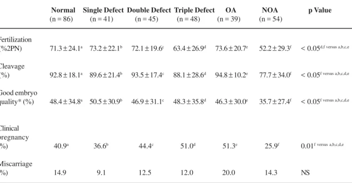
Significantly lower normal fertilization rates were obtained when ejaculated sperm with triple sperm defect (63.4 ± 25.9%) or testicular sperm from pa-tients with non-obstructive azoospermia (52.2 ± 29.3%) were used for ICSI in comparison with other groups (71.3-73.6%) (p < 0.05, Table-2).
There was no difference in fertilization rates when sperm from the epididymis (74.7% ± 21.2%) or
the testicles (69.1% ± 19.6%) of patients with obstruc-tive azoospermia was used for ICSI, as compared with ejaculated sperm with mild (single sperm defect; 73.2 ± 22.1%) to moderate (double sperm defect; 72.1 ± 19.6%) alterations (Tables-2 and 3). Cleavage, pre-embryo quality, clinical pregnancy and miscarriage rates were not statistically different between ejaculated and obstructive azoospermia (OA) groups, independent of whether the spermatozoa from obstructive azoospermic patients was obtained from the epididymis or testicles (Table-3). However, cleavage rates, pre-embryo qual-ity, and clinical pregnancy were significantly lower in non-obstructive azoospermia (NOA) group, when com-pared to the other groups (Table-2).
COMMENTS
Several studies report ICSI results based on the sperm source rather than on sperm defect sever-ity. Sperm source criteria, however, may include sper-matozoa from different etiologies. For instance, ejacu-lated semen may contain slightly abnormal spermato-zoa from a man with a moderate varicocele, but it may also harbor severely defective sperm from men with genetic disorders, such as the Klinefelter syn-drome and AZFc Y chromosome microdeletions. Fur-thermore, in cases of azoospermia, sperm from the epididymis and the testicles can be used for ICSI. However, spermatogenesis in men with obstructive and non-obstructive azoospermia is very distinct. While sperm production is normal in the former, it is severely abnormal, if existing, in the latter, despite the fact that in both cases ICSI may be performed using testicular spermatozoa (16).
se-Table 1 – Descriptive statistics from group 1 (ejaculated sperm subdivided according to the severity of sperm deficiency) and group 2 (testicular/epididymal sperm) including female age, number of mature oocytes (MII) retrieved following follicle aspiration, number of pre-embryos transferred to the uterine cavity. Values are mean ± SD.
Female age (yrs.)
Mature oocytes retrieved (n)
Embryo transfer (n)
Normal
(n = 86)
34.7 ± 4.7
8.6 ± 5.9
3.3 ± 1.4
Single Defect
(n = 41)
32.4 ± 3.8
8.5 ± 4.7
3.5 ± 1.2
Double Defect
(n = 45)
31.6 ± 5.6
9.3 ± 6.2
3.6 ± 1.3
Triple Defect
(n = 48)
32.8 ± 5.1
7.9 ± 4.6
3.2 ± 1.6
OA
(n = 39)
32.6 ± 7.4
9.6 ± 5.5
3.4 ± 1.3
NOA
(n = 54)
33.6 ± 6.5
10.6 ± 5.3
3.0 ±1.6
p Value
NS
NS
NS
Group 1 – Ejaculated Sperm
(n = 220)
Group 2 – Testicular and Epididymal Sperm from Azoospermic Men
(n = 93)
One-way ANOVA was used to compare clinical and laboratorial parameters among groups; p < 0.05 was considered significant; NS = not significant; OA = obstructive azoospermia; NOA = non-obstructive azoospermia, values are mean ± standard deviation.
Table 2 – ICSI outcomes with ejaculated sperm with and without sperm defects, and with epididymal and testicular spermatozoa from men with obstructive and non-obstructive azoospermia.
Fertilization
(%2PN) 71.3 ± 24.1a 73.2 ± 22.1b 72.1 ± 19.6c 63.4 ± 26.9d 73.6 ± 20.7e 52.2 ± 29.3f < 0.05d,f versus a,b,c,e
Cleavage
(%) 92.8 ± 18.1a 89.6 ± 21.4b 93.5 ± 17.4c 88.1 ± 28.6d 94.8 ± 10.2e 77.7 ± 34.0f < 0.05f versus a,b,c,d,e
Good embryo
quality* (%) 48.4 ± 34.8a 50.5 ± 30.9b 46.9 ± 31.1c 48.3 ± 35.8d 46.3 ± 30.0e 35.7 ± 27.4f < 0.05f versus a,b,c,d,e
Clinical pregnancy
(%) 40.9a 36.6b 44.4c 51.0d 51.3e 25.9f 0.01f versus a,b,c,d,e
Miscarriage
(%) 14.9 9.1 12.5 12.0 20.0 14.3 NS
Normal Single Defect Double Defect Triple Defect OA NOA p Value
(n = 86) (n = 41) (n = 45) (n = 48) (n = 39) (n = 54)
Group 1 – Ejaculated Sperm
(n = 220)
Group 2 – Testicular and Epididymal Sperm from Azoospermic Men (n = 93)
verity of sperm abnormalities found in the semen analy-sis. Most studies evaluating ICSI and azoospermia take into account only the sperm source but not the type of azoospermia. These studies tend to have a better out-come when epididymal spermatozoa are used, but these findings can be justified by the fact that epididy-mal sperm are always from obstructive azoospermia, while testicular sperm can be from both types of azoospermia (20,22).
Apart from very few studies, there is no sys-tematic differentiation between obstructive and non-obstructive azoospermia. This prevents consideration of the importance of spermatogenesis defects and of sperm immaturity on fertilization and embryo devel-opment. It is also true for ejaculated sperm in which spermatogenesis can vary greatly in a given ejacu-late. Sperm samples can be within the normal ranges according to the WHO criteria, but can also harbor sperm with defects in count, motility and morphology. Therefore, it is reasonable to speculate that the fertil-izing potential of these gametes may differ. In fact, ICSI outcomes with testicular spermatozoa from non-obstructive azoospermic men have been poorer than ejaculated and epididymal sperm (2-4,21). In addition, miscarriage rates after ICSI with testicular sperma-tozoa have been reported to be higher (5,7), although a clear distinction in the type of azoospermia is not always possible to analyze. These findings strengthen
the importance of the type of azoospermia and not only the sperm source.
Our study took into consideration a different point of view from the previous studies by subdividing sperm deficiencies seen on semen analyses by degree of severity and also by the type of azoospermia. We observed lower fertilization rates when ejaculated sper-matozoa from oligoasthenoteratozoospermic (triple de-fect) men were used for ICSI as compared to the other ejaculated samples, and also in relation to testicular or epididymis spermatozoa from the obstructed azoospermic men. From all subgroups, testicular sper-matozoa from NOA patients had the worst performance after ICSI. We observed significantly lower fertiliza-tion and embryo development as well as pregnancy rates when compared to the other subgroups. How-ever, our study was unable to identify differences in miscarriage among groups as shown by others (5,7). Although there is a tendency for higher miscarriage rates in our group of azoospermic patients, the sample size is limited to allow proper analysis.
Our results indicate that sperm from men with severely altered spermatogenesis, such as ejaculated sperm in OAT and testicular sperm in NOA, have decreased fertility potential after ICSI. We speculate that in terms of severity, non-obstructive azoospermia (NOA) may be a progression of oligoasthenoterato-zoospermia (OAT), thus justifying the diminished per-Table 3 – ICSI outcomes using epididymal and testicular sperm retrieved from obstructive azoospermic men.
Female age (years) 31.5 ± 7.7 36.3 ± 5.1 NS Mature oocytes retrieved (n) 09.4 ± 5.8 09.4 ± 4.9 NS Embryo transfer (n) 03.3 ± 1.3 03.7 ± 1.5 NS Fertilization (% 2PN) 74.7 ± 21.2 69.1 ± 19.6 NS Cleavage (%) 95.5 ± 9.0 92.2 ± 14.4 NS Good embryo quality * (%) 44.6 ± 30.5 52.7 ± 29.6 NS
Clinical pregnancy (%) 51.6 50.0 —
Miscarriage (%) 18.8 25.0 —
Fisher exact test was used to compare clinical and laboratorial parameters between groups. Clinical pregnancy and miscarriage rates were not compared due to the insufficient sample size. P < 0.05 considered significant; NS = not significant; * = 7-9 blastomeres of similar size, and grades I or II cytoplasmic fragmentation on the day of embryo transfer (day 3) (17). OA: obstructive azoospermia, values are mean ± standard deviation.
formance of sperm from both conditions in ICSI. Dif-ferently, in OA cases the absence of sperm in the ejaculate is exclusively due to an obstruction in some point of the ductal system, but the spermatogenesis is normal. ICSI results in this condition is independent of the sperm source, i.e., sperm retrieved from the epididymis or testicles perform similarly and at the same extent of normal to mild/moderate abnormal (single or double defects) ejaculated spermatozoa. Besides, we observed that normal fertilization rates with OA sperm was significantly higher when com-pared to sperm obtained from OAT ejaculated semen, thus reinforcing the hypothesis that the spermatic de-fect severity is more important than the sperm source in ICSI results.
Indeed, sperm from men with severely de-fective spermatogenesis may have a higher tendency to carry deficiencies, such as the ones related to the centrioles and genetic material, which ultimately af-fects the capability of the male gamete to activate the egg and trigger the formation and development of a normal zygote and a viable pre-embryo. A higher chro-mosomal aneuploidy rate and other genetic alterations (9,23) have been found in spermatozoa from NOA and OAT men. Pang et al. (24) in studies of sexual chromosomes and other 12 autosomes showed a higher incidence of chromosome aneuploidy rate in sperm of men with OAT versus fertile controls, varying from 33-74% in the first group versus 4.1-7.7% in the trol. Recent studies (25) tend to differentiate the con-tribution of the male gamete (termed ‘paternal effect’) to early and late embryo development. Lower fertili-zation rates, as observed in our study for OAT sperm and for testicular sperm from NOA men, are explained by early paternal effects, which include alterations in spermatic cytosolic factor and are responsible for the completion of the oocyte meiotic division as well as alterations on sperm centriole, which participate in the formation of embryo mitotic fuses in early cellular di-visions (25). Other alterations such as sperm DNA fragmentation would be associated to the so called late paternal effect that impacts embryo implantation. Recent studies show lower pregnancy rates when the proportion of sperm with fragmented DNA in a given sample is above 10% and absence of pregnancy when the DNA fragmentation is above 25% (26).
CONCLUSION
Adequate fertilization, cleavage and preg-nancy rates are to be expected when ICSI is per-formed with ejaculated spermatozoa from men with mild to moderate sperm alterations or from azoospermic men with normal sperm production, such as the obstructive azoospermic ones. However, lower fertilization rates are achieved when ICSI is performed with sperm from men with oligoasthenoterato-zoospermia and non-obstructive aoligoasthenoterato-zoospermia. Also, embryo development and pregnancy rates are signifi-cantly lower when ICSI is used with testicular sper-matozoa from NOA men. Although ICSI is a formi-dable therapy that trespasses obstacles faced by sperm in its function as a carrier, it cannot alter the message carried by the male gamete.
CONFLICT OF INTEREST
None declared.
REFERENCES
1. Palermo G, Joris H, Devroey P, Van Steirteghem AC: Pregnancies after intracytoplasmic injection of single spermatozoon into an oocyte. Lancet. 1992; 340: 17-8. 2. Friedler S, Raziel A, Strassburger D, Schachter M, Soffer Y, Ron-El R: Factors influencing the outcome of ICSI in patients with obstructive and non-obstructive azoospermia: a comparative study. Hum Reprod. 2002; 17: 3114-21.
3. De Croo I, Van der Elst J, Everaert K, De Sutter P, Dhont M: Fertilization, pregnancy and embryo implantation rates after ICSI in cases of obstructive and non-ob-structive azoospermia. Hum Reprod. 2000; 15: 1383-8. 4. Nicopoullos JD, Gilling-Smith C, Almeida PA, Ramsay JW: The results of 154 ICSI cycles using surgically retrieved sperm from azoospermic men. Hum Reprod. 2004; 19: 579-85.
5. Pasqualotto FF, Rossi LM, Guilherme P, Ortiz V, Iaconelli A Jr, Borges E Jr: Etiology-specific outcomes of intra-cytoplasmic sperm injection in azoospermic patients. Fertil Steril. 2005; 83: 606-11.
sperm injection with testicular spermatozoa is less suc-cessful in men with nonobstructive azoospermia than in men with obstructive azoospermia. Fertil Steril. 2003; 79: 529-33.
7. Buffat C, Patrat C, Merlet F, Guibert J, Epelboin S, Thiounn N: ICSI outcomes in obstructive azoomia: influence of the origin of surgically retrieved sper-matozoa and the cause of obstruction. Hum Reprod. 2006; 21: 1018-24.
8. Balaban B, Urman B, Isiklar A, Alatas C, Mercan R, Aksoy S: Blastocyst transfer following intracytoplas-mic injection of ejaculated, epididymal or testicular spermatozoa. Hum Reprod. 2001; 16: 125-9.
9. Mateizel I, Verheyen G, Van Assche E, Tournaye H, Liebaers I, Van Steirteghem A: FISH analysis of chromo-some X, Y and 18 abnormalities in testicular sperm from azoospermic patients. Hum Reprod. 2002; 17: 2249-57. 10. World Health Organization: Laboratory Manual for the
Examination of Human Semen and Sperm-Cervical Mucus Interaction. 4th edition. Cambridge: Cambridge University Press, 1999.
11. Kruger TF, Menkveld R, Stander FS, Lombard CJ, Van der Merwe JP, van Zyl JA, et al.: Sperm morphologic features as a prognostic factor in in vitro fertilization. Fertil Steril. 1986; 46: 1118-23.
12. Gunalp S, Onculoglu C, Gurgan T, Kruger TF, Lombard CJ: A study of semen parameters with emphasis on sperm morphology in a fertile population: an attempt to develop clinical thresholds. Hum Reprod. 2001; 16: 110-4.
13. Ombelet W, Bosmans E, Janssen M, Cox A, Vlasselaer J, Gyselaers W, et al.: Semen parameters in a fertile versus subfertile population: a need for change in the interpretation of semen testing. Hum Reprod. 1997; 12: 987-93.
14. Lundin K, Soderlund B, Hamberger L: The relationship between sperm morphology and rates of fertilization, pregnancy and spontaneous abortion in an in-vitro fertilization/intracytoplasmic sperm injection programme. Hum Reprod. 1997; 12: 2676-81.
15. Coetzee K, Kruge TF, Lombard CJ: Predictive value of normal sperm morphology: a structured literature re-view. Hum Reprod Update. 1998; 4: 73-82.
16. Schlegel PN, Palermo GD, Goldstein M, Menendez S, Zaninovic N, Veeck LL, et al.: Testicular sperm extrac-tion with intracytoplasmic sperm injecextrac-tion for nonobstructive azoospermia. Urology. 1997; 49: 435-40. 17. Veeck LL: Preembryo Grading. In: Veeck LL (ed.), Atlas of the human oocyte and early conceptus. Baltimore, Williams & Wilkins. 1999; Vol. 2, pp. 62-144.
18. Glantz SA: Primer of Bio-Statistics. 4th Edition. New York, 1997; pp. 323-99.
19. Nicopoullos JD, Gilling-Smith C, Ramsay JW: Does the cause of obstructive azoospermia affect the outcome of intracytoplasmic sperm injection: a meta-analysis. BJU Int. 2004; 93: 1282-6.
20. Ghazzawi IM, Sarraf MG, Taher MR, Khalifa FA: Com-parison of the fertilizing capability of spermatozoa from ejaculates, epididymal aspirates and testicular biop-sies using intracytoplasmic sperm injection. Hum Reprod. 1998; 13: 348-52.
21. Mansour RT, Kamal A, Fahmy I, Tawab N, Serour GI, Aboulghar MA: Intracytoplasmic sperm injection in obstructive and non-obstructive azoospermia. Hum Reprod. 1997; 12: 1974-9.
22. Goker EN, Sendag F, Levi R, Sendag H, Tavmergen E: Comparison of the ICSI outcome of ejaculated sperm with normal, abnormal parameters and testicular sperm. Eur J Obstet Gynecol Reprod Biol. 2002; 104: 129-36. 23. Liu CH, Tsao HM, Cheng TC, Wu HM, Huang CC,
Chen CI, et al.: DNA fragmentation, mitochondrial dys-function and chromosomal aneuploidy in the sperma-tozoa of oligoasthenoteratozoospermic males. J As-sist Reprod Genet. 2004; 21: 119-26.
24. Pang MG, Hoegerman SF, Cuticchia AJ, Moon SY, Doncel GF, Acosta AA, et al.: Detection of aneuploidy for chromosomes 4, 6, 7, 8, 9, 10, 11, 12, 13, 17, 18, 21, X and Y by fluorescence in-situ hybridization in sperma-tozoa from nine patients with oligoasthenoterato-zoospermia undergoing intracytoplasmic sperm injec-tion. Hum Reprod. 1999; 14: 1266-73.
25. Tesarik J: Paternal effects on cell division in the human preimplantation embryo. Reprod Biomed Online. 2005; 10: 370-5.
26. Henkel R, Kierspel E, Hajimohammad M, Stalf T, Hoogendijk C, Mehnert C, et al.: DNA fragmentation of spermatozoa and assisted reproduction technology. Reprod Biomed Online. 2003; 7: 477-84.
Accepted after revision: October 23, 2006
Correspondence address:
Dr. Sandro Esteves
Av. Dr. Heitor Penteado, 1464 Campinas, SP, 13075-460, Brazil Fax: + 55 19 3294-6992