Bacterial Vaginosis
Jacques Pe´pin1*, Sylvie Deslandes1, Genevie`ve Giroux1, Franc¸ois Sobe´la1, Nzambi Khonde1, Soumaila Diakite´2, Sophie Demeule1, Annie-Claude Labbe´3, Nathalie Carrier4, Eric Frost1
1Department of Microbiology and Infectious Diseases, Universite´ de Sherbrooke, Canada,2Unite´ de Sante´ Internationale, Universite´ de Montre´al, Canada,3Department of Microbiology and Infectious Diseases, Hoˆpital Maisonneuve-Rosemont, Montre´al, Canada,4Centre Hospitalier Universitaire de Sherbrooke, Canada
Abstract
Background:The spectrum of bacteria associated with bacterial vaginosis (BV) has recently expanded through taxonomic changes and the use of molecular methods. These methods have yet to be used in large-scale epidemiological studies in Africa where BV is highly prevalent.
Methods:An analysis of samples obtained during a clinical trial of the management of vaginal discharge in four West African countries. Samples were available from 1555 participants; 843 (54%) had BV. Nucleic acids of 13 bacterial genera or species potentially associated with BV were detected through the polymerase chain reaction.
Results:The associations between various components of the vaginal flora were complex. ExcludingLactobacillus, the other 12 micro-organisms were all associated with each other at the p#0.001 level. The prevalence of various bacterial genera or species varied according to age, sexual activity and HIV status. In multivariate analysis, the presence ofGardnerella vaginalis, Bifidobacterium, Megasphaera elsdenii, Dialister, Mycoplasma hominis, Leptotrichia, and Prevotella were independently associated with BV as was the absence of Lactobacillus and Peptoniphilus. However, Mobiluncus, Atopobium vaginae, Anaerococcus, andEggerthellawere not independently associated with BV. Unexpectedly, after treatment with a regimen that included either metronidazole or tinidazole, the proportion of patients with a complete resolution of symptoms by day 14 increased with the number of bacterial genera or species present at enrolment.
Conclusions:Numerous bacterial genera or species were strongly associated with each other in a pattern that suggested a symbiotic relationship. BV cases with a simpler flora were less likely to respond to treatment. Overall, the vaginal flora of West African women with BV was reminiscent of that of their counterparts in industrialized countries.
Citation:Pe´pin J, Deslandes S, Giroux G, Sobe´la F, Khonde N, et al. (2011) The Complex Vaginal Flora of West African Women with Bacterial Vaginosis. PLoS ONE 6(9): e25082. doi:10.1371/journal.pone.0025082
Editor:Rupert Kaul, University of Toronto, Canada
ReceivedMay 19, 2011;AcceptedAugust 25, 2011;PublishedSeptember 20, 2011
Copyright:ß2011 Pepin et al. This is an open-access article distributed under the terms of the Creative Commons Attribution License, which permits unrestricted use, distribution, and reproduction in any medium, provided the original author and source are credited.
Funding:This study was funded by the Canadian International Development Agency through its West Africa Project to Combat AIDS and STI in West Africa. The funder had no role in study design, data collection and analysis, decision to publish, or preparation of the manuscript.
Competing Interests:The authors have declared that no competing interests exist.
* E-mail: [email protected]
Introduction
Bacterial vaginosis (BV) is the most common cause of vaginal discharge, both in industrialized and developing countries and among the HIV-infected and uninfected [1,2]. Its main detrimental effect on pregnancy is preterm delivery [3]. Cross-sectional and cohort studies have revealed a bidirectional association between BV and HIV infection [4–7]. Meta-analyses estimated that BV increases the risk of male-to-female transmission of HIV by 40– 60% [8]. To date, there is no evidence that treatment of BV reduces the risk of HIV, but the high prevalence of BV suggests that its population-attributable fraction of incident HIV among women could be substantial. By increasing genital shedding of HIV, BV might also impact on female-to-male HIV transmission [9].
BV is attributed to a disturbance in the vaginal flora, with fewer lactobacilli and increasing numbers of anaerobic Gram-negative rods. Its etiological agents remain debated, as BV appears to be a polymicrobial process with interrelated organisms leading to a
common outcome.Gardnerella vaginalis is only one among several bacterial genera or species that are more common or present in larger quantities in women with BV compared to healthy controls; others includeMycoplasma hominis, Mobiluncus spp., Prevotella spp., Atopobium vaginae,Eggerthellaspp.,Megasphaeraspp.,Leptotrichiaspp., Peptoniphilusspp.,Anaerococcusspp.,Dialisterspp. and novel bacteria in the orderClostridiales[10–17].
Methods
This study is a sub-analysis of data collected during a randomized controlled trial for the management of symptomatic vaginal discharge. Subjects presenting with symptoms of vaginal discharge were randomized to metronidazole 500 mg twice a day for seven days plus clotrimazole cream for three days versus single-dose treatment with tinidazole (2 g) plus fluconazole (150 mg) (ClinicalTrials.gov NCT00313131) [2].
Ethics statement
The protocol was approved by the Ethical Review Committee of the Ghana Health Service, theComite´ National d’E´thique pour la Recherche en Sante´(Guine´e), theComite´ d’E´thique de l’Institut National de Recherche en Sante´ Publique du Mali, theComite´ d’E´thique sur la Recherche en Sante´(Togo), and theComite´ d’E´thique de la Recherche sur l’Humain du Centre Hospitalier Universitaire de Sherbrooke et de la Faculte´ de Me´decine de l’Universite´ de Sherbrooke(Canada).
Data collection
Between January 2004 and April 2005, women complaining of vaginal discharge were recruited in nine healthcare facilities in four West African countries: i) in Ghana, the sexually transmitted infections (STI) clinics of Accra-Adabraka and Kumasi-Suntreso; ii) in Togo, the STI clinics of Amoutive´, Agoe´-Nyive´ and Adakpame´; iii) in Conakry, Guinea, the Madina, and Carrie`re health centers; iv) in Bamako, Mali, the Korofina, and Soutoura health centers. Pregnant women, those who complained of abdominal pain, those who were not local residents, and those with allergies to one or more study drugs were excluded. After giving written informed consent, participants were identified only by number. Laboratory assays were performed anonymously through an unlinked method. Participants wishing to know their HIV status received pre-test counseling and a duly identified sample was obtained. Processing of this sample, post-test counseling and referral to a treatment facility were performed per clinic routine.
At the initial visit, a questionnaire gathered demographic, behavioral, and clinical information. Samples were obtained from vaginal and cervical secretions. First, a vaginal fluid sample was deposited in a transport medium and used for the detection of pathogens by the polymerase chain reaction (PCR). A second vaginal sample was used to deposit secretions on a slide that was Gram stained to detect the presence of BV, defined as a Nugent score $7 [21]. Yeast, with phenotype compatible with Candida albicans, was noted when present. A cervical sample was tested by PCR for the presence ofNeisseria gonorrhoeae, Chlamydia trachomatis, and Mycoplasma genitalium. HIV serology was performed on capillary blood [2]. All enrolled participants were randomized to one of the treatment regimens and were asked to return 14 days later to document their symptomatic response.
Detection of vaginal pathogens
Methods for PCR assays are described in Text S1 and Table S1 [14,22–27]. Initially, we sought to detect nucleic acids of Trichomonas vaginalis,G. vaginalis,A. vaginae,Prevotellaspp.,Mobiluncus spp.,M. hominisandLactobacillusspp in all vaginal samples obtained upon enrolment. During the course of the study, as the number of putative pathogens reported by other researchers increased, a preliminary screening of additional bacterial genera or species was performed on randomly selected samples with (n = 100) and without BV (n = 100). Testing of all samples was performed only for pathogens that showed an association with BV at the p,0.05 level on this subset, including Leptotrichia spp., Eggerthella spp.,
Megasphaera elsdenii, Dialister spp., Bifidobacterium spp., Peptoniphilus spp. with primers that excludedP. lacrimalis, andAnaerococcusspp.
BVAB-1, BVAB-2, BVAB-3, Peptoniphilus lacrimalis, and the
Clostridium coccoides group were not associated with BV at the p,0.05 level, and therefore, all samples were not tested for these species.
Data analysis
Data were analyzed with Stata 10.0 (StataCorp LP, College Station, Texas). Proportions were compared with thex2 test. In preliminary analyses, we first examined the associations between various bacterial genera or species. We also evaluated the distribution of bacterial species or genera according to age, HIV status, and number of sexual partners. In the main analysis, we examined the association between various bacterial genera or species and the outcome, which was BV defined as per a Nugent score$7. Logistic regression was used for multivariate analysis. Models were built sequentially starting with the variable most strongly associated with the outcome and continuing until no other variable reached significance or altered the odds ratios of variables already in the model. When the final model was reached, each variable was dropped in turn to assess its effect using the likelihood ratio test. Results are presented as adjusted odds ratios (AOR) with their 95% confidence intervals (CI).
Results
Samples were available from 1555 participants, of whom 843 (54%) had BV. As reported elsewhere, 30% had candidiasis, 10% had trichomoniasis, and cervical pathogens (N. gonorrhoeae, C. trachomatis, orM. genitalium) were found in 11%, while 32% of cases had no clear etiology of the vaginal discharge elicited [2]. Table 1 shows the associations between microorganisms among all participants as correlation coefficients, while Table S2 displays the distribution of bacterial species or genera.Lactobacilluswas the most frequently detected, followed byBifidobacterium. Conversely, Mobiluncuswas rarely detected. ExcludingLactobacillus, the other 12 micro-organisms were all associated with each other at the p#0.001 level. In contrast, Lactobacillus poorly correlated with the presence or absence of other microorganisms, and those associations that were significant because of the large sample size were not impressive for their absolute differences in prevalence.
We examined the distribution of various bacterial genera or species among all participants. To simplify the presentation and comparisons between microorganisms, Table 2 displays the results of a common multivariate model, using each micro-organism as the outcome sequentially. More detailed data are available in Tables S3, S4, S5, and S6. In the West African societies where this recruitment occurred, most women (440/472 [93%]) who reported having had more than one sexual partner over the preceding three months were sex workers with numerous partners. Older age was associated with lower likelihood of colonization/ infection with G. vaginalis, M. hominis, A. vaginae, Eggerthella, Leptotrichia,Dialister, and Bifidobacterium. Being sexually active was associated with colonization/infection with G. vaginalis, Prevotella, Mobiluncus, Eggerthella, Leptotrichia, and Bifidobacterium and was inversely associated with Lactobacillus colonization. For some pathogens, there was little difference related to the degree of sexual activity (comparing$2 partners to 1 partner), while others (Prevotella, Eggerthella, Leptotrichia and Bifidobacterium) were more
prevalent among women with$2 partners. HIV infection was
andPeptoniphilusnon-lacrimalis, while it decreased the likelihood of being colonized withLactobacillus.
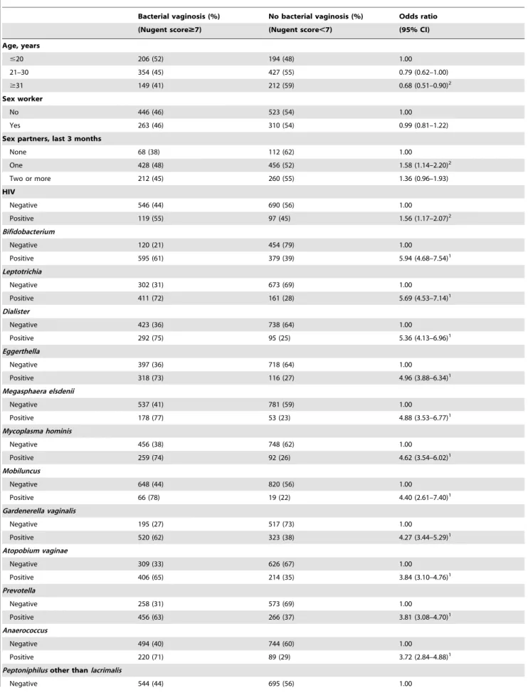
Table 3 shows the correlates of BV in univariate analyses. BV was more frequent in younger women, sexually active women, and HIV-infected women. BV prevalence was equal among sex workers and non-sex workers. BV was strongly associated with G. vaginalis,M. hominis,A. vaginae,Prevotella, Mobiluncus,Leptotrichia, M. elsdenii, Eggerthella, Dialister, Bifidobacterium, and Anaerococcus
(crude odds ratios $3.0) and less strongly associated with
Peptoniphilusnon-lacrimalisand pathogens not thought to be causal agents of BV (T. vaginalis, C. trachomatis, or M. genitalium) (crude odds ratios #2.0). Presence of Lactobacillus was associated with lower odds of BV. Yeast infections were equally frequent in women with or without BV.
Table 4 shows the results of the multivariate analysis of correlates of BV. Number of sexual partners increased the fit of the model even if each category was not significant. Age and HIV status became non-significant and were left out. The presence ofG. vaginalis,Bifidobacterium,M. elsdenii,Dialister,M. hominis,Leptotrichia, and Prevotella was independently associated with BV as was the absence ofLactobacillusandPeptoniphilusnon-lacrimalis. When fitted into this model, the following pathogens were no longer independently associated with BV: A. vaginae (AOR: 1.30; 95% CI: 0.95–1.78; p = 0.10),Mobiluncus (AOR: 1.36; 95% CI: 0.72– 2.56; p = 0.34), Anaerococcus (AOR: 1.17; 95% CI: 0.83–1.66; p = 0.37) andEggerthella(AOR: 1.03; 95% CI: 0.71–1.50; p = 0.86). The presence of T. vaginalis, N. gonorrhoeae, C. trachomatis, or M. genitalium was not independently associated with BV after adjustment for the other correlates of BV (data not shown).
The robustness of our conclusions was tested on alternative models. As it could be argued that the presence ofLactobacillusby PCR was too directly related to the Gram stain definition of BV, this variable was excluded in a subsequent model, which showed that the same eight micro-organisms were associated with BV, while the same four were not significantly associated (data not shown). In models (with or without Lactobacillus) that excluded women with an intermediate Nugent score (4–6), G. vaginalis, Bifidobacterium, Dialister, M. hominis, Leptotrichia, and Prevotella
remained associated with BV, while the presence of M. elsdenii
and the absence of Peptoniphilus non-lacrimalis became
non-significant (Tables S7 and S8). Likewise, A. vaginae, Mobiluncus, Eggerthella, and Anaerococcus were not significantly associated with BV.
Of 349 women without evidence of G. vaginalis, M. hominis, Prevotella, Leptotrichia, M. elsdenii, Dialister, and Bifidobacterium, only 12% (41) had BV as diagnosed by Nugent score. This proportion increased to 25% (54/220), 44% (104/236), 53% (101/189), 68% (126/184), 70% (120/171), 84% (133/158) and 89% (33/37) for women with respectively one, two, three, four, five, six or all of these microorganisms respectively (p,0.001,x2for trend).
In a secondary analysis that included all 570 women with BV who attended their follow-up visit on day 14 (Table S9), the proportion with a complete resolution of symptoms increased with the number of pathogens identified on day 0: 41% of those with a single pathogen, and 57%, 56%, 58%, 70%, 64%, 70%, and 76% of those with one, two, three, four, five, six, or seven pathogens respectively (p,0.001,x2 for trend) had complete resolution of symptoms. This trend was stronger in participants who had been treated with single-dose tinidazole/fluconazole than for those treated with metronidazole/clotrimazole. Age and HIV status had no impact on the likelihood of a complete short-term response to treatment. In a multivariate analysis, complete resolution of symptoms on day 14 was more common in women in whose samplesPrevotella(AOR: 1.91; 95% CI: 1.34–2.74; p,0.001) orM. elsdenii (AOR: 1.54; 95% CI: 1.01–2.34; p = 0.04) had been detected and among those with$2 sex partners (AOR: 1.95; 95% CI: 0.97–3.93; p = 0.06). No other pathogens or the treatment allocation was independently associated with this complete short-term resolution of symptoms.
Discussion
We documented complex interrelationships between several micro-organisms present in the vaginal flora of a large number of women presenting with vaginal discharge in West Africa. These organisms were strongly associated with each other to an extent that can hardly be explained by shared risk factors for transmission
Table 1.Associations between various bacterial species and genera.1
GV MH AV MO PR AN BI DI LE ME PE LA
GV 1 0.320 0.580 0.085 0.394 0.288 0.517 0.304 0.367 0.181 0.206 0.112
MH 1 0.365 0.168 0.331 0.228 0.256 0.339 0.421 0.159 0.195 0.020
AV 1 0.139 0.384 0.417 0.395 0.375 0.388 0.250 0.250 0.084
MO 1 0.207 0.135 0.091 0.221 0.215 0.106 0.107 20.136
PR 1 0.286 0.378 0.401 0.464 0.181 0.275 20.042
AN 1 0.327 0.432 0.343 0.222 0.306 20.054
BI 1 0.397 0.400 0.216 0.236 20.097
DI 1 0.510 0.261 0.400 20.046
LE 1 0.391 0.245 20.093
ME 1 0.086 20.079
PE 1 0.083
LA 1
1Correlation coefficients (phi) were used to measure associations between dichotomic variables and are displayed in the table. All correlations had p-values ,0.001, excepted LA-ME (0.002), LA-AN (0.03), LA-DI (0.07), LA-PR (0.10), and LA-MH (0.43).
Note: GV:Gardnerella vaginalis; AV:Atopobium vaginae; MH:Mycoplasma hominis; PR:Prevotella; MO:Mobiluncus; LE:Leptotrichia; ME:Megasphaera elsdenii;
but likely indicates a symbiotic relationship within the vaginal ecosystem [10,11]. Many organisms were associated with BV in univariate analyses but four became non-significant after adjust-ment for the presence of other bacteria. Previous analyses of the microbiology of BV considered only some of these organisms, used only univariate analyses, or combined all pathogens into a composite variable [11–15,17]. Our sample size was substantially larger than all previous studies and allowed us to adjust for the concomitant presence of multiple bacteria. Amongst our partic-ipants,Mobiluncus,Eggerthella,Anaerococcus, andA. vaginaewere not independently associated with BV but were rather confounded by
their association with other genera or species that themselves were correlates of BV. Our results support the idea that G. vaginalis, Bifidobacterium, M. elsdenii, Dialister, M. hominis, Leptotrichia, and Prevotellaare associated with BV and that its microbiology is the same in Africa as in industrialized countries.
Our interpretation of Table 2 is as follows. Colonization/ infection with several organisms was enhanced by sexual activity and by HIV-associated immunosuppression. Some organisms must be transmitted during intercourse. For others, organisms that are equally prevalent in sex workers and monogamous sexually active women, a non-specific effect of intercourse on the vaginal
Table 2.Correlates of colonization/infection with various bacterial genera or species.
G. vaginalis M. hominis A. vaginae Prevotella Mobiluncus
Age, years
#20 1.00 1.00 1.00 1.00 1.00
21–30 0.82 (0.64–1.06) 0.67 (0.50–0.90)2 0.77 (0.60–0.99)2 0.98 (0.76–1.27) 1.52 (0.83–2.77)
$31 0.59 (0.44–0.81)1 0.46 (0.32–0.67)1 0.73 (0.54–0.99)2 0.91 (0.67–1.24) 1.14 (0.56–2.35) Sex partners, last 3 months
None 1.00 1.00 1.00 1.00 1.00
One 1.37 (0.98–1.91) 1.27 (0.84–1.92) 1.20 (0.86–1.70) 1.04 (0.74–1.46) 3.02(1.08–8.47)2
Two or more 1.45 (1.01–2.07)2 1.14 (0.73–1.76) 1.16 (0.81–1.67) 1.58 (1.10–2.26)2 1.73 (0.58–5.15)
HIV
Negative 1.00 1.00 1.00 1.00 1.00
Positive 1.77 (1.29–2.42)1 2.06 (1.47–2.87)1 1.12 (0.82–1.52) 1.86 (1.37–2.54)1 2.19(1.27–3.80)2
Eggerthella M. elsdenii Leptotrichia Dialister Bifidobacterium
Age, years
#20 1.00 1.00 1.00 1.00 1.00
21–30 0.73 (0.56–0.95)2 0.94 (0.66–1.33) 0.71 (0.55–0.92)2 0.78 (0.59–1.04) 0.74(0.56–0.96)2
$31 0.48 (0.34–0.68)1 0.81 (0.52–1.25) 0.53 (0.38–0.73)1 0.60 (0.42–0.86)2 0.50(0.36–0.68)1
Sex partners, last 3 months
None 1.00 1.00 1.00 1.00 1.00
One 1.29 (0.87–1.91) 1.32 (0.80–2.17) 1.26 (0.88–1.81) 1.10 (0.74–1.64) 1.29 (0.92–1.81)
Two or more 1.59 (1.05–2.40)2 1.21 (0.71–2.06) 1.81 (1.24–2.66)2 1.26 (0.83–1.91) 1.61 (1.11–2.32)2
HIV
Negative 1.00 1.00 1.00 1.00 1.00
Positive 1.41 (1.02–1.96)2 1.03 (0.67–1.57) 1.59 (1.17–2.17)2 1.73 (1.25–2.40)1 2.07(1.46–2.93)1
Anaerococcus Peptoniphilus3 Lactobacillus
Age, years
#20 1.00 1.00 1.00
21–30 0.88 (0.65–1.20) 0.93 (0.68–1.27) 1.07 (0.79–1.44)
$31 0.76 (0.51–1.11) 0.68 (0.46–1.01) 0.99 (0.70–1.42)
Sex partners, last 3 months
None 1.00 1.00 1.00
One 1.06 (0.70–1.62) 0.80 (0.52–1.21) 0.58 (0.37–0.90)2
Two or more 1.01 (0.65–1.58) 1.13 (0.73–1.75) 0.54 (0.34–0.87)2
HIV
Negative 1.00 1.00 1.00
Positive 1.13 (0.78–1.63) 1.88 (1.34–2.65)1 0.66 (0.47–0.92)2
1 #0.001.
2 #0.05.
3other thanlacrimalis.
Table 3.Correlates of bacterial vaginosis in univariate analyses.
Bacterial vaginosis (%) No bacterial vaginosis (%) Odds ratio
(Nugent score$7) (Nugent score,7) (95% CI)
Age, years
#20 206 (52) 194 (48) 1.00
21–30 354 (45) 427 (55) 0.79 (0.62–1.00)
$31 149 (41) 212 (59) 0.68 (0.51–0.90)2
Sex worker
No 446 (46) 523 (54) 1.00
Yes 263 (46) 310 (54) 0.99 (0.81–1.22)
Sex partners, last 3 months
None 68 (38) 112 (62) 1.00
One 428 (48) 456 (52) 1.58 (1.14–2.20)2
Two or more 212 (45) 260 (55) 1.36 (0.96–1.93)
HIV
Negative 546 (44) 690 (56) 1.00
Positive 119 (55) 97 (45) 1.56 (1.17–2.07)2
Bifidobacterium
Negative 120 (21) 454 (79) 1.00
Positive 595 (61) 379 (39) 5.94 (4.68–7.54)1
Leptotrichia
Negative 302 (31) 673 (69) 1.00
Positive 411 (72) 161 (28) 5.69 (4.53–7.14)1
Dialister
Negative 423 (36) 738 (64) 1.00
Positive 292 (75) 95 (25) 5.36 (4.13–6.96)1
Eggerthella
Negative 397 (36) 718 (64) 1.00
Positive 318 (73) 116 (27) 4.96 (3.88–6.34)1
Megasphaera elsdenii
Negative 537 (41) 781 (59) 1.00
Positive 178 (77) 53 (23) 4.88 (3.53–6.77)1
Mycoplasma hominis
Negative 456 (38) 748 (62) 1.00
Positive 259 (74) 92 (26) 4.62 (3.54–6.02)1
Mobiluncus
Negative 648 (44) 820 (56) 1.00
Positive 66 (78) 19 (22) 4.40 (2.61–7.40)1
Gardenerella vaginalis
Negative 195 (27) 517 (73) 1.00
Positive 520 (62) 323 (38) 4.27 (3.44–5.29)1
Atopobium vaginae
Negative 309 (33) 626 (67) 1.00
Positive 406 (65) 214 (35) 3.84 (3.10–4.76)1
Prevotella
Negative 258 (31) 573 (69) 1.00
Positive 456 (63) 266 (37) 3.81 (3.08–4.70)1
Anaerococcus
Negative 494 (40) 744 (60) 1.00
Positive 220 (71) 89 (29) 3.72 (2.84–4.88)1
Peptoniphilusother thanlacrimalis
flora seems plausible and may support a recent description of BV as a ‘‘sexually enhanced disease’’ [28]. For most organisms, prevalence decreased with age, perhaps through a decrease in sexual activity or the progressive development of mucosal immunity, which may wane with HIV infection. The relative protection against BV conferred by age and the higher risk with sexual activity and HIV infection are consistent with findings for some of the organisms individually. In multivariate analysis, HIV and younger age were no longer risk factors for BV, because they were associated with colonization/infection with several bacterial genera or species whose inclusion in the model removed the effect of HIV and age, which lay further away in the causal pathway.
Metronidazole is an imperfect treatment of BV: 10–20% of patients do not respond, 15–30% relapse within 3 months and half relapse within one year [1,29]. Whether relapses may be due to re-infections accompanying intercourse remains controversial [29,30]. Systematically administered metronidazole has little or no activity againstM. hominis, Bifidobacterium, andG. vaginalisand only moderate activity againstDialister[31,32]. Metronidazole has predictable activity against Prevotella, Leptotrichia, and Eggerthella [33,34]. Despite this resistance, metronidazole treatment does reduce vaginal counts of M. hominis, G. vaginalis, Prevotella, Megasphaera, and to a lower extentLeptotrichia, with little difference between oral or intravaginal administration [32,35–37]. Topical clindamycin, the other recommended treatment for BV [38], is not ideal either as one-sixth of vaginal anaerobes at baseline are resistant, and clindamycin resistance can emerge during treat-ment, especially in Prevotella [32,39]. Nevertheless, clindamycin appears equivalent to metronidazole in achieving a clinical cure [40].
Counter-intuitively, we showed that after a regimen that included either metronidazole or tinidazole, the proportion of patients with a complete resolution of symptoms by day 14 increased with the number of pathogens present on day 0 and that the presence ofPrevotellawas strongly associated with a good short-term response. These findings may reflect the symbiotic nature of BV such that the reduction of the metronidazole-susceptible portion of BV pathogens results in a lack of sustainability of the more resistant organisms and a net overall effect of clinical cure. In the presence of multiple pathogens, theG. vaginalis-related biofilm may be less important in the pathogenesis of BV [41].
Our study had limitations. First, we had to be selective in the choice of pathogens to be tested on all samples, for financial and logistical reasons. Additional BV correlates (for instance, the BV-associated bacteria in the Clostridiales order, other Megasphaera species, Papillibacter, or Lachnospiraceae) may have been identified had we not faced this restriction [20,42,43]. Second, the recruitment was performed in West Africa; whether the vaginal flora varies between geographic regions is unknown but it does seem to vary according to ethnicity, and genetically determined factors could be involved in the pathogenesis of BV, as suggested by studies in the United States where the disease is more prevalent among African-American women, even after adjustment for sexual behavior and other confounders [44,45]. Third, all participants, including those that became the control group, had consulted for vaginal discharge. One third of participants without BV had yeast visible on Gram stain, but there is so far little evidence that the bacterial flora of women with vaginal candidiasis is altered as compared to that of healthy women [46]. Fourth, using extremely sensitive molecular methods, we looked for pathogens whose
Bacterial vaginosis (%) No bacterial vaginosis (%) Odds ratio
(Nugent score$7) (Nugent score,7) (95% CI)
Positive 170 (55) 138 (45) 1.58 (1.22–2.02)1
Lactobacillus
Negative 239 (66) 122 (34) 1.00
Positive 476 (40) 718 (60) 0.34 (0.26–0.43)1
Trichomonas vaginals
Negative 622 (44) 778 (56) 1.00
Positive 93 (60) 62 (40) 1.94 (1.39–2.72)1
Neisseria gonorrhoeae
Negative 676 (45) 810 (55) 1.00
Positive 39 (57) 30 (42) 1.52 (0.94–2.48)
Chlamydia trachomatis
Negative 678 (45) 817 (55) 1.00
Positive 37 (62) 23 (38) 2.00 (1.19–3.39)2
Mycoplasma genitalium
Negative 667 (45) 811 (55) 1.00
Positive 48 (62) 29 (38) 2.06 (1.29–3.28)2
Yeasts
Negative 507 (47) 576 (53) 1.00
Positive 208 (44) 264 (56) 0.89 (0.71–1.10)
1 #0.001.
2 #0.05.
doi:10.1371/journal.pone.0025082.t003
presence or absence contributed, to various extents, to the definition of the outcome through the Nugent score. This problem was potentially worse for Lactobacillus, whose morphotype (large Gram-positive rods) accounts for 4/10 points. However, inclusion or exclusion ofLactobacilluspositivity by PCR had no effect on the association of other pathogens with BV. Furthermore,Mobiluncus, a curved Gram-variable rod whose morphotype corresponds to two points, was not associated independently with the outcome. Presumably, the overlap in the Gram stain morphology between the various constituents of the vaginal flora and the large number of organisms that we tested by PCR enabled us to avoid this circular reasoning.
In conclusion, among West African women with BV, numerous bacterial micro-organisms were strongly associated with each other in a pattern that suggested a symbiotic relationship. Overall, the vaginal flora of West African women with BV was reminiscent of that of their counterparts in industrialized countries. Cases
of BV with a simpler flora were less likely to respond to metronidazole or tinidazole. A better understanding of the determinants of therapeutic response is needed before more effective treatments can be developed.
Supporting Information
Table S1 Primers used for various pathogens.
(DOC)
Table S2 Frequency of associations between various bacterial species or genera.
(DOC)
Table S3 Prevalence of micro-organisms according to age.
(DOC)
Table S4 Prevalence of micro-organisms according to self-defined occupation.
(DOC)
Table S5 Prevalence of micro-organisms according to number of sex partners in the last 3 months.
(DOC)
Table S6 Prevalence of micro-organisms according to HIV status.
(DOC)
Table S7 Correlates of bacterial vaginosis (Nugent score $7) in multivariate analysis, excluding patients with an intermediate Nugent score (4–6).
(DOC)
Table S8 Correlates of bacterial vaginosis (Nugent score $7) in multivariate analysis, excluding patients with an intermediate Nugent score (4–6). Presence of Lactobacillus has been removed from this model.
(DOC)
Table S9 Proportion of patients with bacterial vaginosis who reported a complete resolution of vaginal discharge by Day 14.
(DOC)
Text S1 Methods used for nucleic acid amplification testing for various bacterial genus or species.
(DOC)
Acknowledgments
This article is dedicated to the memory of our late colleague Dr Mohamed Sylla. The following persons made important contributions in data acquisition:
Togo: Dr Ke´re´ Banla Abiba, director of the Institut National d’Hygie`ne (INH) and her staff;
Ghana: Mrs Comfort Asamoah-Adu and Dr Thomas Agyarko-Poku; Guine´e: Dr Thierno Mouctar Diallo, Dr Fatoumata Binta Bah, Dr Thierno Amadou Koundouno;
Mali: Dr Sidibe´ Garangue´ Souko and Dr Fatoumata Binta Keita; Canada: Mrs Jose´e Ducharme and Mrs Andre´e Grondin.
Author Contributions
Conceived and designed the experiments: JP A-CL EF. Performed the experiments: S. Deslandes GG S. Demeule A-CL EF. Supervised the data collection: FS NK S. Diakite´ A-CL. Performed statistical analyses: JP NC. Performed laboratory analyses: EF. Wrote the first draft of the manuscript: JP. Wrote subsequent drafts of the manuscript: JP S. Deslandes GG S. Demeule FS NK S. Diakite´ A-CL EF.
Table 4.Correlates of bacterial vaginosis (Nugent score$7) in multivariate analysis.
Adjusted odds ratio (95% CI)
Sex partners, last 3 months
None 1.00
One 1.39 (0.92–2.08)
Two or more 0.85 (0.55–1.31)
Gardenerella vaginalis
Negative 1.00
Positive 2.35 (1.74–3.16)1
Bifidobacterium
Negative 1.00
Positive 2.22 (1.64–3.01)1
Megasphaera elsdenii
Negative 1.00
Positive 2.09 (1.41–3.10)1
Dialister
Negative 1.00
Positive 1.99 (1.39–2.83)1
Mycoplasma hominis
Negative 1.00
Positive 1.91 (1.39–2.64)1
Leptotrichia
Negative 1.00
Positive 1.72 (1.26–2.36)1
Prevotella
Negative 1.00
Positive 1.53 (1.15–2.03)2
Peptoniphilusother thanlacrimalis
Negative 1.00
Positive 0.56 (0.40–0.79)1
Lactobacillus
Negative 1.00
Positive 0.26 (0.19–0.35)1
1 #0.001.
2 #0.05.
References
1. Eckert LO (2006) Acute vulvovaginitis. N Engl J Med 355: 1244–1252. 2. Pepin J, Sobe´la F, Khonde N, Agyarko-Poku T, Diakite´ S, et al. (2006) The
syndromic management of vaginal discharge using single-dose treatments: a randomized controlled trial in West Africa. Bull World Health Organ 84: 729–738.
3. Hillier SL, Nugent RP, Eschenbach DA, Krohn MA, Gibbs RS, et al. (1995) Association between bacterial vaginosis and preterm delivery of a low-birth-weight infant. N Eng J Med 333: 1737–1742.
4. Moodley P, Connolly C, Sturm AW (2002) Interrelationships among human immunodeficiency virus type 1 infection, bacterial vaginosis, trichomoniasis, and the presence of yeasts. J Infect Dis 185: 697–693.
5. Sewankambo N, Gray RH, Wawer MJ, Paxton L, McNaim D, et al. (1997) HIV-1 infection associated with abnormal vaginal flora morphology and bacterial vaginosis. Lancet 350: 546–550.
6. Taha TE, Hoover DR, Dallabetta GA, Kumwenda NI, Mtimavalye LA, et al. (1998) Bacterial vaginosis and disturbances of vaginal flora: association with increased acquisition of HIV. AIDS 12: 1699–1706.
7. Myer L, Denny L, Telerant R, de Souza M, Wright TC, et al. (2005) Bacterial vaginosis and susceptibility to HIV infection in South African women: a nested case-control study. J Infect Dis 192: 1372–1380.
8. Atashili J, Poole C, Ndumbe PM, Adimora AA, Smith JS (2008) Bacterial vaginosis and HIV acquisition: a meta-analysis of published studies. AIDS 22: 1493–1501.
9. Sha BE, Zarrifard MR, Wang QJ, Chen HY, Bremer J, et al. (2005) Female genital-tract HIV load correlates inversely withLactobacillusspecies but positively with bacterial vaginosis andMycoplasma hominis. J Infect Dis 191: 25–32. 10. Thorsen P, Jensen IP, Jeune B, Ebbesen N, Arpi M, et al. (1998) Few
microorganisms associated with bacterial vaginosis may constitute the pathologic core: a population-based microbiologic study among 3596 pregnant women. Am J Obstet Gynecol 178: 580–587.
11. Bradshaw CS, Tabrizi SN, Fairley CK, Morton AN, Rudland E, et al. (2006) The association of Atopobium vaginae and Gardnerella vaginalis with bacterial vaginosis and recurrence after oral metronidazole therapy. J Infect Dis 194: 8288–36.
12. Fredricks DN, Fiedler TL, Marrazzo JM (2005) Molecular identification of bacteria associated with bacterial vaginosis. N Eng J Med 353: 1899–1911. 13. Verstraelen H, Verhelst R, Claeys G, Temmerman M, Vaneechoutte M (2004)
Culture-independent analysis of vaginal microflora: the unrecognized association of Atopobium vaginae with bacterial vaginosis. Am J Obstet Gynecol 191: 1130–1132.
14. Fredricks DN, Fiedler TL, Thomas KK, Oakley BB, Marazzo JM (2007) Targeted PCR for detection of vaginal bacteria associated with bacterial vaginosis. J Clin Microbiol 45: 3270–3276.
15. Marrazzo JM, Thomas KK, Fiedler TL, Ringwood K, Fredricks DN (2010) Risk for acquisition of bacterial vaginosis among women who report sex with women: a cohort study. PLoS One 5: e11139.
16. Marazzo JM, Thomas KK, Fiedler TL, Ringwood K, Fredricks DN (2008) Relationship of specific vaginal bacteria and bacterial vaginosis treatment failure in women who have sex with women. Ann Intern Med 149: 20–28. 17. Haggerty CL, Totten PA, Ferris M, Martin DH, Hoferka S, et al. (2009) Clinical
characteristics of bacterial vaginosis among women testing positive for fastidious bacteria. Sex Transm Dis 85: 242–248.
18. Rottingen JA, Cameron DW, Garnett GP (2001) A systematic review for the epidemiologic interactions between classic sexually transmitted diseases and HIV. How much really is known ? Sex Transm Dis 28: 579–597.
19. Dols JAM, Smit PW, Kort R, Reid GM, Schuren FHJ, et al. (2011) Microarray-based identification of clinically relevant vaginal bacteria in relation to bacterial vaginosis. Am J Obstet Gynecol 204: 305e1–e7.
20. Hummelen R, Fernandes AD, Macklaim JM, Dickson RJ, Changalucha J, et al. (2010) Deep sequencing of the vaginal microbiota of women with HIV. PLoS One 5: e12078.
21. Nugent RP, Krohn MA, Hillier SL (1991) Reliability of diagnosing bacterial vaginosis is improved by a standardised method of Gram stain interpretation. J Clin Microbiol 29: 297–301.
22. Verhelst R, Verstraelen H, Claeys G, Verschraegen G, Delanghe J, et al. (2004) Cloning of 16S rRNA genes amplified from normal and disturbed vaginal microflora suggests a strong association betweenAtopobium vaginae,Gardnerella vaginalisand bacterial vaginosis. BMC Microbiol 4: 16.
23. Matsuki T, Watanabe K, Fujimoto J, Miyamoto Y, Takada T, et al. (2002) Development of 16S rRNA-gene-targeted group-specific primers for the detection and identification of predominant bacteria in human feces. Appl Environ Microbiol 68: 5445–5451.
24. Zariffard MR, Saifuddin M, Sha BE, Spear GT (2002) Detection of bacterial vaginosis-related organisms by real-time PCR for Lactobacilli,Gardnerella vaginalis
andMycoplasma hominis. FEMS Immunol Med Microbiol 34: 277–281. 25. Ferris MJ, Masztal A, Martin DH (2004) Use of species-directed 16S rRNA gene
PCR primers for detection of Atopobium vaginae in patients with bacterial vaginosis. J Clin Microbiol 42: 5892–5894.
26. Blanchard A, Yanez A, Dybvig K, Watson HL, Griffiths G, et al. (1993) Evaluation of intraspecies genetic variation within the 16S rRNA gene of
Mycoplasma hominisand detection by polymerase chain reaction. J Clin Microbiol 31: 1358–1361.
27. Tiveljung A, Forsum U, Monstein HJ (1996) Classification of the genus
Mobiluncusbased on comparative partial 16S rRNA gene analysis. Int J Syst Bacteriol 46: 332–336.
28. Verstraelen H, Verhelst R, Vaneechoutte M, Temmerman M (2010) The epidemiology of bacterial vaginosis in relation to sexual behaviour. BMC Infect Dis 10: 81.
29. Bradshaw CS, Morton AN, Hocking J, Garland SM, Morris MB, et al. (2006) High recurrence rate of bacterial vaginosis over the course of 12 months after oral metronidazole therapy and factors associated with recurrence. J Infect Dis 193: 1478–1486.
30. Schwebke JR, Desmond R (2005) Risk factors for bacterial vaginosis in women at high risk for sexually transmitted diseases. Sex Transm Dis 32: 654–658. 31. Morio F, Jean-Pierre H, Dubreuil L, Jumas-Bilak E, Calvet L, et al. (2007)
Antimicrobial susceptibilities and clinical sources ofDialisterspecies. Antimicrob Agents Chemother 51: 4498–4501.
32. Austin MN, Beigi RH, Meyn LA, Hillier SL (2005) Microbiologic response to treatment of bacterial vaginosis with topical clindamycin or metronidazole. J Clin Microbiol 43: 4492–4497.
33. Liebetrau A, Rodloff AC, Behra-Miellet, Dubreuil L (2003) In vitro activities of a new des-fluoro(6) quinolone, garenoxacin, against clinical anaerobic bacteria. Antimicrob Agents Chemother 47: 3667–3671.
34. Eribe ERK, Olsen I (2008)Leptotrichiaspecies in human infections. Anaerobe 14: 131–137.
35. Hillier SL, Lipinski C, Briselden AM, Eschenbach DA (1993) Efficacy of intravaginal 0.75% metronidazole gel for the treatment of bacterial vaginosis. Obstet Gynecol 81: 963–967.
36. Fredricks DN, Fiedler TL, Thomas KK, Mitchell CM, Marazzo JM (2009) Changes in vaginal bacterial concentrations with intravaginal metronidazole therapy for bacterial vaginosis as assessed by quantitative PCR. J Clin Microbiol 47: 721–726.
37. Mitchell CM, Hitti JE, Agnew KJ, Fredricks DN (2009) Comparison of oral and vaginal metronidazole for treatment of bacterial vaginosis in pregnancy: impact on fastidious bacteria. BMC Infect Dis 9: 89.
38. Centers for Disease Control and Prevention (2006) Sexually Transmitted Diseases treatment guidelines 2006. Morb Mortal Wkly Rep 55(RR-11): 1–64. 39. Beigi RH, Austin MN, Meyn LA, Krohn MA, Hillier SL (2004) Antimicrobial resistance associated with the treatment of bacterial vaginosis. Am J Obstet Gynecol 191: 1124–1129.
40. Oduyebo OO, Anorlu RI, Ogunsola FT (2009) The effects of antimicrobial therapy on bacterial vaginosis in non-pregnant women. Cochrane Database of Systematic Reviews 3: CD006055.
41. Swidsinski A, Mendling W, Loening-Baucke V, Swidsinski S, Dorffel Y, et al. (2008) An adherentGardnerella vaginalisbiofilm persists on the vaginal epidetlium after standard therapy with oral metronidazole. Am J Obstet Gynecol 198: e1–97.e6.
42. Oakley BB, Fiedler TL, Marazzo JM, Fredricks DN (2008) Diversity of human vaginal communities and associations with clinically defined bacterial vaginosis. Appl Environ Microbiol 74: 4898–4909.
43. Ling Z, Kong J, Liu F, Zhu H, Chen X, et al. (2010) Molecular analysis of the diversity of vaginal microbiota associated with bacterial vaginosis. BMC Genomics 11: 488.
44. Koumans EH, Sternberg M, Bruce C, McQuillan G, Kendrick J, et al. (2007) The prevalence of bacterial vaginosis in the United States, 2001–2004; associations with symptoms, sexual behaviors, and reproductive health. Sex Transm Dis 34: 864–869.
45. Ravel J, Gajer P, Abdo Z, Schneider GM, Sara S, et al. (2010) Vaginal microbiome of reproductive-age women. Proc Natl Acad Sci U S A. doi: 10.1073/pnas.10002611107