Activates Inflammasome
in Human and Murine Macrophages
Claire Smalley1, Jeremy Bechelli1, Dedeke Rockx-Brouwer1¤, Tais Saito1, Sasha R. Azar1, Nahed Ismail2, David H. Walker1, Rong Fang1*
1Department of Pathology, University of Texas Medical Branch at Galveston, Galveston, Texas, United States of America,2Department of Pathology, University of Pittsburgh, Pittsburgh, Pennsylvania, United States of America
¤ Current address: Intravacc, Bilthoven, the Netherlands
Abstract
Rickettsiae actively escape from vacuoles and replicate free in the cytoplasm of host cells, where inflammasomes survey the invading pathogens. In the present study, we investi-gated the interactions ofRickettsia australiswith the inflammasome in both mouse and human macrophages.R.australisinduced a significant level of IL-1βsecretion by human macrophages, which was significantly reduced upon treatment with an inhibitor of caspase-1 compared to untreated controls, suggesting caspase-caspase-1-dependent inflammasome activa-tion.Rickettsiainduced significant secretion of IL-1βand IL-18in vitroby infected mouse bone marrow-derived macrophages (BMMs) as early as 8–12 h post infection (p.i.) in a
dose-dependent manner. Secretion of these cytokines was accompanied by cleavage of caspase-1 and was completely abrogated in BMMs deficient in caspase-1/caspase-11 or apoptosis-associated speck-like protein containing a caspase activation and recruitment domain (ASC), suggesting thatR.australisactivate the ASC-dependent inflammasome. Interestingly, in response to the same quantity of rickettsiae, NLRP3-/-BMMs significantly reduced the secretion level of IL-1βcompared to wild type (WT) controls, suggesting that NLRP3 inflammasome contributes to cytosolic recognition ofR.australis in vitro. Rickettsial load in spleen, but not liver and lung, ofR.australis-infected NLRP3-/-mice was significantly greater compared to WT mice. These data suggest that NLRP3 inflammasome plays a role in host control of bacteriain vivoin a tissue-specific manner. Taken together, our data, for the first time, illustrate the activation of ASC-dependent inflammasome byR.australisin macrophages in which NLRP3 is involved.
Introduction
Rickettsial infections pose serious public health problems because of their potential to cause life-threatening human infection and to be used as biological weapons, a situation that is exac-erbated by the lack of a Food and Drug Administration-approved vaccine [1]. Rickettsiae are obligately intracellular bacteria which possess the ability to quickly escape phagosomal
a11111
OPEN ACCESS
Citation:Smalley C, Bechelli J, Rockx-Brouwer D, Saito T, Azar SR, Ismail N, et al. (2016)Rickettsia australisActivates Inflammasome in Human and Murine Macrophages. PLoS ONE 11(6): e0157231. doi:10.1371/journal.pone.0157231
Editor:Gary M. Winslow, Upstate Medical University, UNITED STATES
Received:February 25, 2016
Accepted:May 26, 2016
Published:June 30, 2016
Copyright:© 2016 Smalley et al. This is an open access article distributed under the terms of the
Creative Commons Attribution License, which permits unrestricted use, distribution, and reproduction in any medium, provided the original author and source are credited.
Data Availability Statement:All relevant data are within the paper.
Funding:This work was supported by grant AI101413 from the National Institute of Allergy and Infectious Diseases. Claire Smalley is supported by Pre-Doctoral McLaughlin Fellowship Award from the University of Texas Medical Branch at Galveston. The funders had no role in study design, data collection and analysis, decision to publish, or preparation of the manuscript.
vacuoles and replicate within the cytosol of host cells. However, the interactions of rickettsiae with cytosolic sensors, such as nucleotide binding and oligomerization domain (NOD)-like receptors (NLRs) in innate immune cells, have never been investigated. This is a gap in our knowledge that impedes the development of new therapeutic approaches and vaccine develop-ment strategies.
The inflammasome is a large multi-protein complex consisting of NLRs and the protease, caspase-1 [2]. Inflammasome activation by pathogens hinges upon violation of the host cell cytosol by activities such as those of pore-forming toxins, specialized microbial secretion sys-tems, or the cytosolic presence of the pathogen itself [2]. In response to these stimulants and/or danger signals (e.g., ATP), activation of NLRs can oligomerize ASC, which in turn activates caspase-1 to trigger its protease activity. Caspase-1 then mediates cleavage of pro-IL-1βand pro-IL-18 and secretion of IL-1βand IL-18 and/or inflammatory cell death, known as pyropto-sis [3]. Among NLRs that have been described as critical components of inflammasomes, NLRP3 plays a critical role in adjuvant-driven cellular immunity and, as such, exploitation of this pathway by vaccines may enhance efficacy, thus reinforcing the importance of investigat-ing inflammasome activation and understandinvestigat-ing the underlyinvestigat-ing mechanisms [4].
By using murine models of rickettsioses, we have identified the critical roles of IFN-γ, den-dritic cells (DCs), NK cells, TLRs and effector CD8+T cells in host protective immunity against rickettsial infection [5–12]. AlthoughR.conorii-infected C3H/HeN mice provide an excellent mouse model of human rickettsial infection [13], we utilizedR.australis-infected C57BL/6 (B6) mice in the present study because of the availability of various gene-deficient mice on the B6 background.Rickettsia australisis the etiologic agent of Queensland tick typhus [14]. Infec-tion of B6 mice withR.australisprovides an excellent murine model of rickettsial disease that targets endothelial cells (ECs) and macrophages and mimics the pathological findings of spot-ted fever group (SFG) rickettsioses in humans [15–18]. Using this model, we investigated whether rickettsiae are recognized in the cytosol by inflammasome and the mechanisms involvedin vitroandin vivo.
Although ECs are the primary target cells for rickettsial infection, pathogenic rickettsiae also invade macrophages as observed in established animal models and in the arthropod feed-ing inoculation site [19,20]. In response to IFN-γand TNF-α, macrophages are activated and serve as crucial effector cells mediating clearance of intracellular pathogens. Upon infection, perivascular infiltration of macrophages, together with lymphocytes and other cells, is a com-ponent of rickettsial vasculitis [21]. Therefore, understanding the interactions of rickettsiae with macrophages will greatly increase our knowledge regarding the pathogenesis of rickettsial infections and immunity against rickettsiae. In the present study, we focused on interactions of inflammasome withR.australisin mouse and human macrophages. We hypothesize thatR.
australisare recognized by cytosolic sensors, ASC-dependent inflammasome involving NLRP3, in macrophages leading to secretion of IL-1βand IL-18.
Materials and Methods
Rickettsia
Rickettsia australis(Cutlack strain) were cultivated in Vero cells and purified as previously described with modifications [6,22]. Briefly, infected cells were collected and suspended in SPG buffer (218 mM sucrose, 3.76 mM KH2PO4, 7.1 mM K2HPO4, 4.9 mM potassium
pathogen free embryonated chicken eggs. Yolk sacs from infected eggs were homogenized in a Waring blender and diluted to a 10% suspension in SPG buffer. All of these rickettsial stocks were quantified by plaque assay before use in experiments, as previously described [15]. The rickettsial stock was stored at -80°C until use. All the experiments described in this study were performed in a certified biosafety level 3 (BSL3) laboratory at UTMB.
Generation of human macrophages
THP-1 cells were purchased from ATCC and cultured as previously described without antibi-otics [23]. THP-1 cells were differentiated in 100μg/μl PMA (Sigma-Aldrich), reconstituted in
DMSO (Sigma-Aldrich) for 16 h, followed by 24 h recovery in fresh medium. Cells were plated at a density of 1 × 10⁶cells per well in a 6-well-plate, and infected withR.australisat an MOI of 5.
Human peripheral blood monocytes (PBMC) were isolated from buffy coats obtained from the UTMB blood bank. Cells were isolated as previously described [24]. Cells were plated at a density of 1 × 10⁶cells in each well in a 12-well-plate, and infected withR.australisat an MOI of 2 or 5.
Confocal immunofluorescence microscopy
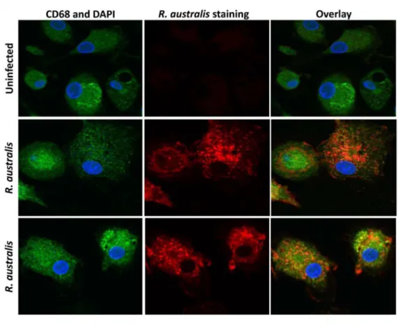
For immunofluorescence detection ofR.australisin human PBMC-derived macrophages, cells were first seeded on glass coverslips in 12-well plates one day before infection. At 24 h p.i., cells were washed with PBS, fixed with 4% paraformaldehyde in PBS for 20 min, permeabilized with 0.5% Triton-X in PBS for 20 min and blocked with 3% BSA in PBS for 30 min. Samples were incubated with rabbit polyclonal antibodies directed againstR.australis, goat anti-human CD68 (BioLegend, San Diego CA, #Y1/82A) followed by appropriate secondary antibodies including Alexa Fluor488- conjugated chicken anti-goat IgG (Life Technologies, NY, #A21467). The anti-CD68 antibody (Ab) preferentially labels human macrophages. Nuclei were stained with DAPI in ProLong1Gold Antifade Mountant (Life Technology, NY, P-36931). Coverslips were sealed with nail polish, and visualized by confocal microscopy with a 60 × water immersion lens (Olympus Fluoview 1000) using FV10-ASW software (Olympus, PA).
Measurements of cytokines
Supernatants of cell cultures were collected and filtered to be rickettsiae-free before removal from the BSL3 laboratory. Cytokine concentrations in the culture supernatant were measured by using Quantikine enzyme-linked immunosorbent assay (ELISA) kits. Detection of cytokines in murine samples were performed using the ELISA kit from eBioscience (San Diego CA). The limits of detection of the cytokines were as follows: IL-1β, 16 pg/ml; 18, 25.6 pg/ml; and IL-10, 62.5 pg/ml. Measurement of IL-1βand IL-18 in human samples was performed using ELISA kits from R&D Systems (IL-18, limit of detection: 12.5 pg/ml) and eBioscience (IL-1β, limit of detection: 1pg/ml).
Caspase-1 inhibition
Mice and generation of bone marrow-derived macrophages
Wild type (WT) B6 mice, caspase-1/11-double knockout mice and C57BL/6N (B6N) mice were purchased from Jackson Laboratories (Bar Harbor, Maine). ASC-/-and NLRP3-/-mice were gifts of Dr. Vishva Dixit at Genentech (California, USA). All mice were maintained and manipulated in an animal biosafety level-3 (ABSL3) facility at the University of Texas Medical Branch, Galveston, TX. This study was carried out in strict accordance with the recommenda-tions in the guidelines of the National Institutes of Health Guide for the Care and Use of Labo-ratory Animals. All experiments and procedures were approved by the Institutional Animal Care and Use Committee (IACUC) and Institutional Biosafety Committee of the University of Texas Medical Branch at Galveston. Forin vivoexperiments, mice were inoculated intrave-nously (i.v.) through the tail vein withR.australisat the dose indicated. After infection, mice were monitored daily for signs of illness until day 20 post infection (p.i.). No animal death was observed. Some of the mice were euthanized on days 2 and 4 p.i.. Mice were first anesthetized by inhalational isoflurane (Isoflurane1USP, Piramal Healthcare Limited, 502321 Andhra Pra-desh, India) and then euthanized by CO2narcosis and asphyxia followed by cervical
disloca-tion. After the death of animals, mouse tissues including lung, liver and spleen were isolated for evaluation of the bacterial replication and pathology by quantitative real-time PCR and his-topathological analysis. All necessary precautions were taken to minimize the discomfort and pain to animals used in the experiments.
Generation of primary bone marrow-derived macrophages (BMMs) from 6–8 week old female WT mice, ASC-/-mice and NLRP3-/-mice was performed as previously described [25]. Briefly, after femurs and tibias were dissected, bone marrows were flushed, and cells were culti-vated in low-endotoxin DMEM/F12 containing 10% (v/v) newborn calf serum (Thermoscienti-fic, Gibco, CA, 16010159) supplemented with either 20% supernatant from L929 cell culture or recombinant M-CSF (PeproTech, NJ, 315–02) at 37°C in 5% CO2. On day 6 of culture, cells
were harvested and characterized by flow cytometric analysis after staining with anti-F4/80 and anti-CD11b antibodies. Cells were used if they contained85 to 90% F4/80 (+) and CD 11b (+) cells. These cells were plated in 24-well plates at a density of 1 × 10⁶cells/ well in DMEM/F12 containing 10% newborn calf serum and used for experiments within 24 hrs.
In vitro
infections
Forin vitrorickettsial infection, macrophages were infected withR.australisat a multiplicity of infection (MOI) of 10:1, 6:1, 2:1 or as indicated. To synchronize bacterial internalization, rick-ettsiae were centrifuged onto the cells at 560 ×gfor 5 min. Cells were incubated continuously at 37°C in 5% CO2. Uninfected cells served as negative controls. Positive controls were the cells
primed with LPS (200 ng/ml) for 4 hours followed by incubation with ATP (5 mM) for 45 min. The viability of infected mouse BMMs was examined by trypan blue staining and LIVE/ DEAD1Fixable Near-IR Dead Cell Stain Kit (Life Technologies, Grand Island, NY) in accor-dance with the manufacturer’s protocol. Positive control was composed of half of them alive and half of them dead. Flow cytometry was performed on 30,000 cells with a FACSCalibur laser cytometer (Becton-Dickinson, BD Biosciences, San Jose, CA). Data analysis on the entire ungated cell population was performed using FlowJo software.
Western immunoblotting
(3K) (Amicon) as described by the manufacturer. Briefly, 2 ml of supernatant were loaded onto columns and centrifuged at 7000 ×gfor 60 min at 4°C. Protein concentration was determined by BCA Assay (Pierce Biotechnology). The cell lysates and concentrated supernatant were sep-arated by SDS-PAGE, transferred to a polyvinylidene difluoride (PVDF) membrane and probed with caspase-1 p20 antibody (EMD Millipore, MA, 06-503-I) for lysates, and anti-caspase-1 p10 antibody (sc-514, Santa Cruz Biotechnology) for concentrated supernatants. Immunoreactive bands were visualized using an appropriate secondary antibody and electro-chemiluminescence detection reagents (Thermoscientific, Pierce, IL, 32106). Equal protein loading of the gels was controlled by detectingβ-actin with mouse monoclonal Ab (Sigma, MO, A1978) in the cellular lysates. The detection of pro-caspase-1 (45 kDa) and activated cas-pase-1 (10 or 20 kDa) is indicative of activation of inflammasome as described previously [2].
Quantification of bacterial loads in infected tissues
in vivo
and NLRP3
transcripts in infected macrophages
in vitro
by quantitative real-time
PCR
To determine the rickettsial load in infected tissues, mouse lung, liver and spleen were isolated from infected animals. Rickettsial loads were quantified using quantitative PCR following DNA extraction as described in our previous studies with modifications [8,16]. Briefly, tissues were first placed in RNA later (Thermo Fisher Scientific, Waltham, MA). Total DNA was extracted using a DNeasy tissue kit (Qiagen, CA, 69506), and rickettsial burdens were deter-mined using an iCycler IQ from Bio-Rad (Hercules, CA). The following primers (Sigma-Genosys, St. Louis, MO) and probes (Biosearch Technologies, Novato, CA) targeting Rickett-sia-specific citrate synthase (CS) gene (gltA) as described previously [8,16] (gltAforward,
GAGAGAAAATTATATCCAAATGTTGAT;gltAreverse,AGGGTCTTCGTGCATTTCTT;gltA
probe,CATTGTGCCATCCAGCCTACGGT). The results were normalized to the weight of the same sample and expressed as copy number of CS genes per ng of tissue. Tissues from unin-fected mice served as a negative control.
Uninfected andR.australis-infected WT BMMs were collected in RNA later at the indicated time points. Total RNA was prepared using Qiagen RNeasy Mini kit (Valencia, CA) following the manufacturer’s recommendations. Reverse transcription (RT) was performed using iso-lated and DNase-treated RNA with Bio-Rad iScript cDNA synthesis kit (Hercules, CA). The resulting cDNAs were used as template for quantitative reverse transcription–polymerase chain reaction (RT-PCR). Gene expression of NLRP3 was determined using SYBR Green PCR Master Mix on an iCycler IQ (Bio-Rad, Hercules, CA) using primers targeting NLRP3 (For-ward 5’-CCT TCA GGC TGA TCC AAG AG-3’, Reverse 5’-GCC AAA GAG GAA TCG GAC AAC-3’) and GAPDH (Forward 5’-ATGGTGAAGGTCGGTGTGAA-3’, Reverse 5’-CTCCT TGGAGGCCATGTA-3’) as described previously [26,27]. Quantitative results were expressed as the mRNA relative ratio (2-ΔΔCt) normalized to the housekeeping gene as previously described [28].
Statistical analysis
the statistical analyses were performed using GraphPad Prism software version 5.01.Pvalues of 0.05 or less were the threshold for statistical significance.
Results
R
.
australis
infects human macrophages and activates inflammasome
So far, it remains elusive whether human macrophages can be infected byR.australis in vitro. To investigate the activation of inflammasome by rickettsiae in human macrophages, we first examined whetherR.australisinfects PBMC-derived macrophages by confocal immunofluo-rescence microscopy.R.australis(red) was detected in the cytosol (nucleus as blue) of infected human PBMC-derived macrophages, suggesting that rickettsiae were effectively taken up by and established infection in these cells (Fig 1A). Interestingly, human PBMC-derived macro-phages secreted a significantly higher level of IL-1βas early as 3 h p.i. compared to uninfected controls (Fig 1B). The levels of IL-1βsecreted by these infected macrophages were greater at 24 h p.i. compared to 3 h p.i. (Fig 1B). At 24 h p.i.,R.australisalso induced a significantly increased level of IL-18 in human PBMC-derived macrophages (Fig 1C). These results suggest thatR.australisinfects primary human macrophages and promotes secretion of the inflamma-some-derived cytokines including IL-1βand IL-18.
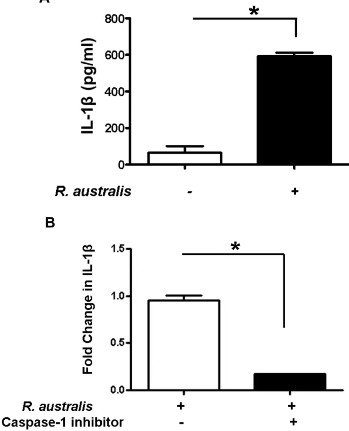
To further study the mechanisms involved in secretion of inflammasome-derived IL-1βin human macrophages, we examined the dependence of IL-1βsecretion on caspase-1 inR. aus-tralis-infected THP-1 derived macrophages. Consistent with the results from human PBMC-derived macrophages (Fig 1B), a significantly higher level of IL-1βwas observed inR.australis -infected THP-1 derived macrophages compared to un-infected controls at 24 h p.i. (Fig 2A). Interestingly,Rickettsia-infected THP-1 derived macrophages treated with an inhibitor of cas-pase-1 produced a significantly less fold change in IL-1βcompared to untreated controls (Fig 2B), suggesting that caspase-1 is critical for secretion of inflammasome-derived IL-1βin human macrophages upon rickettsial infection. Therefore, these results suggest thatR.australis
infects both human PBMC-derived macrophages and THP-1 cells, and promotes caspase-1-dependent secretion of IL-1βmost likely via activating inflammasome pathway.
R
.
australis
activates inflammasome in mouse macrophages
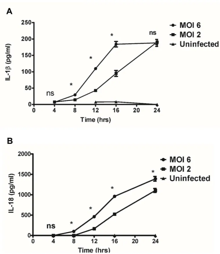
Activation of inflammasomes by microbes promotes the cleavage and maturation of pro-inflammatory cytokines IL-1βand IL-18 in a caspase-1/caspase-11-dependent manner. To investigate whetherR.australisactivates inflammasome in mouse macrophages, we first deter-mined the secretion of inflammasome-derived IL-1 family cytokines including IL-1βand IL-18 byR.australis-infected mouse macrophages as well as investigating the kinetics- and dose-dependent mechanisms involved. As positive controls, WT BMMs stimulated with LPS plus ATP produced significantly higher levels of IL-1βand IL-18 compared to unstimulated con-trols. As shown inFig 3A,R.australisinduced significant secretion of IL-1βat 8 h p.i. upon infection at a high dose (MOI of 6) and at 12 h p.i. at a low dose (MOI of 2). The levels of IL-1β
induced by a high dose ofR.australiswere significantly higher than those with the low dose infection at 8 h, 12 h and 16 h p.i.. Interestingly, although the production levels of IL-1β
Fig 1. Infection of human PBMC-derived macrophages withR.australisand activation of inflammasome.Human PBMC-derived macrophages were prepared and infected withR.australis. Cells and culture supernatant were collected at 24 h p.i.. A, Cytosolic rickettsiae were detected by confocal immunofluorescence microscopy after infection withR.australisat an MOI of 5. Images were acquired using
infection withR.australisat 24 h p.i.. We also confirmed these results with LIVE/DEAD1 Fix-able Dead Cell Stain by flow cytometric analysis (S1 Fig).
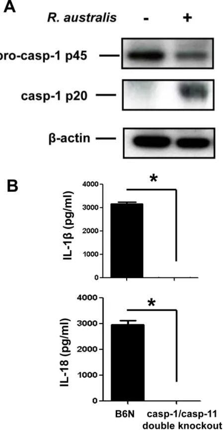
To further determine whether the secretion of IL-1βand IL-18 upon rickettsial infection is mediated by inflammasome, we investigated the activation of caspase-1 and the role of casp-1/ 11 in this process. The cell lysates of uninfected and infected BMMs showed expression of pro-caspase-1 (p45) while activated pro-caspase-1 (p20) was detected only in infected samples (Fig 4A). To confirm that the inflammasome pathway accounts for the production of cytokines such as IL-1βand IL-18 inR.australis-infected macrophages, we employed caspase-1/11-double knockout mice and the corresponding controls, B6N mice. B6N mice have the same genetic background as the caspase-1/11-double knockout mice. In response toR.australisinfection, BMMs from B6N mice produced significant levels of IL-1βand IL-18 (Fig 4B). However, we did not detect any production of these cytokines in caspase-1/caspase-11-double knockout mice. These results confirmed that caspase-1 and/or caspase-11 are essential for production of IL-1βand IL-18, suggesting thatR.australisactivates inflammasome in mouse macrophages.
ASC inflammasome is required for recognition of cytosolic
R
.
australis
Next, we aimed to identify the NLRs involved in the recognition ofR.australisin the host cell cytosol. Except NLRC4, most of the inflammasome NLRs signal through a critical adaptor pro-tein, ASC. Upon rickettsial infection, BMMs of ASC-/-mice failed to produce significant levels of IL-1βand IL-18 (Fig 5A and 5B). To exclude the possibility of unresponsiveness of ASC -/-BMMs upon stimulation, we determined the production of IL-10, an inflammasome-indepen-dent cytokine. Interestingly, bothR.australis-infected and LPS plus ATP-stimulated ASC -/-BMMs produced significantly higher levels of IL-10 than WT -/-BMMs (Fig 5C), suggesting that ASC-/-BMMs were responsive to rickettsial infection. These results also exclude the possibility that the incapability of ASC-/-BMMs to produce inflammasome-derived IL-1 family cytokines upon rickettsial infection was due to the failure of taking upR.australisintracellularly. To con-firm that the abolished secretion of IL-1βand IL-18 in ASC-/-BMM is not due to mechanisms other than inactivation of caspase-1, we determined the cleavage of caspase-1 in the superna-tant of infected WT and ASC-/-BMM by immunoblotting. As shown inFig 5D, at 24 h p.i., activated caspase-1 p10 was only detected in infected WT but not ASC-/-BMMs. Therefore, ASC, or ASC-dependent inflammasomes, were essential for the recognition ofR.australisin the cytosol of mouse macrophages. Our results also suggest that ASC may suppress the produc-tion of IL-10 in response to infectious stimuli including rickettsial antigen.
NLRP3 mediates the recognition of
R
.
australis
by inflammasome in
BMMs
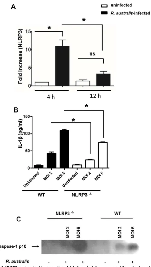
To further investigate the NLR inflammasome(s) responsible for recognition of rickettsiae in the cytosol, we first examined the transcriptional expression levels of NLRP3 in WT BMMs upon rickettsial infection. As early as 4 h p.i., infection withR.australisat an MOI of 6 signifi-cantly increased NLRP3 transcripts in WT macrophages (Fig 6A). As infection progressed, the quantity of NLRP3 mRNA progressively decreased at 12 h p.i. compared to 4 h p.i.. The tran-scriptional levels of NLRP3 were not significantly increased at 12 h p.i. compared to uninfected controls (Fig 6A). To further investigate whether NLRP3 is responsible for recognition of rick-ettsiae in the cytosol, we infected NLRP3-/-and WT BMMs withR.australisat both high (MOI determined by ELISA. Data represent two independent experiments with consistent results. Each experiment included at least 4 replicates.*, p<0.05.
Fig 2. Activation of inflammasome by rickettsiae in THP-1 derived macrophages and caspase-1-dependent secretion of IL-1β.Human THP-1 cells were differentiated to macrophages using PMA, and infected withR.australisat an MOI of 5. A, Infection with R.australisinduced a significant increase in IL-1βproduction compared to uninfected cells at 24 h p.i.. B, Inhibition of caspase-1 significantly reduced the secretion levels of IL-1βbyR.autralis-infected THP-1 derived macrophages. The fold change in IL-1βby treated cells vs. untreated controls was calculated and compared. Data represent two independent experiments with consistent results. Each experiment included at least 4 replicates.*,p<0.05.
Fig 3. Kinetics and dose-dependent mechanisms of secretion of IL-1βand IL-18 byRickettsia-infected BMMs.WT BMMs were isolated, cultivated, and infected withR.australisat MOI of 2 or 6. Cell culture supernatants were harvested at 4 hour intervals over 24 h p.i.. Secretion of IL-1β(A) and IL-18 (B) was determined by ELISA. Data represent two independent experiments with consistent results. Each experiment included at least 4 replicates.*,p<0.05; ns, not significantly different.
Fig 4.R.australisactivated inflammasome in BMMs.A, WT BMMs were isolated, cultivated and infected withR.australisat an MOI of 10. At 24 h p.i., culture supernatant and cell lysates were collected. Cell lysates were processed for detection of activation of caspase-1. B, BMMs of B6N and caspase-1/11-double knockout mice were isolated and infected withR.australisas described above. The secretion levels of IL-1βand IL-18 were determined by ELISA.*,p<0.05.
Fig 5. ASC-dependent inflammasome recognized cytosolic rickettsiae.BMMs of WT and ASC-/-mice were isolated,
of 6) and low (MOI of 2) doses. At 12 h p.i., the secretion levels of IL-1βby NLRP3-/-BMMs were significantly decreased compared to WT counterparts in response to both low and high doses ofR.australis(Fig 6B). To further confirm that NLRP3 is involved in inflammasome activation byR.australis, we determined the cleavage of caspase-1 in the processed superna-tant of WT and NLRP3-/-BMMs by immunoblotting. As shown inFig 6C, at 12 h p.i., acti-vated caspase-1 p10 was detected in the supernatant of infected WT BMM. The density of caspase-1 p10 was correlated with the dose of rickettsial infection, suggesting a dose-depen-dent inflammasome activation mechanism. In line withFig 6B, the cleaved caspase-1 was detected in NLRP3-/-BMMs infected withR.australisat a high dose infection (Fig 6C). These results suggest that NLRP3 mediates the secretion of IL-1βby inflammasome pathway upon rickettsial infection and that there is an alternative NLRP3-independent pathway for inflammasome activation. These data also suggest that NLRP3 inflammasome contributes to recognition ofR.australisin mouse macrophages. Thus, our results demonstrate that NLRP3 is activated byR.australisin BMMs.
NLRP3 inflammasome contributes to the
in vivo
control of
R
.
australis
in
spleen
To further explore the role of NLRP3 inflammasome in host defense against rickettsial infec-tionsin vivo, we measured rickettsial loads in infected tissues and survival of infected NLRP3 -/-and WT mice. Interestingly, the concentrations ofR.australisin the spleen of NLRP3-/-mice were significantly higher than those in WT mice on day 4 p.i., but not at day 2 p.i., suggesting that NLRP3 contributes to rickettsial elimination in spleen (S2 Fig). On days 2 and 4 p.i., rick-ettsial loads in liver and lung of NLRP3-/-mice were not significantly different from those in infected WT mice (S2 Fig). Furthermore, compared to day 2 p.i., bacterial loads in tissues of NLRP3-/-mice on day 4 p.i. were greater, particularly in spleen, suggesting that rickettsial infec-tion progresses in NLRP3-/-mice (S2 Fig). We did not find any significant difference in the sur-vival of NLRP3-/-and WT mice upon infection withR.australisat a dose of 2.8 × 10⁵ plaque-forming units (PFUs) (S3 Fig). Furthermore, histopathological analysis did not show any sig-nificant difference in inflammatory infiltrations in infected lung, liver and spleen of infected NLRP3-/-and WT mice in either day 2 or day 4 p.i. (S4 Fig). These data suggest that the contri-bution of NLRP3 inflammasome to host control ofR.australis in vivois tissue- or cell type-spe-cific. Taken together, these data suggest that NLRP3 inflammasome is not crucial to control rickettsial infectionin vivoand only contributes to host control of rickettsiae in a tissue- or cell-specific mechanism.
Discussion
In this study, we have demonstrated that cytosolic-replicatingR.australisinfects human pri-mary and THP-1-derived macrophages, and induces the secretion of caspase-1-dependent cytokines, most likely through inflammasome pathway, which had never been reported previ-ously.R.australisactivated inflammasome in mouse macrophages via time- and dose-depen-dent mechanisms. ASC-dependose-depen-dent inflammasomes were responsible for recognition ofR.
australisin host cytosol while NLRP3 inflammasome significantly contributed to this process. Thein vivorole of NLRP3 inflammasome in host immune responses toR.australiswas tissue-determined by detection of the active unit p10 in the processed supernatant at 12 h p.i. (C). Data represent mean±SD for at least 3 replicates each group.*,p<0.05 for a significant difference between WT and
NLRP3-/-mice; ns, not significantly different.
specific as evidenced by significantly increased bacterial loads in spleen, but not liver and lung, of NLRP3-/-mice compared to WT mice. More importantly, for the first time, we demon-strated thatR.australisactivated inflammasome in human macrophages with kinetics that dif-fered from mouse macrophages. Our findings have provided novel knowledge of the
mechanisms by which the host immune surveillance system interacts withRickettsiavia macrophages.
Rickettsia australisactivated ASC-dependent inflammasome in murine BMMs as indicated by the following evidence: 1) Secretion of IL-1βand IL-18 upon infection was completely abro-gated in cells deficient in ASC and caspase-1/caspase-11-double knockout cells (Figs4and5); 2)Rickettsia australisinfection induced cleavage of caspase-1 in the cell lysates and supernatant (Figs4A,5Dand6C). Although neutrophil-dependent, inflammasome-independent process-ing of IL-1βhas been described recently [30], our current data excluded the possibility of inflammasome-independent processing and secretion of IL-1βand IL-18 and demonstrated inflammasome activation byR.australisin macrophages. ASC serves as the essential adaptor molecule for several NLRs including NLRP1, NLRP3 and absent in melanoma 2 (AIM2) [31,
32]. Our results showed that NLRP3 contributed significantly to the activation of inflamma-some byR.australisin BMMs. Future investigations are required to reveal the upstream signals mediating the activation of NLRP3 inflammasome byR.australis, such as potassium efflux [33,
34], lysosomal degradation [35], and ROS production [36]. Interestingly, we also found that a significant level of inflammasome-derived IL-1βsecretion was NLRP3-independent (Fig 6B), particularly at a high dose of infection, which suggests that ASC-dependent NLR inflamma-somes other than NLRP3, potentially NLRP1 and/or AIM2, coordinate with NLRP3 or also play a significant role in the recognition of cytosolicR.australis. Furthermore, we did not find significant difference in IL-1βsecretion by infected BMMs of NLRP3-/-mice compared to WT mice at 24 h p.i., a time at which the levels of IL-1βsecretion reached a peak in macrophages of WT mice (Fig 3A). The differential contributions of NLRP3 to inflammasome activation byR.
australisat early versus late time points are likely explained by two possibilities: 1) Inflamma-somes other than NLRP3 play a major role in recognizingR.australisand the related danger signals at the late stage of infection; 2) NLRP3 inflammasome is down-regulated by other immune mechanisms such as caspase-11, autophagy, or cytokines specifically suppressive for NLRP3. Thesein vitrofindings may account for the dispensable role of NLRP3 inflammasome in host control ofR.australis in vivo. Although it is less likely that a higher dose ofR.australis
could lead to differential host susceptibilities of NLRP3-/-mice compared to WT mice (S3 Fig), our data clearly illustrated the significant contribution of NLRP3 inflammasome to host defense in a tissue-specific manner (S2 Fig). Considering the different proportions of cell types in spleen compared to liver and lung,R.australismay mainly activate NLRP3 inflammasome in leukocytes such as macrophages. Interestingly, we found significantly enhanced host suscep-tibilities and increased rickettsial loads in tissues of mice deficient in ASC during rickettsial infection compared to WT mice in our preliminaryin vivostudies. Our previous studies have clearly demonstrated that thein vivoproduction of IL-10 in murine models of fatal rickett-sioses is associated with the severity of disease [37]. As shown inFig 5C, ASC significantly sup-pressed the secretion of IL-10 byR.australis-infected macrophages, implying that ASC may contribute to host resistance against rickettsiae. Although further investigations are required to completely understand how inflammasome contributes to host immunityin vivoagainst these intracellular bacteria, our data suggest that ASC/NLRP3 inflammasome plays a role in host defense.
Inflammasome activation by rickettsial infection in macrophages was both time-and dose-dependent. Distinct from other facultatively cytosolic bacteria includingListeria,Shigella, Bur-kholderiaandFrancisella, rickettsiae are obligately cytosolic bacteria which quickly escape pha-gosomal vacuoles and replicate within the cytosol of host cells including macrophages. Mouse macrophages secrete IL-1βand IL-18 in response toListeriaat 5 h p.i. [38],Shigellaat 6 h p.i. [39],Burkholderiaat 4 h p.i.[40], andFrancisellaat 5 h p.i.[41]. Our data suggest that the kinet-ics and possibly the mechanisms of inflammasome activation byR.australisare distinct from other cytosolic bacteria. In response toR.australis, mouse macrophages secrete IL-1βand IL-18 as late as at 8 h p.i. after a high dose of infection and at 12 h after a low dose of infection. The levels of these inflammasome-derived cytokines increased progressively as the infection progressed and reached a peak at 24 h p.i. regardless of the dose. The delayed activation of inflammasome byR.australisin mouse macrophages compared with several facultatively cyto-solic bacteria mentioned above suggests that this intracellular bacterium may initiate an eva-sion mechanism to escape inflammasome assembly at the early stage of infection. The dose-independent secretion of IL-1βat 24 h p.i. by infected mouse macrophages suggests that inflammasomes responsible for recognizing these intracellular bacteria at the late stage of infec-tion are very sensitive to the activainfec-tion of ligand(s) generated during rickettsial infecinfec-tion, and could be an ideal candidate for vaccine development targeting inflammasome activation in the future.
While we have shown thatR.australisactivated caspase-1-dependent inflammasome in both murine and human macrophages, it remains unclear whether canonical or noncanonical inflammasomes are activated by rickettsiae. Kayagaki et al. demonstrated that the non-canoni-cal inflammasome pathway engages caspase-11 to activate caspase-1 and the subsequent release of IL-1βand IL-18 [42].Ehrlichia, another obligately intracellular bacterial species closely related to rickettsiae, triggers cleavage of caspase-1 and IL-18 secretion in BMMs [43]. Interestingly, type I interferon signaling promotes host susceptibility to fatal ehrlichiosis poten-tially via activation of non-canonical inflammasomes [43]. Thus, it is an attractive hypothesis that caspase-11 mediates caspase-1 activation, which further processes IL-1βsecretion inR.
australis-infected murine macrophages. Recently several intracellular bacterial pathogens, includingLegionella pneumophila,Yersinia pseudotuberculosis, andSalmonella entericaserovar Typhimurium(S.Typhimurium), were reported to activate both canonical caspase-1-depen-dent and non-canonical caspase-1-indepencaspase-1-depen-dent inflammasomes in primary human macro-phages [44]. Considering the fact that we have not examined caspase-11-dependent
inflammatory cell death, pyroptosis, in infected human macrophages, our data suggest thatR.
australisat least activates canonical inflammasome in human macrophages. Furthermore, NLRP3 inflammasome has been described to be involved in both canonical and non-canonical inflammasome pathways [42,45]. Therefore, our future studies will investigate whether the canonical or non-canoncial pathway accounts for NLRP3 activation by rickettsial infection.
In agreement with our data in murine macrophages,R.australisnot only infects both human primary and THP-1 derived macrophages, but also activates the inflammasome in human macrophages. It is worth noting that: 1) The kinetics of secretion of IL-1βbyR. austra-lis-infected macrophages may differ from IL-18. We observed a significant difference in secre-tion levels of IL-18, but not IL-1β, in mouse macrophages at high and low doses after rickettsial infection at 24 h p.i. (Fig 3). Furthermore,R.australisinitiated significant secretion of IL-1β, but not IL-18, as early as 3 h p.i. in human primary macrophages (Fig 1); 2) Induction of IL-1β
are potentially different from the mouse counterparts. Future studies focusing on these inter-esting points will not only provide information critical for further understanding the pathogen-esis of severe rickettsioses but also will enable us to uncover new mechanisms involved in activation of inflammasomes.
The present study investigated the essential and dispensable components in the inflamma-some pathway in mouse and human macrophages, which have never been previously examined during rickettsial infection.Rickettsia australisactivated ASC-dependent inflammasomes in which NLRP3 contributed significantly to recognition of the bacteria. Our data suggest inflam-masomes other than NLRP3 might play a critical role in the cytosolic surveillance system at the late stage of rickettsial infection. Our investigations also point out the potentially important role of macrophages in human rickettsioses.
Supporting Information
S1 Fig.R.australisinfection did not cause significant cell death in mouse macrophages.
BMMs were isolated and infected withR.australis. At 24 h p.i., uninfected and infected macro-phage viability was examined as described in Materials and Methods.
(TIF)
S2 Fig. NLRP3 inflammasome contributed to thein vivocontrol of rickettsial infection in
spleen.NLRP3-/-and WT mice were infected i.v. withR.australisat a dose of 1 × 10⁵PFU per mouse. On days 2 and 4 p.i., bacterial loads in spleen, liver and lung of infected mice were determined by quantitative real-time PCR. Results are means ± SE of data from two indepen-dent experiments with consistent results, where each experimental group included 4 mice.
,
p<0.05. (TIF)
S3 Fig. Host susceptibility of NLRP3-/-mice to infection withR.australiswas not
signifi-cantly different compared to WT mice.NLRP3-/-and WT mice were infected i.v. withR. aus-tralisat a dose of 2.8 × 10⁵PFUs per mouse. Mice were monitored daily until day 20 p.i.. (TIF)
S4 Fig. Histopathological analysis did not show any significant difference in inflammatory infiltrations in lung, liver and spleen of infected WT and NLRP3-/-mice.NLRP3-/-and WT mice were infected i.v. withR.australisat a dose of 1 × 10⁵PFU per mouse. On days 2 and 4 p. i., mice were sacrificed and tissues were processed for histopathological analysis.
(TIF)
Acknowledgments
This work was supported by grant AI101413 from the National Institute of Allergy and Infec-tious Diseases. Claire Smalley is supported by a Pre-Doctoral McLaughlin Fellowship Award from the University of Texas Medical Branch at Galveston.
We sincerely thank Dr. Vishva Dixit for kindly providing ASC-/-and NLRP3-/-mice for our research.
Author Contributions
References
1. Walker DH. Rickettsiae and rickettsial infections: the current state of knowledge. Clin Infect Dis. 2007; 45 Suppl 1:S39–44. PMID:17582568
2. Schroder K, Tschopp J. The inflammasomes. Cell. 2010; 140(6):821–32. doi:10.1016/j.cell.2010.01. 040PMID:20303873
3. Casson CN, Shin S. Inflammasome-mediated cell death in response to bacterial pathogens that access the host cell cytosol: lessons fromLegionella pneumophila. Front Cell Infect Microbiol. 2013; 3:111. doi:10.3389/fcimb.2013.00111PMID:24409420
4. Li H, Willingham SB, Ting JP, Re F. Cutting edge: inflammasome activation by alum and alum's adju-vant effect are mediated by NLRP3. J Immunol. 2008; 181(1):17–21. PMID:18566365
5. Feng HM, Popov VL, Walker DH. Depletion of gamma interferon and tumor necrosis factor alpha in mice withRickettsia conorii-infected endothelium: impairment of rickettsicidal nitric oxide production resulting in fatal, overwhelming rickettsial disease. Infect Immun. 1994; 62(5):1952–60. PMID: 8168962
6. Fang R, Ismail N, Soong L, Popov VL, Whitworth T, Bouyer DH et al. Differential interaction of dendritic cells withRickettsia conorii: impact on host susceptibility to murine spotted fever rickettsiosis. Infect Immun. 2007; 75(6):3112–23. PMID:17403875
7. Jordan JM, Woods ME, Feng HM, Soong L, Walker DH. Rickettsiae-stimulated dendritic cells mediate protection against lethal rickettsial challenge in an animal model of spotted fever rickettsiosis. J Infect Dis. 2007; 196(4):629–38. PMID:17624851
8. Fang R, Ismail N, Walker DH. Contribution of NK cells to the innate phase of host protection against an intracellular bacterium targeting systemic endothelium. Am J Pathol. 2012; 181(1):185–95. doi:10. 1016/j.ajpath.2012.03.020PMID:22617213
9. Jordan JM, Woods ME, Soong L, Walker DH. Rickettsiae stimulate dendritic cells through toll-like receptor 4, leading to enhanced NK cell activationin vivo. J Infect Dis. 2009; 199(2):236–42. doi:10. 1086/595833PMID:19072551
10. Walker DH, Olano JP, Feng HM. Critical role of cytotoxic T lymphocytes in immune clearance of rickett-sial infection. Infect Immun. 2001; 69(3):1841–6. PMID:11179362
11. Walker DH, Popov VL, Feng HM. Establishment of a novel endothelial target mouse model of a typhus group rickettsiosis: evidence for critical roles for gamma interferon and CD8 T lymphocytes. Lab Invest. 2000; 80(9):1361–72. PMID:11005205
12. Feng H, Popov VL, Yuoh G, Walker DH. Role of T lymphocyte subsets in immunity to spotted fever group Rickettsiae. J Immunol. 1997; 158(11):5314–20. PMID:9164951
13. Walker DH, Popov VL, Wen J, Feng HM.Rickettsia conoriiinfection of C3H/HeN mice. A model of endothelial-target rickettsiosis. Lab Invest. 1994; 70(3):358–68. PMID:7511715
14. Graves S, Stenos J. Rickettsioses in Australia. Ann N Y Acad Sci. 2009; 1166:151–5. doi:10.1111/j. 1749-6632.2009.04530.xPMID:19538275
15. Feng HM, Wen J, Walker DH.Rickettsia australisinfection: a murine model of a highly invasive vasculo-pathic rickettsiosis. Am J Pathol. 1993; 142(5):1471–82. PMID:8494048
16. Xin L, Shelite TR, Gong B, Mendell NL, Soong L, Fang R, et al. Systemic treatment with CpG-B after sublethal rickettsial infection induces mouse death through indoleamine 2,3-dioxygenase (IDO). PLoS One. 2012; 7(3):e34062. doi:10.1371/journal.pone.0034062PMID:22470514
17. Walker DH, Olano JP, Feng HM. Critical role of cytotoxic T lymphocytes in immune clearance of rickett-sial infection. Infect Immun. 2001; 69(3):1841–6. PMID:11179362
18. de Sousa R, Ismail N, Nobrega SD, França A, Amaro M, Anes M, et al. Intralesional expression of mRNA of interferon-gamma, tumor necrosis factor-alpha, interleukin-10, nitric oxide synthase, indolea-mine 2,3-dioxygenase, and RANTES is a major immune effector in Mediterranean spotted fever rickett-siosis. J Infect Dis. 2007; 196(5):770–81. PMID:17674321
19. Walker DH, Hudnall SD, Szaniawski WK, Feng HM. Monoclonal antibody-based immunohistochemical diagnosis of rickettsialpox: the macrophage is the principal target. Mod Pathol. 1999; 12(5):529–33. PMID:10349992
20. Cragun WC, Bartlett BL, Ellis MW, Hoover AZ, Tyring SK, Mendoza N, et al. The expanding spectrum of eschar-associated rickettsioses in the United States. Arch Dermatol. 2010; 146(6):641–8. doi:10. 1001/archdermatol.2010.48PMID:20404224
22. Sears KT, Ceraul SM, Gillespie JJ, Allen ED Jr, Popov VL, Ammerman NC et al. Surface proteome analysis and characterization of surface cell antigen (Sca) or autotransporter family ofRickettsia typhi. PLoS Pathog. 2012; 8(8):e1002856. doi:10.1371/journal.ppat.1002856PMID:22912578
23. Melehani JH, James DB, DuMont AL, Torres VJ, Duncan JA.Staphylococcus aureusleukocidin A/B (LukAB) kills human monocytes via host NLRP3 and ASC when extracellular, but not intracellular. PLoS Pathog. 2015; 11(6):e1004970 doi:10.1371/journal.ppat.1004970PMID:26069969 24. Johnston L, Harding SA, La Flamme AC. Comparing methods forex vivocharacterization of human
monocyte phenotypes andin vitroresponses. Immunobiology. 2015; 220(12):1305–10. doi:10.1016/j. imbio.2015.07.014PMID:26256247
25. Zhang X, Goncalves R, Mosser DM. The isolation and characterization of murine macrophages. Curr Protoc Immunol. 2008; Chapter 14:Unit 14.1.
26. Tsuchiya K, Hara H, Kawamura I, Nomura T, Yamamoto T, Daim S et al. Involvement of absent in mela-noma 2 in inflammasome activation in macrophages infected withListeria monocytogenes. J Immunol. 2010; 185(2):1186–95. doi:10.4049/jimmunol.1001058PMID:20566831
27. Valbuena G, Bradford W, Walker DH. Expression analysis of the T-cell-targeting chemokines CXCL9 and CXCL10 in mice and humans with endothelial infections caused by rickettsiae of the spotted fever group. Am J Pathol. 2003; 163(4):1357–69. PMID:14507644
28. Livak KJ, Schmittgen TD. Analysis of relative gene expression data using real-time quantitative PCR and the 2 (-delta delta C(T)) method. Methods 2001; 25(4):402–8. PMID:11846609
29. Radulovic S, Price PW, Beier MS, Gaywee J, Macaluso JA, Azad A. Rickettsia-macrophage interac-tions: host cell responses toRickettsia akariandRickettsia typhi. Infect Immun. 2002; 70(5):2576–82. PMID:11953398
30. Netea MG, van de Veerdonk FL, van der Meer JW, Dinarello CA, Joosten LA. Inflammasome-indepen-dent regulation of IL-1-family cytokines. Annu Rev Immunol. 2015; 33:49–77. doi: 10.1146/annurev-immunol-032414-112306PMID:25493334
31. Rathinam VA, Jiang Z, Waggoner SN, Sharma S, Cole LE, Waggoner L, et al. The AIM2 inflammasome is essential for host defense against cytosolic bacteria and DNA viruses. Nat Immunol. 2010; 11 (5):395–402. doi:10.1038/ni.1864PMID:20351692
32. Rathinam VA, Vanaja SK, Fitzgerald KA. 2012. Regulation of inflammasome signaling. Nat Immunol. 13(4):333–42. doi:10.1038/ni.2237PMID:22430786
33. Matsuo J, Nakamura S, Takeda S, Ishida K, Yamazaki T, Yoshida M. et al. Synergistic costimulatory effect ofchlamydia pneumoniaewith carbon nanoparticles on NLRP3 inflammasome-mediated inter-leukin-1βsecretion in macrophages. Infect Immun. 2015; 83(7):2917–25. doi:10.1128/IAI.02968-14 PMID:25939513
34. Petrilli V, Papin S, Dostert C, Mayor A, Martinon F, Tschopp J. Activation of the NALP3 inflammasome is triggered by low intracellular potassium concentration. Cell Death Differ. 2007; 14:1583–1589. PMID:17599094
35. Hornung V, Latz E. Critical functions of priming and lysosomal damage for NLRP3 activation. Eur J Immunol. 2010; 40:620–623. doi:10.1002/eji.200940185PMID:20201015
36. Tschopp J, Schroder K. NLRP3 inflammasome activation: the convergence of multiple signalling path-ways on ROS production? Nat Rev Immunol. 2010; 10:210–215. doi:10.1038/nri2725PMID: 20168318
37. Fang R, Ismail N, Shelite T, Walker DH. CD4+CD25+Foxp3-T-regulatory cells produce both gamma
interferon and interleukin-10 during acute severe murine spotted fever rickettsiosis. Infect Immun. 2009; 77(9):3838–49. doi:10.1128/IAI.00349-09PMID:19564386
38. Wu J, Fernandes-Alnemri T, Alnemri ES. Involvement of the AIM2, NLRC4, and NLRP3 inflamma-somes in caspase-1 activation byListeria monocytogenes. J Clin Immunol. 2010; 30(5):693–702. doi: 10.1007/s10875-010-9425-2PMID:20490635
39. Cai S, Batra S, Wakamatsu N, Pacher P, Jeyaseelan S. NLRC4 inflammasome-mediated production of IL-1βmodulates mucosal immunity in the lung against Gram-negative bacterial infection. J Immunol. 2012; 188(11):5623–35. doi:10.4049/jimmunol.1200195PMID:22547706
40. Ceballos-Olvera I, Sahoo M, Miller MA, Del Barrio L, Re F. Inflammasome-dependent pyroptosis and IL-18 protect againstBurkholderia pseudomalleilung infection while IL-1βis deleterious. PLoS Pathog. 2011; 7(12):e1002452. doi:10.1371/journal.ppat.1002452PMID:22241982
42. Kayagaki N, Warming S, Lamkanfi M, Vande Walle L, Louie S, Dong J et al. Non-canonical inflamma-some activation targets caspase-11. Nature. 2011; 479(7371):117–21. doi:10.1038/nature10558 PMID:22002608
43. Yang Q, Stevenson HL, Scott MJ, Ismail N. Type I interferon contributes to noncanonical inflamma-some activation, mediates immunopathology, and impairs protective immunity during fatal infection with lipopolysaccharide-negative ehrlichiae. Am J Pathol. 2014; 185(2):446–61. doi:10.1016/j.ajpath. 2014.10.005PMID:25481711
44. Casson CN, Yu J, Reyes VM, Taschuk FO, Yadav A, Copenhaver AM et al. Human caspase-4 medi-ates noncanonical inflammasome activation against gram-negative bacterial pathogens. Proc Natl Acad Sci U S A. 2015; 112(21):6688–93. doi:10.1073/pnas.1421699112PMID:25964352