Systems that Access the Host Cytosol
Cierra N. Casson1, Alan M. Copenhaver1, Erin E. Zwack2, Hieu T. Nguyen1, Till Strowig3, Bahar Javdan1, William P. Bradley1, Thomas C. Fung1, Richard A. Flavell3, Igor E. Brodsky2*, Sunny Shin1*
1Department of Microbiology, Perelman School of Medicine, University of Pennsylvania, Philadelphia, Pennsylvania, United States of America, 2Department of Pathobiology, School of Veterinary Medicine, University of Pennsylvania, Philadelphia, Pennsylvania, United States of America,3Department of Immunobiology and Howard Hughes Medical Institute, Yale University School of Medicine, New Haven, Connecticut, United States of America
Abstract
Inflammasome activation is important for antimicrobial defense because it induces cell death and regulates the secretion of IL-1 family cytokines, which play a critical role in inflammatory responses. The inflammasome activates caspase-1 to process and secrete IL-1b. However, the mechanisms governing IL-1arelease are less clear. Recently, a non-canonical inflammasome was described that activates caspase-11 and mediates pyroptosis and release of IL-1aand IL-1b. Caspase-11 activation in response to Gram-negative bacteria requires Toll-like receptor 4 (TLR4) and TIR-domain-containing adaptor-inducing interferon-b(TRIF)-dependent interferon production. Whether additional bacterial signals trigger caspase-11 activation is unknown. Many bacterial pathogens use specialized secretion systems to translocate effector proteins into the cytosol of host cells. These secretion systems can also deliver flagellin into the cytosol, which triggers caspase-1 activation and pyroptosis. However, even in the absence of flagellin, these secretion systems induce inflammasome activation and the release of IL-1aand IL-1b, but the inflammasome pathways that mediate this response are unclear. We observe rapid IL-1a and IL-1brelease and cell death in response to the type IV or type III secretion systems of Legionella pneumophilaand Yersinia pseudotuberculosis. Unlike IL-1b, IL-1a secretion does not require caspase-1. Instead, caspase-11 activation is required for both IL-1asecretion and cell death in response to the activity of these secretion systems. Interestingly, whereas caspase-11 promotes IL-1b release in response to the type IV secretion system through the NLRP3/ASC inflammasome, caspase-11-dependent release of IL-1ais independent of both the NAIP5/NLRC4 and NLRP3/ASC inflammasomes as well as TRIF and type I interferon signaling. Furthermore, we find both overlapping and non-redundant roles for IL-1aand IL-1bin mediating neutrophil recruitment and bacterial clearance in response to pulmonary infection by L. pneumophila. Our findings demonstrate that virulent, but not avirulent, bacteria trigger a rapid caspase-11-dependent innate immune response important for host defense.
Citation:Casson CN, Copenhaver AM, Zwack EE, Nguyen HT, Strowig T, et al. (2013) Caspase-11 Activation in Response to Bacterial Secretion Systems that Access the Host Cytosol. PLoS Pathog 9(6): e1003400. doi:10.1371/journal.ppat.1003400
Editor:Ralph R. Isberg, Tufts University School of Medicine, United States of America
ReceivedDecember 3, 2012;AcceptedApril 19, 2013;PublishedJune 6, 2013
Copyright:ß2013 Casson et al. This is an open-access article distributed under the terms of the Creative Commons Attribution License, which permits unrestricted use, distribution, and reproduction in any medium, provided the original author and source are credited.
Funding:Supported in part by grants from the National Institutes of Health (R00-AI087963 to SS; T32-GM007229 to AMC; T32-AI060516 to EEZ; T32-AI055400 to WPB), pilot grants from the University Research Foundation, McCabe Fund, NIH/NIDDK Center for Molecular Studies in Digestive and Liver Diseases (P30-DK050306), The Mari Lowe Center for Comparative Oncology (IEB), and a Benjamin Franklin Scholars grant (BJ). This material is based upon work supported by the National Science Foundation under Grant No. DGE-0822 (CNC, graduate research fellowship). The funders had no role in study design, data collection and analysis, decision to publish, or preparation of the manuscript.
Competing Interests:The authors have declared that no competing interests exist. * E-mail: [email protected] (IEB); [email protected] (SS)
Introduction
Antibacterial defense is initiated by germline-encoded pattern recognition receptors (PRRs), which detect conserved pathogen-associated molecular patterns (PAMPs) [1–3]. Plasma membrane-bound PRRs, such as the Toll-like receptors (TLRs), detect PAMPs present in the extracellular space and endosomal compartments, whereas cytosolic PRRs, such as the NOD-like receptors (NLRs), survey the host cytosol for the presence of invasive pathogens [3–7]. Invasive microorganisms or other cellular stresses induce assembly of cytosolic protein complexes known as inflammasomes, which play a critical role in host defense [8–11]. Inflammasomes respond to a wide variety of activators, including bacterial pore-forming toxins and bacterial PAMPS, such as flagellin or RNA [12–18]. Particular NLRs respond to their cognate stimuli and recruit the adapter protein ASC and pro-caspase-1 through homotypic protein-protein
interactions between pyrin domains and caspase recruitment domains (CARD), leading to autoprocessing and activation of caspase-1 [19–23]. Caspase-1 is responsible for processing and secreting IL-1 family cytokines and mediates a proinflammatory cell death termed pyroptosis [9,11,24,25].
Caspase-11 participates in the activation of a non-canonical inflammasome that induces cell death and the secretion of IL-1a and IL-1b in response to Gram-negative pathogens, such as
upregulation and activation, but how type I IFN mediates activation of caspase-11 is not well-defined [27–29]. Caspase-11 contributes to NLRP3-dependent activation of caspase-1 and subsequent caspase-1-dependent IL-1b secretion and cell death. Caspase-11 also facilitates an NLRP3- and caspase-1-independent pathway that results in cell death and release of IL-1a [26–29]. This caspase-11-dependent, caspase-1-independent pathway is responsible for LPS-induced septic shockin vivo[26,30]. Although caspase-11 is activated in response to signals from Gram-negative pathogens and certain pore-forming toxins, whether caspase-11 contributes to inflammasome activation in response to virulence-associated secretion systems that deliver bacterial ligands into host cytosol is unknown.
Bacterial pathogens use evolutionarily conserved secretion systems, such as type III or type IV secretion systems (T3SS or T4SS), to translocate effector proteins into the cytosol of host cells [31,32]. In addition to bona fide virulence factors, these secretion systems also translocate bacterial molecules such as flagellin or structural components of the secretion machinery itself, which results in inflammasome activation [14,16,33–36]. Legionella pneumophila, an opportunistic pathogen that causes a severe pneumonia known as Legionnaires’ disease [37,38], utilizes its
dot/icm-encoded T4SS as a virulence factor to translocate bacterial effector proteins into the host cell cytosol and establish a replicative vacuole [39–46].L. pneumophila induces T4SS-depen-dent inflammasome activation through two genetically distinct pathways [47]. T4SS-mediated translocation of flagellin into the cytosol triggers caspase-1 activation and pyroptosis through the NLR NAIP5 in conjunction with another NLR, NLRC4 [16,36,47–50]. Caspase-1 activation is also triggered independent-ly of the NLRC4/flagellin pathway through the adaptor protein ASC, but the bacterial factor that is recognized and the upstream proteins that regulate this pathway remain unknown [47,51]. However, although ASC is necessary for robust secretion of IL-1b in response to L. pneumophilaas well as a number of pathogens, such asSalmonellaorYersiniaspecies which employ T3SSs, ASC is
dispensable for induction of pyroptosis that is rapidly triggered in response to these infections. We therefore considered the possibility that in addition to its role in delayed inflammasome activation in response to Gram-negative bacteria, caspase-11 might participate in rapid cell death and release of IL-1a in response to the presence of bacterial pathogens that access the host cell cytosol by means of type IV and type III secretion systems.
Here, we demonstrate that IL-1aand IL-1bare rapidly released in response to bacterial T4SS activity independently of bacterial flagellin. In this system, we find IL-1bsecretion requires caspase-1, but caspase-1 is dispensable for cell death and IL-1a release in response to a functionalL. pneumophilaT4SS. Instead, caspase-11 is required for both IL-1a release and cell death in response toL. pneumophilaT4SS activity. Consistent with recent findings, caspase-11 contributes to optimal NLRP3-mediated caspase-1 activation and IL-1b secretion in response to L. pneumophila. However, caspase-11-dependent IL-1a release and cell death in L. pneumophila-infected cells are independent of the NAIP5/NLRC4 and NLRP3/ASC inflammasomes. In contrast to the role of TRIF and IFNAR in the response against Gram-negative bacteria, caspase-11 activation and cytokine release in response to the T4SS of L. pneumophila are independent of both TRIF and IFNAR signaling. We further demonstrate that T3SS activity of the unrelated pathogen Yersinia pseudotuberculosis induces a similarly rapid caspase-11-dependent response that also leads to cell death and release of IL-1aand IL-1b. Finally, we find that both IL-1a and IL-1b are critical in vivo for neutrophil recruitment and bacterial clearance. Overall, our data show that caspase-11 is poised to respond robustly to a conserved feature of pathogenic bacteria, bacterial access to the host cytosol through specialized secretion systems. This establishes caspase-11 as a critical regulator of immune system-mediated discrimination of pathogenic and nonpathogenic bacteria.
Results
LPS priming induces rapid IL-1aand IL-1bsecretion in response toL. pneumophilaT4SS activity
L. pneumophilainfection induces IL-1aand IL-1bsecretion that requires T4SS activity [47,52]. IL-1bsecretion is regulated by a flagellin-dependent NAIP5/NLRC4 inflammasome and a poorly defined ASC inflammasome that both activate caspase-1 [47,51]. The mechanisms underlying IL-1asecretion are less clear, but IL-1a secretion is still robustly induced by flagellin-deficient L. pneumophila, which do not activate the NAIP5/NLRC4 inflamma-some [52]. Recent studies have described a non-canonical inflammasome triggered in response to Gram-negative bacteria. This non-canonical inflammasome requires lipopolysaccharide (LPS) for the upregulation and activation of caspase-11 and subsequent IL-1aand IL-1brelease [26–29]. Whether caspase-11 is also activated in response to bacteria that use specialized secretion systems to translocate bacterial molecules into the host cytosol is unknown. We thus hypothesized that LPS priming would upregulate caspase-11, pro-IL-1a, and pro-IL-1b and allow for more robust and rapid IL-1a and IL-1bsecretion in response to T4SS activity. To test this, we first compared IL-1a and IL-1b release in unprimed and LPS-primed bone marrow-derived macrophages (BMDMs). As shown previously [48,52], unprimed BMDMs secrete robust levels of IL-1aand IL-1bby 20 hours post-infection with wild-typeL. pneumophila(WT Lp) (Figure 1A). Slightly attenuated levels of secreted IL-1a and IL-1b are observed with flagellin-deficientL. pneumophila(DflaALp), which do not activate the NAIP5/NLRC4 inflammasome [17,18]. Secretion of both Author Summary
cytokines is significantly diminished during infection with L. pneumophila lacking DotA, an essential component of the T4SS (DdotALp), and is significantly diminished in caspase-1/caspase-11-deficient (Casp12/2Casp112/2) macrophages as well (Figure 1A). The diminished IL-1 secretion induced by DdotA Lp is not due
to a lack of pro-IL-1 production, as DdotA Lp and WT Lp induce robust levels of pro-IL-1b (Figure S1A). At 4 hours post-infection, unprimed macrophages do not secrete IL-1 (Figure 1B). However, LPS-primed cells rapidly secrete IL-1a and IL-1b, and this secretion is abrogated inCasp12/2Casp112/2
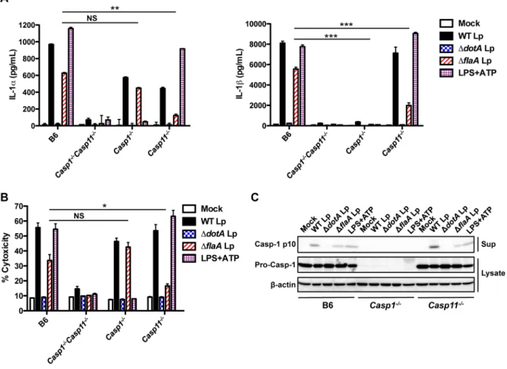
Figure 1. LPS priming induces rapid IL-1aand IL-1bsecretion in response toL. pneumophilaT4SS activity.(A) Unprimed B6 orCasp12/2
Casp112/2BMDMs were infected with WTL. pneumophila(WT Lp),DdotLp,DflaALp, or PBS (mock infection) for 20 hours. (B) B6 orCasp12/2
Casp112/2BMDMs were either unprimed or primed with 0.5mg/mL LPS for 2.5 hours and infected with WT Lp,DdotALp,DflaALp, or PBS for 4 hours. (C) B6 BMDMs were pretreated with either 20mM or 40mM of the caspase-1 inhibitor YVAD-cmk or DMSO vehicle control for 0.5 hours and infected
with WT Lp,DdotALp,DflaALp, or PBS for 20 hours. Levels of IL-1aand IL-1bin the supernatants were measured by ELISA. Graphs show the mean6 SEM of triplicate wells. Data are representative of three or four independent experiments. *** is p,0.001 by 2-way ANOVA with Bonferroni post-test. NS is not significant.
macrophages (Figure 1B). Secretion of IL-18, another IL-1 family cytokine, also requires T4SS activity and is eliminated inCasp12/2 Casp112/2cells (Figure S1B). Comparable levels of the caspase-1/ caspase-11-independent cytokines IL-12 and TNF-aare secreted in the absence and presence of LPS priming (Figure S1C–D). These data suggest that LPS priming upregulates a factor required for rapid IL-1aand IL-1brelease in response toL. pneumophilaT4SS activity.
Caspase-1 catalytic activity is required for IL-1b but not IL-1asecretion
Secretion of IL-1b in response to both canonical and non-canonical inflammasome activation requires caspase-1 [26,53,54]. In contrast, IL-1a release downstream of the non-canonical inflammasome depends on caspase-11, and does not require caspase-1 [26]. To test if the catalytic activity of caspase-1 is required for IL-1a secretion in response to L. pneumophila, we inhibited caspase-1 catalytic activity with the pharmacological inhibitor YVAD-cmk (YVAD). Consistent with previous studies [53], IL-1bsecretion in response toL. pneumophilais substantially inhibited by YVAD. However, YVAD has no effect on IL-1a
secretion, indicating that IL-1arelease in response toL. pneumophila
does not require caspase-1 catalytic activity (Figure 1C), as has been shown for other inflammasome activators [55]. Given that IL-1a secretion occurs more rapidly upon LPS priming, is abrogated in Casp12/2Casp112/2 macrophages, and does not require caspase-1 catalytic activity, we considered the possibility that caspase-11 might participate in inflammasome activation duringL. pneumophilainfection.
Caspase-11 contributes to inflammasome activation in response to flagellin-deficientL. pneumophila
To test the genetic requirement for caspase-11 in the inflammasome response to L. pneumophila, we infected BMDMs from either caspase-1-deficient (Casp12/2) or caspase-11-deficient (Casp112/2) mice. In the absence of flagellin, caspase-11 is required for IL-1asecretion, whereas it is not essential for IL-1b secretion but contributes to maximal secretion (Figure 2A). These data suggest that caspase-11 is activated in response to L. pneumophila infection independently of flagellin. Indeed, there is robust processing and secretion of caspase-11 in response to WT
Figure 2. Caspase-11 controls the release of IL-1aand IL-1band pyroptosis in response to flagellin-deficientL. pneumophila.B6,
Casp12/2Casp112/2,Casp12/2, orCasp112/2BMDMs were primed with 0.5mg/mL LPS for 2.5 hours and infected with WTL. pneumophila(WT Lp), DdotALp,DflaALp, or PBS (mock infection) or treated with 2.5 mm ATP for 1(C) or 4(A,B) hours. (A) Levels of IL-1aand IL-1bin the supernatants were measured by ELISA. Graphs show the mean6 SEM of triplicate wells. (B) Cell death (% cytotoxicity) was measured by LDH release into the supernatants relative to Triton X-100-lysed cells. Graphs show the mean6SEM of triplicate wells. (C) Levels of processed caspase-1 (casp-1 p10) in the supernatants and full-length caspase-1 (pro-casp-1) and b-actin in the cell lysates were determined by immunoblot analysis. Data are representative of three independent experiments. *** is p,0.001 by two-way ANOVA with Bonferroni post-test, ** is p,0.01 by two-way ANOVA with Bonferroni post-test, and * is p,0.05 by unpaired t-test. NS is not significant.
and DflaA Lp (Figure S2). In accordance with previous findings [26,53], caspase-1 is absolutely required for IL-1b secretion. In contrast, we observe robust IL-1arelease even in the absence of caspase-1. Both IL-1aand IL-1brelease in response toDflaALp are caspase-11-dependent in both primed and unprimed macro-phages (Figures 2, S3A–B), making L. pneumophila distinct from other Gram-negative bacteria that require priming to induce robust caspase-11 upregulation and activation [27]. Thus, while caspase-11 contributes to maximal caspase-1-dependent IL-1b secretion, it is both necessary and sufficient for IL-1a release in response to flagellin-deficientL. pneumophila.
Cell death in B6 BMDMs is partially flagellin-dependent, but is flagellin-independent in Casp12/2 BMDMs (Figure 2B). Impor-tantly, cell death in response to flagellin-deficient L. pneumophila
requires caspase-11, thus correlating caspase-11-dependent cell death with IL-1a release from host cells. In contrast, and consistent with previous findings [26], LPS+ATP induces canon-ical caspase-1-dependent pyroptosis and secretion of IL-1aand IL-1bthat is independent of caspase-11. Because caspase-1 must be processed to mediate IL-1bsecretion [53], we examined whether caspase-1 processing is decreased in the absence of caspase-11, which could account for the decreased IL-1bsecretion in response toDflaA Lp. Caspase-1 processing is slightly attenuated but not abrogated in response to DflaA Lp in Casp112/2 macrophages, consistent with the slight decrease in IL-1bsecretion (Figures 2C, S3C). Thus, flagellin-deficient L. pneumophila trigger a canonical caspase-1-dependent inflammasome as well as a non-canonical caspase-11-dependent inflammasome.
Caspase-11 activation is independent of ASC and NLRC4 The ASC and NAIP5/NLRC4 inflammasomes are required for caspase-1 activation and IL-1b secretion in response to
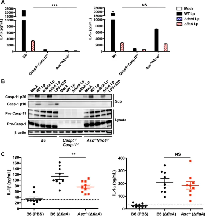
L. pneumophila [47]. To determine if these inflammasomes are also required for caspase-11 activation and IL-1a release, we infected ASC/NLRC4-deficient (Asc2/2Nlrc42/2) BMDMs withL. pneumophila. Asc2/2Nlrc42/2 BMDMs do not secrete IL-1b in response to either WT Lp,DflaALp, or LPS+ATP. However,Asc2/
2Nlrc42/2BMDMs still release IL-1a in response toDflaALp in primed and unprimed macrophages (Figures 3A, S4). Thus, unlike IL-1b, IL-1a is released independently of flagellin, ASC, and NLRC4. Accordingly, despite an absence of processed caspase-1 p10, robust levels of processed caspase-11 p26 are detected in the supernatants of Asc2/2Nlrc42/2cells infected with either WT or DflaALp but not in response to LPS+ATP (Figure 3B).
We next sought to determine whether IL-1a is also released independently of ASC and NLRC4 during in vivo infection. Because flagellin-deficient L. pneumophila do not activate the NLRC4 inflammasome [16,17,47], infecting Asc2/2 mice with DflaA Lp eliminates both the ASC and NLRC4 inflammasome pathways. Importantly, the level of IL-1b in the bronchoalveolar lavage fluid (BALF) 24 hours post-infection is significantly attenuated in Asc2/2 mice infected with DflaA Lp (Figure 3C). In contrast, the level of IL-1ain the BALF is unaffected even in the absence of both the ASC and NLRC4 inflammasomes. Both IL-1aand IL-1brelease are significantly diminished in caspase-1/ caspase-11-deficient mice (Figure S5). Collectively, our data indicate thatL. pneumophilatriggers caspase-11 activation and IL-1arelease independently of the ASC and NLRC4 inflammasomes during bothin vitroandin vivoinfection.
Caspase-11 mediates both NLRP3-dependent and NLRP3-independent inflammasome responses
L. pneumophilainduces caspase-1 activation and IL-1band IL-18 secretion through two genetically distinct pathways, one
involving ASC and one involving NLRC4 (Figures 4A, S6A–B) [47]. The upstream host and bacterial components of the ASC-dependent response to L. pneumophila are still unknown, but are independent of the flagellin/NAIP5/NLRC4 pathway (Figures 4A, S6B) [47]. Because caspase-11 contributes to maximal IL-1b secretion in response toDflaA Lp, we further investigated the ASC-dependent mechanism of inflammasome activation. NLRP3, an NLR involved in inflammasome-dependent responses to a wide variety of pathogens, requires ASC to mediate caspase-1 processing during both canonical and non-canonical inflammasome activation [9,12,18,26,56]. We therefore investigated the role of NLRP3 in the response toDflaA
Lp. Notably, IL-1b and IL-18 secretion are abrogated during infection of NLRP3-deficient (Nlrp32/2) BMDMs with DflaA Lp in both primed and unprimed macrophages (Figures 4B, S7A– C). Consistently, we do not detect processed caspase-1 p10 in the supernatants of Nlrp32/2 macrophages infected with DflaA Lp (Figure 4C). Thus, NLRP3 functions together with ASC, caspase-1, and caspase-11 to control IL-1bsecretion in response to flagellin-deficient L. pneumophila. However, IL-1a release and cell death following infection with flagellin-deficientL. pneumophila
are independent of NLRP3 (Figures 4B, S7A), indicating that caspase-11 also mediates an NLRP3-independent response towards flagellin-deficient L. pneumophila. Accordingly, NLRP3-dependent IL-1b secretion in response to flagellin-deficient L. pneumophila was inhibited by extracellular potassium, whereas NLRP3-independent caspase-11-dependent IL-1a secretion and cell death were not affected (Figure S7D–E).
Non-canonical inflammasome responses toL. pneumophilaoccur independently of TRIF and IFNAR
pneumophila is TRIF-independent, we investigated whether the TLR signaling adaptor MyD88 contributes to caspase-11 upregulation. When immortalized macrophages deficient for both MyD88 and Trif (iMyd882/2Trif2/2) were infected, caspase-11 upregulation was abrogated in response to both WT
andDflaA Lp (Figure S11A–B), and we were unable to detect caspase-11 activation (data not shown). Thus, although TRIF is not required for caspase-11 activation, a TLR-dependent signal is likely required as the loss of both MyD88 and TRIF eliminates caspase-11 upregulation and activation.
Figure 3. Caspase-11 activation is independent of ASC and NLRC4.(A) Unprimed B6,Casp12/2Casp112/2, orAsc2/2Nlrc42/2BMDMs were infected with WTL. pneumophila(WT Lp),DdotALp,DflaALp, or PBS (mock infection) for 20 hours, and levels of IL-1aand IL-1bin the supernatants were measured by ELISA. Graphs show the mean6SEM of triplicate wells. (B) Unprimed B6,Casp12/2Casp112/2, orAsc2/2Nlrc42/2BMDMs were infected with WT Lp,DdotALp,DflaALp, or PBS (mock infection) for 20 hours or treated with LPS+ATP for 1 hour. Levels of processed caspase-1
(casp-1 p10) and caspase-11 (casp-11 p26) in the supernatants, and pro-caspase-1, pro-caspase-11, andb-actin (loading control) in the cell lysates were determined by immunoblot analysis. (C) 8–12 week old B6 andAsc2/2mice were infected intranasally with either 1
6106DflaALp or PBS. Bronchoalveolar lavage fluid (BALF) was collected 24 hours post-infection, and levels of IL-1aand IL-1bwere measured by ELISA. Graphs show the mean6SEM of 9 mice per group. Dashed line represents the limit of detection. Data are representative of three independent experiments (A,B) or are displayed as the pooled results of two independent experiments (C). *** is p,0.001 by two-way ANOVA with Bonferroni post-test. ** is p,0.01 by unpaired t-test. NS is not significant.
Caspase-11 mediates inflammasome activation in response toYersinia pseudotuberculosistype III secretion system activity
Because caspase-11 activation in response to L. pneumophila
expressing a functional T4SS is so rapid and robust, we sought to test whether this robust caspase-11-dependent inflammasome activation might be a general response to the activity of specialized secretion systems that allow for bacterial access to the host cytosol. TheYersinia pseudotuberculosistype III secretion system (T3SS) induces inflammasome activation independently of bacterial flagellin and the known secreted effector proteins, and this inflammasome activation is important for bacterial clearance [57]. Since wild-type
Yersiniainduces cell death that is independent of both caspase-1 and -11 and requires the secreted effector YopJ [57,58], we instead infectedCasp12/2Casp112/2,Casp12/2, andCasp112/2BMDMs with a strain ofY. pseudotuberculosisthat expresses a T3SS but lacks the six known secreted effectors (D6 Yp). Similarly toL. pneumophila
infection, both IL-1aand IL-1brelease in response toD6 Yp are caspase-11-dependent (Figure 6A). Again, caspase-1 is absolutely
required for IL-1b secretion, whereas IL-1a is released indepen-dently of caspase-1. Secretion of IL-12, an inflammasome-independent cytokine, is unaffected (Figure S12). Cell death in response to D6 Yp is both caspase-1 and caspase-11-dependent, with a more dramatic reduction in death inCasp112/2 BMDMs (Figure 6B). Furthermore, Y. pseudotuberculosis-induced release of both IL-1aand IL-1brequires the presence of a functional T3SS, as
Y. pseudotuberculosisunable to form a functional T3SS pore in the host cell plasma membrane (DyopBYp) do not induce secretion of either cytokine. These data indicate a general role for caspase-11 in the induction of rapid cell death and robust release of IL-1aand IL-1b in response to bacterial secretion systems that are capable of accessing the host cell cytosol, but may be independent of the activities of specific virulence factors per se.
IL-1a and IL-1b control bacterial burden and neutrophil recruitmentin vivo
As caspase-11 contributes to flagellin-independent IL-1aand IL-1brelease from infected macrophagesin vitroand IL-1aand IL-1b
Figure 4. Caspase-11 mediates both NLRP3-dependent and NLRP3-independent immune responses.(A) B6,Casp12/2Casp112/2,Asc2/2,
Nlrc42/2, orAsc2/2Nlrc42/2BMDMs were primed with 0.5mg/mL LPS for 2.5 hours and infected with WTL. pneumophila(WT Lp),DdotALp,DflaALp, or PBS (mock infection) or treated with 2.5 mm ATP for 4 hours. Levels of IL-1aand IL-1bin the supernatants were measured by ELISA and cell death (% cytotoxicity) was measured by LDH release into the supernatants relative to Triton X-100-lysed cells. Graphs show the mean6SEM of triplicate wells. (BandC) B6 orNlrp32/2BMDMs were primed with 0.5
mg/mL LPS for 2.5 hours and infected with WT Lp,DdotALp,DflaALp, or PBS (mock
infected) or treated with 2.5 mm ATP for 1 hour (C) or 4 hours (B). (B) Levels of IL-1aand IL-1bin the supernatants were measured by ELISA and cell death (% cytotoxicity) was measured by LDH release into the supernatants relative to Triton X-100-lysed cells. Graphs show the mean6SEM of triplicate wells. (C) Levels of processed caspase-1 (casp-1 p10) in the supernatants and pro-caspase-1 in the cell lysates were determined by immunoblot analysis. Data are representative of two (A,C) or three (B) independent experiments. *** is p,0.001 by one-way ANOVA with Tukey post-test. NS is not significant.
secretion is flagellin-independent in vivo, we wanted to determine the contribution of IL-1a and IL-1b to host defense against
L. pneumophila in vivo. IL-1a and IL-1b both bind the IL-1 receptor (IL-1R), which signals through the MyD88 adaptor protein [59–61]. As MyD88 is critical for control of L. pneumophilareplication during in vivoinfection but deletion of an
individual MyD88-dependent TLR or a combination of TLRs does not recapitulate MyD88 deficiency, it is likely that other MyD88-dependent receptors, including the IL-1R, may play a role [62–66]. IL-1R signaling contributes to chemokine production by non-hematopoietic cells during infection with wild-type, flagellin-expressing L. pneumophila [67]. However, the role of IL-1R
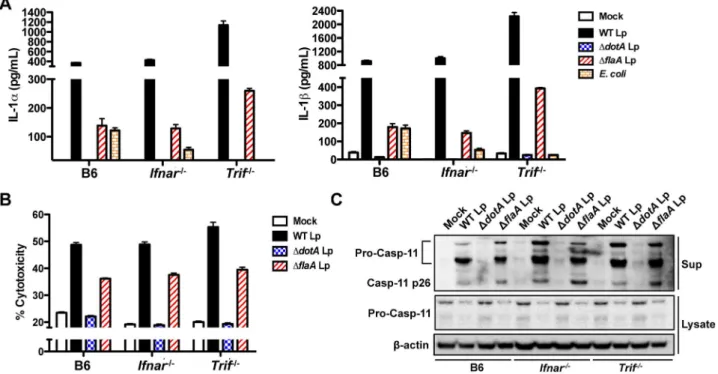
Figure 5. Non-canonical inflammasome responses toL. pneumophilaoccur independently of TRIF and IFNAR.(A) Unprimed B6,Ifnar2/2, orTrif2/2BMDMs were infected with WTL. pneumophila(WT Lp),DdotALp,DflaALp,E. coli, or PBS (mock infection) for 16 hours. Levels of IL-1aand IL-1bin the supernatants were measured by ELISA. (B) Unprimed B6,Ifnar2/2, orTrif2/2BMDMs were infected with WT Lp,DdotALp,DflaALp, or PBS (mock infection) for 16 hours. Cell death (% cytotoxicity) was measured by LDH release into the supernatants relative to Triton X-100-lysed cells. Graphs show the mean6SEM of triplicate wells. (C) B6,Ifnar2/2, orTrif2/2BMDMs were primed with 0.4
mg/mL Pam3CSK4 for 4 hours and infected
with WT Lp,DdotALp,DflaALp, or PBS for 16 hours. Levels of full-length caspase-11 (pro-casp-11) and processed caspase-11 (casp11 p26) in the supernatants and pro-casp-11 andb-actin (loading control) in the cell lysates were determined by immunoblot analysis. Data are representative of two independent experiments.
doi:10.1371/journal.ppat.1003400.g005
Figure 6. Caspase-11 mediates inflammasome activation in response to a functionalYersiniatype III secretion system.BMDMs from B6,Casp12/2Casp112/2,Casp12/2, orCasp112/2mice were primed with 0.05mg/mL LPS for 4 hours and infected with type III secretion system-deficientY. pseudotuberculosis(DyopBYp), effectorlessY. pseudotuberculosisDHOJMEK (D6 Yp), or PBS (mock infection) or treated with 2.5 mm ATP for 4 hours. (A) Levels of IL-1aand IL-1bin the supernatants were measured by ELISA. (B) Cell death (% cytotoxicity) was measured by lactate dehydrogenase (LDH) release relative to Triton X-100-lysed cells. Graphs show the mean6SEM of triplicate wells. Data are representative of two independent experiments. *** is p,0.001 and ** is p,0.01 by two-way ANOVA with Bonferroni post-test. NS is not significant.
signaling during infection with flagellin-deficient L. pneumophila, which do not activate the NAIP5/NLRC4 inflammasome, has not been investigated. We therefore infected B6 and IL-1R-deficient (Il1r12/2) mice intranasally withDflaALp and measured bacterial burden in the lung over the course of seven days. Though both B6 and Il1r12/2 mice received similar initial bacterial burdens, Il1r12/2 mice show a defect in bacterial clearance as early as 24 hours post-infection (Figure 7A). Bacterial burden remains elevated in the absence of IL-1R signaling, with the Il1r12/2 mice still exhibiting a log-increase in bacterial load at 120 hours
post-infection. Since IL-1R signaling is important for neutrophil recruitment [68], we examined whether Il1r12/2 mice have a defect in neutrophil recruitment to the pulmonary airway during
L. pneumophilainfection. Indeed,Il1r12/2mice exhibit a significant decrease in neutrophil recruitment to the airway 24 hours post-infection, possibly contributing to their inability to efficiently clear the pathogen (Figure 7B–C).
The IL-1R signals in response to both IL-1a and IL-1b; however, these cytokines can play non-redundant roles in anti-bacterial defense [69]. To determine the relative contributions of
Figure 7. IL-1aand IL-1bcontrol bacterial burden and neutrophil recruitmentin vivo.(A) 8–12 week old B6 orIl1r12/2mice were infected with 16106DflaA L. pneumophilaintranasally (IN). Lungs were plated to quantify CFU per gram. Graph shows the mean6SEM of three or four infected mice per group. Dashed line represents the limit of detection. (BandC) B6 orIl1r12/2mice were infected with 1
6106DflaALp IN. 24 hours post-infection, bronchoalveolar lavage fluid (BALF) was collected and the percentage of neutrophils in the BALF was quantified by flow cytometry. Percentages are reported as the frequency of live cells in the BALF. (B) Representative flow cytometry plots showing the percentage of Gr-1+Ly6G+ neutrophils. (C) Graph showing the percentage of neutrophils. Each point represents an individual mouse and lines indicate the mean of 4 mice per group. (D, E, andF) B6 mice were injected intraperitoneally (IP) with either PBS, 100mg isotype control antibody (iso), 100mg anti-IL-1aantibody, 100mg anti-IL-1bantibody, or 100mg each of anti-IL-1aand anti-IL-1b(anti-IL-1a/b) 16 hours before infection. The mice were then intranasally
infected with either 16106DflaALp or mock infected with PBS. (D and E) 24 hours post-infection, BALF was collected and flow cytometry was performed to quantify the percentage of neutrophils. (D) Representative flow cytometry plots showing the percentage of Gr-1+
Ly6G+
IL-1aand IL-1bto neutrophil recruitment and bacterial clearance duringL. pneumophilainfection, we utilized neutralizing antibodies to selectively block either IL-1a or IL-1b prior to infection. Specific cytokine neutralization in the BALF could be observed 24 hours post-infection (Figure S13). Critically, IL-1a neutraliza-tion alone significantly diminishes the percentage of neutrophils recruited to the BALF at 24 hours post-infection and results in a half-log increase in bacterial CFUs, in marked contrast to isotype control antibody or neutralization of IL-1b, which on its own did not have a significant effect (Figure 7D–F). However, neutraliza-tion of both IL-1aand IL-1bfully recapitulates the magnitude of neutrophil reduction and defect in bacterial clearance observed in theIl1r12/2mice. Collectively, these data indicate that although there are some overlapping roles for these cytokines during L.
pneumophila infection, IL-1a plays a distinct role from IL-1b in driving neutrophil recruitment to the airway and mediating bacterial clearance.
Discussion
Inflammasomes respond robustly to conserved features of pathogenic microbes, such as pore-forming toxins or specialized secretion systems that access the host cytosol. Inflammasomes therefore play a central role in enabling the immune system to discriminate between virulent and avirulent bacteria [70]. Recent reports show a role for caspase-11 in regulating the activation of a non-canonical inflammasome that promotes cell death as well as IL-1a and IL-1b secretion. This non-canonical inflammasome
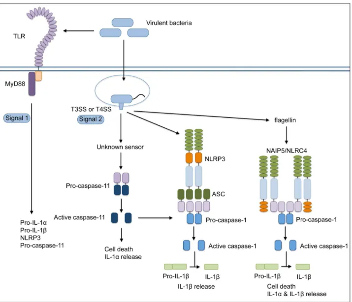
responds to both pathogenic and non-pathogenic Gram-negative bacteria independently of specialized secretion systems that translocate bacterial molecules into the host cytosol [26–29]. This pathway involves the TRIF- and IFNAR-dependent upregulation and activation of caspase-11 and occurs with relatively delayed kinetics in comparison to the response to pathogenic bacteria. Intriguingly, we find that the activity of theL. pneumophilaDot/Icm T4SS leads to rapid and robust caspase-11 activation indepen-dently of the TRIF-IFNAR axis, and this activation triggers rapid cell death and release of both IL-1a and IL-1b (Figure 8). We extend these results to show that the evolutionarily distinct T3SS of another pathogen, Y. pseudotuberculosis, also rapidly triggers caspase-11-dependent responses. Collectively, our findings dem-onstrate that caspase-11 is critical for inflammasome activation in response to the secretion systems of virulent bacteria that enable bacterial molecules to access the host cell cytosol and demonstrate that IL-1aand IL-1btogether play a crucial protective role during acute infectionin vivo.
We demonstrate that in response to the activity of bacterial secretion systems that enable cytosolic access, caspase-11 contrib-utes to NLRP3-mediated inflammasome activation and caspase-1-dependent IL-1b secretion and to a second ASC and NLRC4-independent pathway that does not require caspase-1 and leads to cell death as well as robust IL-1a release. These L. pneumophila -induced pathways are similar to recent findings with a number of Gram-negative bacterial pathogens, includingC. rodentium, E. coli, andS. typhimurium[26–29]. However, we observe rapid and robust T4SS-dependent activation of these two caspase-11-mediated pathways byL. pneumophila, whereas the response to Gram-negative bacteria lacking specialized secretion systems occurs less robustly and with much slower kinetics. Intriguingly, we observe a similarly rapid caspase-11-dependent induction of cell death and IL-1 release in response to the structurally and evolutionarily unrelated T3SS of
Y. pseudotuberculosis. Importantly, this pathway is independent of host sensing of flagellin, as it is triggered by flagellin-deficient L. pneumophila, andY. pseudotuberculosisdownregulates flagellin expres-sion when the T3SS is expressed [71]. Thus, our data suggest that the caspase-11 inflammasome is poised to respond robustly and rapidly to the activity of bacterial secretion systems that are capable of delivering microbial products to the host cell cytosol and may enable the host to respond to pathogens that evade flagellin-dependent responses. This could have significance for understand-ing the role of caspase-11 activation at mucosal sites colonized by large numbers of commensal bacteria. At mucosal barriers, it would be expected that the non-canonical inflammasome pathway would not be robustly activated by commensal bacteria but could respond rapidly to the presence of bacterial secretion systems that enable pathogen access to the host cytosol.
Our findings are consistent with recent observations that theL. pneumophilaDot/Icm T4SS triggers the caspase-11-dependent non-canonical inflammasome [72], as well as the finding that bacteria that enter the cytosol either due to failure to maintain integrity of their replicative vacuoles or natural entry into the cytoplasm also trigger rapid caspase-11 activation [73]. Thus the pathway that leads to caspase-11 activation appears to be particularly sensitive to pathogens that ‘violate the sanctity of the cytosol’ [74], either through the activity of specialized secretion systems that translo-cate bacterial molecules into the cytosol or through their direct entry into the host cell cytosol. Whether other pathogens that replicate within the cytosol, such asListeriaorShigella, or cytosolic viruses possess mechanisms to evade this pathway remains to be determined.
L. pneumophilaT4SS-mediated activation of caspase-11 differs from the other pathways of non-canonical inflammasome
activa-tion in several ways. First, L. pneumophila-mediated activation of caspase-11 does not require TRIF or IFNAR signaling. We observe a moderate dependence on TRIF and IFNAR signaling when macrophages are primed with LPS prior to infection, consistent with LPS-dependent upregulation of caspase-11 expres-sion through the TLR4-TRIF-IFNAR axis [27–29]. However, in the absence of LPS priming, TRIF and IFNAR signaling are dispensable forL. pneumophila-dependent caspase-11 activation. In this context, it is likely that MyD88 compensates for the absence of TRIF, as cells deficient for both MyD88 and TRIF failed to activate caspase-11 in response toL. pneumophila. Thus, although the TLR4-TRIF-IFNAR axis is required for caspase-11 activation in response to Gram-negative bacteria, a MyD88-dependent signal is sufficient for caspase-11 activation in response to pathogens that utilize virulence-associated secretion systems to translocate bacte-rial molecules into the host cytosol. It is possible that different signals are capable of activating caspase-11 through distinct pathways, but these pathways occur with distinct kinetics because they may indicate distinct levels of pathogenicity. Thus, while caspase-11 is robustly upregulated by LPS priming, this upregula-tion alone is insufficient for rapid activaupregula-tion in response to bacteria that lack specialized secretion systems, asDdotAorDyopBbacteria do not induce rapid cell death even in primed cells. Collectively, these data indicate a two-signal model for rapid caspase-11 activation during infection with virulent bacteria, where bacterial PAMPs induce caspase-11 upregulation, but rapid caspase-11 activation requires a second, secretion system-dependent signal (Figure 8).
The specific secretion system-dependent signals responsible for caspase-11 activation are currently unknown. While rapid activation of caspase-11 requires the presence of a functional type III or type IV secretion system or cytosolic access of the bacteria, whether the signal is an as-yet-undefined translocated bacterial molecule or a cellular response to the pore forming activity of these systems remains to be determined. The delayed NLRP3- and caspase-11-dependent response to Gram-negative bacteria sug-gests that in addition to LPS-induced upregulation of inflamma-some components, bacterial mRNA provides an additional signal for activating the NLRP3 inflammasome [18,75], although the role of caspase-11 in this response has not been formally demonstrated. Activity of the type III or IV secretion systems may bypass the need for bacterial mRNA. Alternatively, these secretion systems may translocate bacterial RNA [70,76,77], and the rapid caspase-11-dependent response they induce could be due to more rapid delivery of bacterial mRNA into the host cell cytosol.
Furthermore, the host factors required for activation of the NLRP3-independent caspase-11-dependent inflammasome also remain to be identified. As this pathway is independent of flagellin sensing, NLRP3, ASC, and NLRC4, an unknown upstream sensor and/or adaptor may be involved in caspase-11 activation in response to a translocated bacterial substrate or an endogenous signal induced by infection. This sensor may also be upregulated by type I IFN signaling itself [27–29].
data are consistent with a model in which IL-1ais an endogenous alarmin that is released during cell death [79].
Interestingly, caspase-11 also contributes to control of flagellin-expressing L. pneumophila by serving as a component of an NLRC4-dependent inflammasome that promotes trafficking of the L. pneumophila-containing vacuole to lysosomes [80]. Thus, caspase-11 may function in multiple ways to controlL. pneumophila
infection. Importantly, we find that IL-1a, IL-1b, and IL-1R signaling play an important role in the control of L. pneumophila
infection through efficient neutrophil recruitment to the airway. IL-1a and IL-1b play both distinct and overlapping roles in mediating neutrophil recruitment and controlling bacterial repli-cation, as depletion of IL-1a alone showed a more pronounced defect in neutrophil recruitment and bacterial clearance than depletion of IL-1balone, but loss of both cytokines resulted in a further reduction of neutrophil recruitment and an increased defect in bacterial clearance. Further analysis is required to define the relative contributions of the various caspase-11-mediated effector functions to the control ofL. pneumophilareplicationin vivo. In conclusion, these studies demonstrate that T3SS and T4SS activities trigger rapid and robust activation of caspase-11. This activation contributes to maximal NLRP3-dependent IL-1b secretion as well as to NLRP3-independent IL-1a release and host cell death. The downstream effector functions of these pathways are important for host defense againstL. pneumophila in vivo, as IL-1a and IL-1b promote neutrophil recruitment to L. pneumophila-infected lungs and control pulmonary bacterial repli-cation. Our results highlight the contribution of caspase-11 to rapid inflammasome activation and discrimination between pathogenic and nonpathogenic bacteria.
Materials and Methods
Ethics statement
This study was carried out in strict accordance as defined in the federal regulations set forth in the Animal Welfare Act (AWA), the recommendations in the Guide for the Care and Use of Laboratory Animals of the National Institutes of Health, and the guidelines of the University of Pennsylvania Institutional Animal Use and Care Committee. The protocols were approved by the Institutional Animal Care and Use Committee at the University of Pennsylvania (protocols#803465 and#803459).
Bacterial strains
Legionella pneumophila serogroup 1 strains were used in all experiments. Macrophages were infected with Lp02 (thyA), a thymidine auxotroph derived from strain Lp01 [40], orDdotA[81] and DflaA [16] isogenic mutant strains. Forin vivo studies, mice were infected with the Lp02DflaAor the JR32 [82]DflaAisogenic mutant strain where indicated. For in vitroand in vivostudies, L. pneumophila were cultured on charcoal yeast extract agar for 48 hours at 37uC prior to infection.Escherichia coliBL21 strain was cultured in LB broth for 16 hours at 37uC prior to infection. The
Yersinia pseudotuberculosis strains used were IP2666 DyopHOJEMK
(D6) [58] and DyopB [83]. Yersinia were grown overnight with aeration in 26YT broth at 26uC. The bacteria were diluted into fresh 26YT containing 20 mM sodium oxalate and 20 mM MgCl2. Bacteria were grown with aeration for 1 hour at 26uC followed by 2 hours at 37uC prior to infection.
Mice
C57BL/6 mice were purchased from Jackson Laboratories.
Casp12/2Casp112/2[84],Casp12/2(unpublished data, T.S. and R.A.F.), Casp112/2 [30], Asc2/2 [23], Nlrc42/2 [85], Asc2/2
Nlrc42/2 [47], Ifnar2/2 [86], Trif2/2 [87], Il1r12/2 [88], and Nalp32/2[89] mice are all on the C57BL/6 background.Asc2/2, Nlrc42/2, and Nlrp32/2 mice were originally generated by Millenium Pharmaceuticals and were a kind gift of Richard Flavell. Animals were maintained in accordance with the guidelines of the University of Pennsylvania Institutional Animal Use and Care Committee.
In vivoinfection studies
8–12 week-old mice were anesthetized by intraperitoneal injection of a ketamine/xylazine/PBS solution at a dose of 100 mg/kg ketamine and 10 mg/kg xylazine. Mice were infected intranasally with 40ml of a bacterial suspension containing 16106 CFU L. pneumophila or PBS vehicle control. For antibody neutralization experiments, mice were injected intraperitoneally with 100mg anti-IL-1aantibody (clone ALF-161), 100mg anti-IL-1bantibody (clone B122), 100mg of each anti-IL-1aand anti-IL-1b antibody, or 100mg Armenian hamster IgG1isotype control antibody (eBioscience) 16 hours prior to intranasal infection. At the indicated timepoints after infection, mice were sacrificed, and the bronchoalveolar lavage fluid (BALF) and lungs were harvested. To determine bacterial load, the lungs were mechanically homogenized in sterile distilled H2O and a portion of the lysate was spread onto CYE plates. Animal experiments were performed in accordance with approved University of Pennsylvania Institu-tional Animal Care and Use Committee protocols and procedures.
Macrophage experiments
Bone marrow was collected from the femurs and tibiae of mice. Bone marrow cells were differentiated into macrophages by culturing the cells in RPMI containing 30% L929 cell supernatant and 20% FBS at 37uC in a humidified incubator. The macrophages were replated one day prior to infection in RPMI containing 15% L929 cell supernatant and 10% FBS. For experiments involving LPS-primed macrophages, macrophages in 48-well plates (2.06105cells/well) were pretreated with 0.5mg/ mL LPS for 2.5 hours and either mock-infected with PBS, infected withL. pneumophilaat an MOI = 10 for 4 hours, or treated with 2.5 mM ATP for 1 or 4 hours. For experiments performed in the absence of LPS priming, macrophages in 48-well plates (2.06105 cells/well) were either mock-infected with PBS, infected withL. pneumophilaat an MOI = 10 for 16 or 20 hours, or infected withE. coliat an MOI = 25 for 1 hour followed by gentamycin treatment for 15 hours. To assess the involvement of caspase-1 catalytic activity, macrophages were treated with 20mM or 40mM of the
caspase-1 inhibitor YVAD-cmk (Bachem) or an equivalent volume of dimethyl sulfoxide (vehicle control) 0.5 hours prior to infection. ForL. pneumophilaandE. coliinfections, bacteria were centrifuged down onto the macrophages at 1200 RPM for ten minutes prior to incubation. ForY. pseudotuberculosisinfection, bacteria were washed three times with pre- warmed DMEM, added to the cells at an MOI = 20, and centrifuged down onto the macrophages at 1000 rpm for 5 min. Cells were incubated at 37uC for 1 hour post-infection followed by addition of 100mg/mL gentamicin. Supernatants were harvested 4 hours post infection for ELISA and LDH analysis.
Cytotoxicity assays
Immunoblotting
Supernatants from infected cells were mixed 1:1 with 2 X SDS-PAGE sample buffer or infected BMDMs were directly lysed in 1 X SDS-PAGE sample buffer. Samples were boiled, separated by SDS-PAGE, and transferred to Immobilon P membranes (Millipore). Primary antibodies against caspase-1 p10 (Santa Cruz Biotechnology), caspase-11 (Sigma, clone 17D9), IL-1b (R&D systems), and b-actin (Sigma) were used. Detection was performed with HRP-conjugated anti-rabbit IgG (Cell Signaling Technology) or anti-rat IgG (Santa Cruz Biotechnology or Jackson Immuno).
ELISA
Harvested supernatants from infected macrophages or the BALF from infected mice were assayed using capture and detection antibodies specific for IL-18 (MBL), IL-1a, IL-1b, and IL-12p40 (BD Biosciences).
Flow cytometry
To determine neutrophil recruitment to the airway, BALF cells were stained with Live/Dead Fixable Dead Cell Stain (Invitrogen), and antibodies specific for CD45, Gr-1 (eBioscience), and Ly6G (Biolegend). Data were collected with an LSRII flow cytometer (BD Biosciences) and post-collection data was analyzed using FlowJo (Treestar). Cells were gated on singlets and live cells. Neutrophils were identified as being CD45+
, Gr-1+
, and Ly6G+
.
Statistical analysis
Plotting of data and statistical analysis were performed using Graphpad Prism software, and statistical significance was deter-mined by the unpaired two-tailed Student’s t test, one-way ANOVA with Tukey post-test, or two-way ANOVA with Bonferroni post-test. Differences were considered statistically significant if thePvalue was,0.05.
Supporting Information
Figure S1 Caspase-1/caspase-11-deficient cells do not have a gross defect in cytokine secretion.(A) Unprimed B6 or Casp12/2Casp112/2 BMDMs were infected with WT L. pneumophila(Lp),DdotALp, DflaALp, or PBS (mock infection) for 20 hours. Levels of full-length IL-1b (pro-IL-1b) and b-actin (loading control) in the cell lysates were determined by immunoblot analysis. (B) B6 or Casp12/2Casp112/2 BMDMs were primed with 0.5mg/mL LPS for 2.5 hours and infected with
WT Lp,DdotALp,DflaALp, or PBS for 4 hours. The level of IL-18 in the supernatants was measured by ELISA. (C) Unprimed B6 or Casp12/2Casp112/2 BMDMs were infected with WT Lp, DdotALp,DflaALp, or PBS for 20 hours. Levels of IL-12 p40 and TNF-ain the supernatants were measured by ELISA. (D) B6 or
Casp12/2Casp112/2BMDMs were primed with 0.5mg/mL LPS for 2.5 hours and infected with WT Lp,DdotA Lp,DflaALp, or PBS for 4 hours. Levels of IL-12 p40 and TNF-a in the supernatants were measured by ELISA. Graphs show the mean 6SEM of triplicate wells. Data are representative of two (B) or three (A, C, and D) independent experiments.
(TIF)
Figure S2 Caspa11 is rapidly upregulated and se-creted in response to L. pneumophila. B6 or Casp12/2 Casp112/2 BMDMs were primed with 0.5mg/mL LPS for 2.5 hours and infected with WT L. pneumophila(Lp), DdotA Lp, DflaALp, or PBS (mock infection) for 4 hours. Levels of full-length caspase-11 (pro-casp-11) and active caspase-11 (casp11 p26) in the
supernatants, and pro-casp-11 andb-actin (loading control) in the cell lysates were determined by immunoblot analysis.
(TIF)
Figure S3 Caspase-11 is activated in response to L. pneumophilaindependently of macrophage priming.(A) Unprimed B6 and Casp12/2 BMDMs or (B) B6, Casp12/2 Casp112/2, and Casp112/2 BMDMs were infected with WT L. pneumophila(Lp),DdotALp,DflaALp, or PBS (mock infection) for 20 hours. Levels of IL-1a and IL-1b in the supernatants were measured by ELISA. Graphs show the mean6SEM of triplicate wells. (C) B6,Casp12/2, orCasp112/2BMDMs were primed with 0.5mg/mL LPS for 2.5 hours and infected with WT Lp,DdotALp,
DflaALp, or PBS for 4 hours or treated with LPS+2.5 mm ATP for 1 hour. Levels of mature IL-1b in the supernatant were determined by immunoblot analysis. Data are representative of two independent experiments.
(TIF)
Figure S4 IL-1a release is ASC/NLRC4-independent.
B6, Casp12/2Casp112/2, or Asc2/2Nlrc42/2 BMDMs were primed with 0.5mg/mL LPS for 2.5 hours and infected with WT L. pneumophila (Lp), DdotA Lp, DflaA Lp, or PBS (mock infection) or treated with 2.5 mm ATP for 4 hours. Levels of IL-1a and IL-1b in the supernatants were measured by ELISA. Graphs show the mean 6 SEM of triplicate wells. Data are representative of three independent experiments.
(TIF)
Figure S5 Both IL-1aand IL-1bsecretion are caspase-1/ caspase-11-dependentin vivo.8–12 week old B6 orCasp12/2 Casp112/2 mice were infected intranasally with 1
6106DflaALp. BALF was collected 24 hours post-infection, and levels of IL-1aand IL-1bwere measured by ELISA. Graphs show the mean6SEM of three mice per group. Dashed line represents the limit of detection. * is p,0.05 by unpaired t-test.
(TIF)
Figure S6 Mature IL-1bsecretion is not always concor-dant with cell death.B6,Casp12/2Casp112/2,Asc2/2,Nlrc42/2, orAsc2/2Nlrc42/2BMDMs were primed with 0.5mg/mL LPS for 2.5 hours and infected with WTL. pneumophila(Lp),DdotALp,DflaA
Lp or PBS (mock infection) for 4 hours or treated with 2.5 mm ATP for 1 hour. (A) Levels of mature IL-1bin the supernatants, and full-length IL-1b (pro-IL-1b) and b-actin (loading control) in the cell lysates were determined by immunoblot analysis. Data are representative of two independent experiments. (B) The level of IL-18 in the supernatants was measured by ELISA. Graphs show the mean6SEM of triplicate wells.
(TIF)
Figure S7 Flagellin-independent, NLRP3-dependent IL-1bsecretion occurs independently of macrophage prim-ing.(A) Unprimed B6 orNlrp32/2BMDMs were infected with WT L. pneumophila (Lp), DdotA Lp, DflaA Lp, or PBS (mock infection) for 16 hours. Levels of IL-1a and IL-1b in the supernatants were measured by ELISA. (B) B6 or Nlrp32/2 BMDMs were primed with 0.5mg/mL LPS for 2.5 hours and infected with WT Lp,DdotALp,DflaALp or PBS (mock infection) or treated with 2.5 mM ATP for 4 hours. The level of IL-18 in the supernatants was measured by ELISA. (C) B6 or Nlrp32/2 BMDMs were infected with WT Lp,DdotALp,DflaALp, or PBS (mock infection) for 16 hours. The level of IL-18 in the supernatants was measured by ELISA. (D and E) B6 BMDMs were primed with 0.5mg/mL LPS for 2.5 hours and infected with
50 mM KCl, or 50 mM NaCl were added prior to infection. (D) Levels of IL-1aand IL-1bin the supernatants were measured by ELISA. (E) Cell death was measured by LDH release. Graphs show the mean6SEM of triplicate wells. Data are representative of two independent experiments (A–C).
(TIF)
Figure S8 TRIF/IFNAR-independent IL-1 release occurs with Pam3CSK4-primed macrophages.(A) B6,Ifnar2/2, or Trif2/2 BMDMs were primed with 0.4mg/mL Pam3CSK4 for 4 hours and infected with WTL. pneumophila(Lp),DdotALp,DflaA
Lp,E. coli, or PBS (mock infection) for 16 hours. The levels of IL-1a and IL-1bin the supernatants were measured by ELISA. (B) B6,
Ifnar2/2, or Trif2/2 BMDMs were primed with 0.4mg/mL Pam3CSK4 for 4 hours and infected with WT Lp,DdotALp,DflaA
Lp, or PBS for 16 hours. Cell death (% cytotoxicity) was measured by LDH release. (C) B6,Ifnar2/2, orTrif2/2BMDMs were primed with 0.5mg/mL LPS for 2.5 hours and infected with WT Lp,DdotA
Lp,DflaALp, or PBS for 4 hours. Cell death was measured by LDH release. (D) B6,Ifnar2/2, orTrif2/2 BMDMs were primed with 0.5mg/mL LPS for 2.5 hours and infected with WT Lp,DdotALp, DflaALp, or PBS for 4 hours. Levels of IL-1a and IL-1b in the supernatants were measured by ELISA. Graphs show the mean6 SEM of triplicate wells. Data are representative of two independent experiments.
(TIF)
Figure S9 Caspase-11 is upregulated and secreted in an IFNAR- and TRIF-independent manner. Unprimed B6,
Ifnar2/2, or Trif2/2BMDMs were infected with WT Lp,DdotA Lp,DflaALp, or PBS for 16 hours. Levels of full-length caspase-11 (pro-casp-11) and active caspase-11 (casp11 p26) in the superna-tants, and pro-casp-11 and b-actin (loading control) in the cell lysates were determined by immunoblot analysis.
(TIF)
Figure S10 Detection of caspase-11 protein upregula-tion in cell lysates is moderate in response to L. pneumophila.(A) Unprimed B6,Ifnar2/2, orTrif2/2BMDMs were infected with WT Lp, DdotALp, DflaALp, E. coli, or PBS (mock infection) for 16 hours. (B) B6, Ifnar2/2, or Trif2/2 BMDMs were primed with 0.4mg/mL Pam3CSK4 for 4 hours
and infected with WT Lp,DdotALp,DflaALp,E. coli, or PBS for 16 hours. (C) B6,Ifnar2/2, orTrif2/2BMDMs were primed with 0.5mg/mL LPS for 2.5 hours and infected with WT Lp,DdotALp,
DflaALp,E. coli, or PBS for 4 hours. Levels of full-length caspase-11 (pro-casp-caspase-11) and b-actin (loading control) in the cell lysates were determined by immunoblot analysis.
(TIF)
Figure S11 Caspase-11 is not upregulated in the ab-sence of both MyD88 and Trif.(A) Immortalized B6 (iB6) or MyD88/Trif-deficient (iMyd882/2Trif2/2) BMDMs were primed
with 0.4mg/mL Pam3CSK4 for 4 hours and infected with WT
Lp, DdotA Lp, DflaA Lp, E. coli, or PBS (mock infection) for 16 hours. (B) iB6 or iMyd882/2Trif2/2macrophages were primed with 0.5mg/mL LPS for 4 hours and infected with WT Lp,DdotA
Lp,DflaALp, or PBS (mock infection) for 4 hours. Levels of full-length caspase-11 (pro-casp-11) andb-actin (loading control) were determined by immunoblot analysis.
(TIF)
Figure S12 Caspase-11-deficient cells secrete compa-rable amounts of IL-12 in response toY. pseudotuber-culosis. B6, Casp12/2Casp112/2, Casp12/2, or Casp112/2 mice were primed with 0.05mg/mL LPS for 2.5 hours and infected with type III secretion system-deficient Y. pseudotuber-culosis (DyopB Yp), effectorless Y. pseudotuberculosis DHOJMEK (D6 Yp), or PBS (mock infection) or treated with 2.5 mm ATP for 4 hours. The level of IL-12 p40 in the supernatants was measured by ELISA. Graphs show the mean 6 SEM of triplicate wells. Data are representative of two independent experiments.
(TIF)
Figure S13 Intraperitoneally injected antibodies neu-tralize cytokine in the BALF.8–12 week old B6 mice were injected intraperitoneally (IP) with either PBS, 100mg anti-IL-1a
antibody, 100mg anti-IL-1bantibody, or 100mg each of anti-IL-1a and anti-IL-1b (anti-IL-1a/b) 16 hours before infection. The mice were then infected with either 16106 DflaA Lp or mock infected with PBS intranasally (IN). 24 hours post-infection, bronchoalveolar lavage fluid (BALF) was collected and the levels of IL-1a and IL-1b were measured by ELISA. Labels indicate what was received intraperitoneally (IP) and what was received intranasally (IN). Graphs show the mean6 SEM of 8 mice per group and represent the pooled results of two independent experiments.
(TIF)
Acknowledgments
We are grateful to Junying Yuan (Harvard University) for permission to utilize Casp112/2 mice, Tiffany Horng (Harvard University School of Public Health) for providing Casp112/2bone marrow, and Susan Weiss (University of Pennsylvania Perelman School of Medicine) for providing Ifnar2/2 bone marrow. We also thank the Brodsky lab for valuable feedback on experimental design.
Author Contributions
Conceived and designed the experiments: CNC AMC EEZ IEB SS. Performed the experiments: CNC AMC EEZ HTN BJ WPB TCF IEB SS. Analyzed the data: CNC AMC EEZ HTN BJ IEB SS. Contributed reagents/materials/analysis tools: TS RAF IEB SS. Wrote the paper: CNC IEB SS.
References
1. Janeway CA, Jr. (1989) Approaching the asymptote? Evolution and revolution in immunology. Cold Spring Harb Symp Quant Biol 54 Pt 1: 1–13. 2. Medzhitov R (2007) Recognition of microorganisms and activation of the
immune response. Nature 449: 819–826.
3. Janeway CA, Jr., Medzhitov R (2002) Innate immune recognition. Annu Rev Immunol 20: 197–216.
4. Fritz JH, Ferrero RL, Philpott DJ, Girardin SE (2006) Nod-like proteins in immunity, inflammation and disease. Nat Immunol 7: 1250–1257.
5. Ting JP, Kastner DL, Hoffman HM (2006) CATERPILLERs, pyrin and hereditary immunological disorders. Nature reviews Immunology 6: 183–195. 6. Harton JA, Linhoff MW, Zhang J, Ting JP (2002) Cutting edge:
CATER-PILLER: a large family of mammalian genes containing CARD, pyrin, nucleotide-binding, and leucine-rich repeat domains. Journal of immunology 169: 4088–4093.
7. Inohara Chamaillard, McDonald C, Nunez G (2005) NOD-LRR proteins: role in host-microbial interactions and inflammatory disease. Annu Rev Biochem 74: 355–383.
8. Davis BK, Wen H, Ting JP (2011) The inflammasome NLRs in immunity, inflammation, and associated diseases. Annual review of immunology 29: 707– 735.
9. Martinon F, Burns K, Tschopp J (2002) The inflammasome: a molecular platform triggering activation of inflammatory caspases and processing of proIL-beta. Molecular cell 10: 417–426.
10. Miao EA, Leaf IA, Treuting PM, Mao DP, Dors M, et al. (2010) Caspase-1-induced pyroptosis is an innate immune effector mechanism against intracellular bacteria. Nature immunology 11: 1136–1142.
12. Mariathasan S, Weiss DS, Newton K, McBride J, O’Rourke K, et al. (2006) Cryopyrin activates the inflammasome in response to toxins and ATP. Nature 440: 228–232.
13. Boyden ED, Dietrich WF (2006) Nalp1b controls mouse macro-phage susceptibility to anthrax lethal toxin. Nature genetics 38: 240– 244.
14. Miao EA, Alpuche-Aranda CM, Dors M, Clark AE, Bader MW, et al. (2006) Cytoplasmic flagellin activates caspase-1 and secretion of interleukin 1beta via Ipaf. Nature immunology 7: 569–575.
15. Franchi L, Amer A, Body-Malapel M, Kanneganti TD, Ozoren N, et al. (2006) Cytosolic flagellin requires Ipaf for activation of caspase-1 and interleukin 1beta in salmonella-infected macrophages. Nat Immunol 7: 576–582.
16. Ren T, Zamboni DS, Roy CR, Dietrich WF, Vance RE (2006) Flagellin-deficient Legionella mutants evade caspase-1- and Naip5-mediated macrophage immunity. PLoS Pathog 2: e18.
17. Molofsky AB, Byrne BG, Whitfield NN, Madigan CA, Fuse ET, et al. (2006) Cytosolic recognition of flagellin by mouse macrophages restricts Legionella pneumophila infection. The Journal of experimental medicine 203: 1093–1104. 18. Kanneganti TD, Ozoren N, Body-Malapel M, Amer A, Park JH, et al. (2006) Bacterial RNA and small antiviral compounds activate caspase-1 through cryopyrin/Nalp3. Nature 440: 233–236.
19. Kofoed EM, Vance RE (2011) Innate immune recognition of bacterial ligands by NAIPs determines inflammasome specificity. Nature 477: 592–595. 20. Hornung V, Ablasser A, Charrel-Dennis M, Bauernfeind F, Horvath G, et al.
(2009) AIM2 recognizes cytosolic dsDNA and forms a caspase-1-activating inflammasome with ASC. Nature 458: 514–518.
21. Dostert C, Petrilli V, Van Bruggen R, Steele C, Mossman BT, et al. (2008) Innate immune activation through Nalp3 inflammasome sensing of asbestos and silica. Science 320: 674–677.
22. Srinivasula SM, Poyet JL, Razmara M, Datta P, Zhang Z, et al. (2002) The PYRIN-CARD protein ASC is an activating adaptor for caspase-1. The Journal of biological chemistry 277: 21119–21122.
23. Sutterwala FS, Ogura Y, Szczepanik M, Lara-Tejero M, Lichtenberger GS, et al. (2006) Critical role for NALP3/CIAS1/Cryopyrin in innate and adaptive immunity through its regulation of caspase-1. Immunity 24: 317–327. 24. Cookson BT, Brennan MA (2001) Pro-inflammatory programmed cell death.
Trends in microbiology 9: 113–114.
25. Fink SL, Cookson BT (2005) Apoptosis, pyroptosis, and necrosis: mechanistic description of dead and dying eukaryotic cells. Infection and immunity 73: 1907–1916.
26. Kayagaki N, Warming S, Lamkanfi M, Vande Walle L, Louie S, et al. (2011) Non-canonical inflammasome activation targets caspase-11. Nature 479: 117– 121.
27. Rathinam VA, Vanaja SK, Waggoner L, Sokolovska A, Becker C, et al. (2012) TRIF licenses caspase-11-dependent NLRP3 inflammasome activation by gram-negative bacteria. Cell 150: 606–619.
28. Gurung P, Malireddi RK, Anand PK, Demon D, Walle LV, et al. (2012) Toll or interleukin-1 receptor (TIR) domain-containing adaptor inducing interferon-beta (TRIF)-mediated caspase-11 protease production integrates Toll-like receptor 4 (TLR4) protein- and Nlrp3 inflammasome-mediated host defense against enteropathogens. The Journal of biological chemistry 287: 34474–34483. 29. Broz P, Ruby T, Belhocine K, Bouley DM, Kayagaki N, et al. (2012) Caspase-11 increases susceptibility to Salmonella infection in the absence of caspase-1. Nature 490: 288–291.
30. Wang S, Miura M, Jung YK, Zhu H, Li E, et al. (1998) Murine caspase-11, an ICE-interacting protease, is essential for the activation of ICE. Cell 92: 501–509. 31. Juhas M, Crook DW, Hood DW (2008) Type IV secretion systems: tools of bacterial horizontal gene transfer and virulence. Cellular microbiology 10: 2377–2386.
32. Cornelis GR (2006) The type III secretion injectisome. Nature reviews Microbiology 4: 811–825.
33. Brodsky IE, Monack D (2009) NLR-mediated control of inflammasome assembly in the host response against bacterial pathogens. Seminars in immunology 21: 199–207.
34. Miao EA, Mao DP, Yudkovsky N, Bonneau R, Lorang CG, et al. (2010) Innate immune detection of the type III secretion apparatus through the NLRC4 inflammasome. Proceedings of the National Academy of Sciences of the United States of America 107: 3076–3080.
35. Sun YH, Rolan HG, Tsolis RM (2007) Injection of flagellin into the host cell cytosol by Salmonella enterica serotype Typhimurium. Journal of Biological Chemistry 282: 33897–33901.
36. Molofsky AB, Byrne BG, Whitfield NN, Madigan CA, Fuse ET, et al. (2006) Cytosolic recognition of flagellin by mouse macrophages restricts Legionella pneumophila infection. J Exp Med 203: 1093–1104.
37. Fraser DW, Tsai TR, Orenstein W, Parkin WE, Beecham HJ, et al. (1977) Legionnaires’ disease: description of an epidemic of pneumonia. The New England journal of medicine 297: 1189–1197.
38. McDade JE, Shepard CC, Fraser DW, Tsai TR, Redus MA, et al. (1977) Legionnaires’ disease: isolation of a bacterium and demonstration of its role in other respiratory disease. N Engl J Med 297: 1197–1203.
39. Marra A, Blander SJ, Horwitz MA, Shuman HA (1992) Identification of a Legionella pneumophila locus required for intracellular multiplication in human macrophages. Proc Natl Acad Sci U S A 89: 9607–9611.
40. Berger KH, Isberg RR (1993) Two distinct defects in intracellular growth complemented by a single genetic locus in Legionella pneumophila. Mol Microbiol 7: 7–19.
41. Roy CR, Berger KH, Isberg RR (1998) Legionella pneumophila DotA protein is required for early phagosome trafficking decisions that occur within minutes of bacterial uptake. Mol Microbiol 28: 663–674.
42. Segal G, Purcell M, Shuman HA (1998) Host cell killing and bacterial conjugation require overlapping sets of genes within a 22-kb region of the Legionella pneumophila genome. Proc Natl Acad Sci U S A 95: 1669–1674. 43. Vogel JP, Andrews HL, Wong SK, Isberg RR (1998) Conjugative transfer by the
virulence system of Legionella pneumophila. Science 279: 873–876. 44. Nagai H, Kagan JC, Zhu X, Kahn RA, Roy CR (2002) A bacterial guanine
nucleotide exchange factor activates ARF on Legionella phagosomes. Science 295: 679–682.
45. Ensminger AW, Isberg RR (2009) Legionella pneumophila Dot/Icm translo-cated substrates: a sum of parts. Current opinion in microbiology 12: 67–73. 46. Hubber A, Roy CR (2010) Modulation of host cell function by Legionella
pneumophila type IV effectors. Annual review of cell and developmental biology 26: 261–283.
47. Case CL, Shin S, Roy CR (2009) Asc and Ipaf Inflammasomes direct distinct pathways for caspase-1 activation in response to Legionella pneumophila. Infection and immunity 77: 1981–1991.
48. Zamboni DS, Kobayashi KS, Kohlsdorf T, Ogura Y, Long EM, et al. (2006) The Birc1e cytosolic pattern-recognition receptor contributes to the detection and control of Legionella pneumophila infection. Nat Immunol 7: 318–325. 49. Amer A, Franchi L, Kanneganti TD, Body-Malapel M, Ozoren N, et al. (2006)
Regulation of Legionella phagosome maturation and infection through flagellin and host Ipaf. J Biol Chem 281: 35217–35223.
50. Lightfield KL, Persson J, Brubaker SW, Witte CE, von Moltke J, et al. (2008) Critical function for Naip5 in inflammasome activation by a conserved carboxy-terminal domain of flagellin. Nature immunology 9: 1171–1178.
51. Case CL, Roy CR (2011) Asc modulates the function of NLRC4 in response to infection of macrophages by Legionella pneumophila. mBio 2: e001117–11. 52. Shin S, Case CL, Archer KA, Nogueira CV, Kobayashi KS, et al. (2008) Type
IV secretion-dependent activation of host MAP kinases induces an increased proinflammatory cytokine response to Legionella pneumophila. PLoS Pathog 4: e1000220.
53. Broz P, von Moltke J, Jones JW, Vance RE, Monack DM (2010) Differential requirement for Caspase-1 autoproteolysis in pathogen-induced cell death and cytokine processing. Cell host & microbe 8: 471–483.
54. Thornberry NA, Bull HG, Calaycay JR, Chapman KT, Howard AD, et al. (1992) A novel heterodimeric cysteine protease is required for interleukin-1 beta processing in monocytes. Nature 356: 768–774.
55. Gross O, Yazdi AS, Thomas CJ, Masin M, Heinz LX, et al. (2012) Inflammasome activators induce interleukin-1alpha secretion via distinct pathways with differential requirement for the protease function of caspase-1. Immunity 36: 388–400.
56. Mariathasan S, Newton K, Monack DM, Vucic D, French DM, et al. (2004) Differential activation of the inflammasome by caspase-1 adaptors ASC and Ipaf. Nature 430: 213–218.
57. Brodsky IE, Palm NW, Sadanand S, Ryndak MB, Sutterwala FS, et al. (2010) A Yersinia effector protein promotes virulence by preventing inflammasome recognition of the type III secretion system. Cell host & microbe 7: 376–387. 58. Lilo S, Zheng Y, Bliska JB (2008) Caspase-1 activation in macrophages infected
with Yersinia pestis KIM requires the type III secretion system effector YopJ. Infection and immunity 76: 3911–3923.
59. Dower SK, Kronheim SR, Hopp TP, Cantrell M, Deeley M, et al. (1986) The cell surface receptors for interleukin-1 alpha and interleukin-1 beta are identical. Nature 324: 266–268.
60. Burns K, Martinon F, Esslinger C, Pahl H, Schneider P, et al. (1998) MyD88, an adapter protein involved in interleukin-1 signaling. The Journal of biological chemistry 273: 12203–12209.
61. Adachi O, Kawai T, Takeda K, Matsumoto M, Tsutsui H, et al. (1998) Targeted disruption of the MyD88 gene results in loss of IL-1- and IL-18-mediated function. Immunity 9: 143–150.
62. Hawn TR, Smith KD, Aderem A, Skerrett SJ (2006) Myeloid differentiation primary response gene (88)- and toll-like receptor 2-deficient mice are susceptible to infection with aerosolized Legionella pneumophila. J Infect Dis 193: 1693– 1702.
63. Archer KA, Roy CR (2006) MyD88-dependent responses involving toll-like receptor 2 are important for protection and clearance of Legionella pneumophila in a mouse model of Legionnaires’ disease. Infect Immun 74: 3325–3333.
64. Sporri R, Joller N, Albers U, Hilbi H, Oxenius A (2006) MyD88-dependent IFN-gamma production by NK cells is key for control of Legionella pneumophila infection. J Immunol 176: 6162–6171.
65. Hawn TR, Berrington WR, Smith IA, Uematsu S, Akira S, et al. (2007) Altered Inflammatory Responses in TLR5-Deficient Mice Infected with Legionella pneumophila. J Immunol 179: 6981–6987.