The Utility of Sentinel Lymph Node Biopsy in
Papillary Thyroid Carcinoma with Occult
Lymph Nodes
Xingqiang Yan1,2, Ruichao Zeng2, Zhaosheng Ma1, Chengze Chen2, Endong Chen2, Xiaohua Zhang2*, Feilin Cao1*
1Department of Surgical Oncology, Taizhou Hospital, Wenzhou Medical University, Linhai, Zhejiang Province, People’s Republic of China,2Department of Surgical Oncology, The First Affiliated Hospital, Wenzhou Medical University, Wenzhou, Zhejiang Province, People’s Republic of China
*[email protected](XHZ);[email protected](FLC)
Abstract
Background
The sentinel lymph node (SLN) is defined as the first draining node from the primary lesion, and it has proven to be a good indicator of the metastatic status of regional lymph nodes in solid tumors. The aim of this study was to evaluate the clinical application of SLN biopsy (SLNB) in papillary thyroid carcinoma (PTC) with occult lymph nodes.
Methods
From April 2006 to October 2012, 212 consecutive PTC patients were treated with SLNB using carbon nanoparticle suspension (CNS). Then, the stained nodes defined as SLN were collected, and prophylactic central compartment neck dissection (CCND) followed by total thyroidectomy or subtotal thyroidectomy were performed. All the samples were sent for pathological examination.
Results
There were 78 (36.8%) SLN metastasis (SLNM)-positive cases and 134 (63.2%) SLNM-negative cases. The sensitivity, specificity, positive and SLNM-negative predictive values, and false-positive and false-negative rates of SLNB were 78.8%, 100%, 100%, 84.3%, 0%, and 21.2%, respectively. The PTC patients with SLNM were more likely to be male (48.2% vs. 32.7%, p = 0.039) and exhibited multifocality (52.6% vs. 33.3%, p = 0.025) and extrathyroi-dal extension (56.7% vs. 33.5%, p = 0.015). A greater incidence of non-SLN metastases in the central compartment was found in patients with SLNM (41/78, 52.6%) than in those with-out SLNM (21/134, 15.7%; p<0.05). However, the SLNM-negative PTC patients with non-SLN metastases were more likely to be male (37.9% vs. 9.5%, p<0.05).
OPEN ACCESS
Citation:Yan X, Zeng R, Ma Z, Chen C, Chen E, Zhang X, et al. (2015) The Utility of Sentinel Lymph Node Biopsy in Papillary Thyroid Carcinoma with Occult Lymph Nodes. PLoS ONE 10(6): e0129304. doi:10.1371/journal.pone.0129304
Academic Editor:Peyman Björklund, Uppsala University, SWEDEN
Received:November 24, 2014
Accepted:May 7, 2015
Published:June 5, 2015
Copyright:© 2015 Yan et al. This is an open access article distributed under the terms of theCreative Commons Attribution License, which permits unrestricted use, distribution, and reproduction in any medium, provided the original author and source are credited.
Data Availability Statement:All relevant data are within the manuscript and its supporting information file.
Funding:The authors have no support or funding to report.
Conclusions
The application of SLNB using CNS is technically feasible, safe, and useful, especially for male patients with co-existing multifocality and extrathyroidal extension. However, the sen-sitivity of SLNB must be improved and its false-negative rate reduced before it can be a rou-tine procedure and replace prophylactic CCND. More attention should be paid to PTC patients (especially males) without SLNM for signs of non-SLN metastases.
Introduction
Sentinel lymph nodes (SLNs), the first station in the lymphatic drainage basin, receive lymph flow from primary tumors and reflect the status of the remaining lymph nodes. In surgery, SLN biopsy (SLNB) has become a common method for treating several types of human malig-nant tumors, especially melanoma and breast cancer[1,2]. However, SLNB application to the treatment of papillary thyroid carcinoma (PTC) patients has not been thoroughly investigated.
PTC, including papillary microcarcinoma (PTMC, tumor size1 cm), is the most common type of differentiated thyroid cancer and spreads predominantly via the lymphatics to the local draining lymph nodes. The metastases of occult lymph nodes are detected in approximately 27–90% of PTC cases after surgery and histologic examination[3–5]. Although PTC has a high
rate of regional lymph node metastasis (LNM), the impact of regional LNM on the prognosis is unclear. Recent studies have indicated that regional LNMs increase the risk of locoregional re-currence and have an adverse effect on survival, especially in older patients (age>45 years) [4,6–8]. A study of 9,904 patients with PTC or follicular thyroid carcinoma indicated that
LNM is a significant independent factor of poor prognosis based on multivariate analysis[9]. By contrast, other studies have demonstrated that occult LNM increases locoregional recur-rence but does not affect disease-specific survival[10–12]. An investigation revealed that
palpa-ble lymph nodes in PTC patients should undergo therapeutic node dissection, whereas patients with occult lymph nodes do not benefit from prophylactic node dissection[13]. Therefore, ap-propriate management of PTC with overt or occult lymph nodes is very important for improv-ing survival, decreasimprov-ing regional recurrence and avoidimprov-ing over-treatment.
In recent years, with the development of nanotechnology, nanocarbons have been widely used as a lymph node tracer in malignant tumors. An injection of carbon nanoparticle suspen-sion (CNS) consists of nanosized carbon particles with an average diameter of 150 nm. Upon injection into the tissues around the tumor, nanocarbon particles are rapidly engulfed by mac-rophages. The particles then enter the lymphatic vessels and accumulate in the lymph nodes, staining them black. This technique has facilitated the vital staining of tumor-draining lymph nodes, and has been applied in the detection of SLNs in breast and gastric cancers[14,15].
Patients and Methods
Ethics Statement
This study was approved by the Ethics Committee of The First Affiliated Hospital of Wenzhou Medical University, and written informed consent was obtained from all study subjects prior to enrollment. All data analyzed were anonymized.
Patients
This prospective study was conducted on 212 patients with incipient thyroid cancer and a diag-nosis of PTC. All patients underwent surgical treatment by SLNB and prophylactic CCND at our hospital from 1 April 2006 to 31 October 2012. They were diagnosed with PTC based on preoperative fine-needle aspiration cytology and postoperative pathology. We excluded pa-tients who had preoperative evidence of cervical LNM based on physical examination or fine-needle aspiration cytology, ultrasound, and computed tomography. Patients with previous neck surgery or preoperative detectable lymph nodes were also excluded from our study.
Surgical Procedure
All the surgery operations were performed by three experienced surgeons, who had performed more than one hundred thyroid surgeries per year within the past decade and had experience with thyroid SLN procedures. A transverse low-collar skin incision was followed by separation of the skin flap and a longitudinal incision in the linea alba cervicalis. Then, the thyroid capsule was carefully opened to completely expose the thyroid gland without injury to the capsule. The thyroid gland and the ipsilateral jugular vein were exposed. SLNB was performed according to the method described by Hao et al.[17]. Then, approximately 1 ml of a CNS (Chongqing Lummy Pharmaceutical Co., Ltd.) was injected using a 27-gauge needle into the parenchyma surrounding the primary tumor. For bilateral tumors or multiple tumors, the CNS was injected into those tumors that were suspected to be malignant. Within minutes, the stained lymphatic vessels from the primary tumor became apparent, and black-stained lymph nodes were identi-fied via tracing the stained lymphatic vessels (Fig 1). We focused on black-stained lymph nodes in the central compartment. These black-stained lymph nodes, defined as SLNs, were carefully collected and sent to the pathology department for frozen and routine pathology. Subsequently, prophylactic CCND followed by total thyroidectomy or subtotal thyroidectomy were per-formed. The remaining stained lymph nodes in the central compartment, defined as non-SLNs, were sent for routine pathology. Each SLN was cut in half: one half was used for frozen pathology and the other half for routine pathology. All the samples were cut into 5-um-thick slices and stained by hematoxylin-eosin. LNM was defined as tumor tissue found in the slice. Several tumor markers were tested by an immunohistochemistry assay to improve the diagno-sis. Criteria to identify malignant keratin-positive cells included strong cytokeratin immunore-activity, anatomic location in nodal sinuses, and cytologic atypia similar to the primary carcinoma. All the diagnoses were made by two experienced pathologists, and all the final diag-noses were based on the routine pathology findings.
Data Analysis
data, respectively. SPSS version (SPSS Inc., Chicago, IL, USA), version 19, was used in all of the statistical analyses, and statistical significance was defined as a p-value less than 0.05.
Results
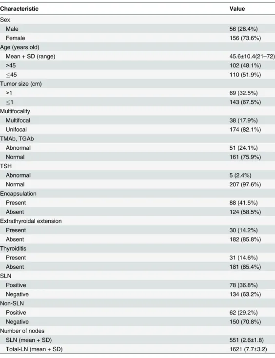
The characteristics of all patients are shown inTable 1(Details are shown inS1 Table). In total, 212 patients underwent SLNB and prophylactic CCND; the mean age was 45.6 years (ranging from 21 to 72 years). The majority of the patients were female (156/212, 73.6%) and diagnosed with PTMC (143/212, 67.5%). Most PTCs do not present multifocality (38/212, 17.9%) and extrathyroidal extension (30/212, 14.2%). Nearly half of the patients were diagnosed with tumor encapsulation (88/212, 41.5%). A minority of patients presented high levels of serum TMAb and TGAb (51/212, 24.1%) and TSH (5/212, 2.4%) and developed thyroiditis (31/212, 14.6%). In the final pathologic reports, SLN metastases (SLNMs) were found in 78 (36.8%) pa-tients and non-SLNs were found in 62 (29.2%) papa-tients; 551 lymph nodes were detected in the SLNBs and 1621 lymph nodes were removed from the central compartment.
The SLNB results are illustrated inFig 2. In the present study, 78 patients were diagnosed with SLNM, and in 41 patients, further metastases in the non-SLN samples were detected. The remaining 134 patients were not diagnosed with SLNM, whereas 21 patients exhibited further metastases in the non-SLN samples. The SLNB results are shown inTable 2Part A. The sensi-tivity, specificity, positive and negative predictive values, and false-positive and false-negative rates of SLNB were 78.8%, 100%, 100%, 84.3%, 0%, and 21.2%, respectively (Table 2Part B). Among these patients, males had a significantly higher rate of SLNM compared with females (48.2% vs. 32.7%, p = 0.039), and multifocal PTC had a higher probability of SLNM compared with unifocal PTC (52.6% vs. 33.3%, p = 0.025). More SLNMs were found in PTC with extra-thyroidal extension than in PTC without extraextra-thyroidal extension (56.7% vs. 33.5%, p = 0.015). However, other factors such as patient age; tumor size; serum TMAb, TGAb and TSH levels; encapsulation; and thyroiditis did not show statistical significance for SLNM (Table 3). A mul-tivariate analysis was performed to determine whether these factors were independently corre-lated with SLNM. Male gender (OR = 1.939, p = 0.043), multifocality (OR = 2.204, p = 0.035) and extrathyroidal extension (OR = 2.624, p = 0.033) were independently predictive of SLNM
Fig 1. SLNB using CNS. The white arrow indicates the black-stained SLN in the central compartment.
SLNB = sentinel lymph node biopsy; SLN = sentinel lymph node; CNS = carbon nanoparticle suspension.
(Table 4). In addition, patients with SLNM (41/78) had a greater incidence of non-SLN com-pared to those without SLNM (21/134) (52.6% vs. 15.7%, p = 0.000) (Table 3).
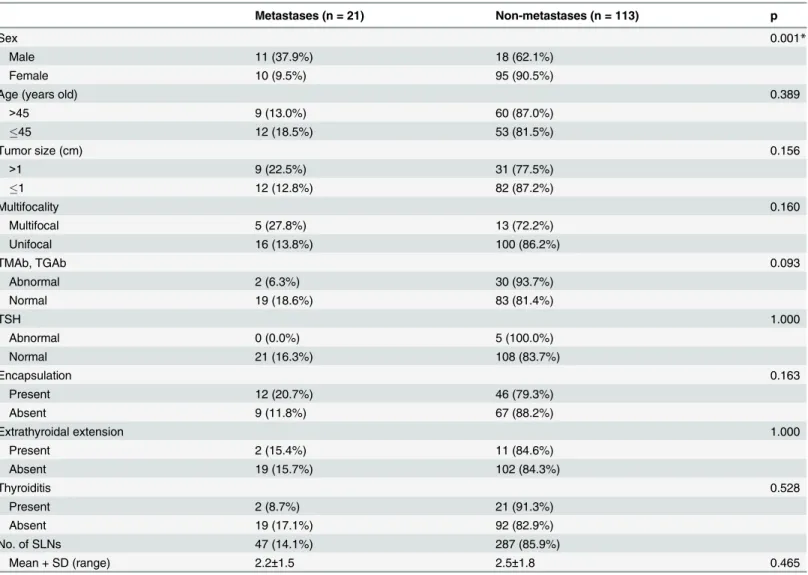
Among the 134 patients who were not diagnosed with SLNM, 21 exhibited further metastases in the non-SLN samples. Male patients also had a significantly higher rate of non-SLN metasta-ses compared with female patients (37.9% vs. 9.5%, p = 0.001). Age; tumor size; multifocality;
Table 1. Patient demographics and tumor characteristics (n = 212).
Characteristic Value
Sex
Male 56 (26.4%)
Female 156 (73.6%)
Age (years old)
Mean + SD (range) 45.6±10.4(21–72)
>45 102 (48.1%)
45 110 (51.9%)
Tumor size (cm)
>1 69 (32.5%)
1 143 (67.5%)
Multifocality
Multifocal 38 (17.9%)
Unifocal 174 (82.1%)
TMAb, TGAb
Abnormal 51 (24.1%)
Normal 161 (75.9%)
TSH
Abnormal 5 (2.4%)
Normal 207 (97.6%)
Encapsulation
Present 88 (41.5%)
Absent 124 (58.5%)
Extrathyroidal extension
Present 30 (14.2%)
Absent 182 (85.8%)
Thyroiditis
Present 31 (14.6%)
Absent 181 (85.4%)
SLN
Positive 78 (36.8%)
Negative 134 (63.2%)
Non-SLN
Positive 62 (29.2%)
Negative 150 (70.8%)
Number of nodes
SLN (mean + SD) 551 (2.6±1.8)
Total-LN (mean + SD) 1621 (7.7±3.2)
TMAb = thyroid microsomal antibody; TGAb = thyroglobulin antibody; TSH = thyroid stimulating hormone; SLN = sentinel lymph node; Non-SLN = non-sentinel lymph node; Total-LN = total lymph nodes
serum TMAb, TGAb and TSH levels; encapsulation; extrathyroidal extension; thyroiditis; and mean number of SLNs were not significantly related to the non-SLNs (Table 5).
There were two types of postoperative complications in this study, including 2 hematomas and 20 cases of transient hypoparathyroidism. No cases of permanent recurrent nerve damaged and no complications or side effects related to the CNS were detected.
Discussion
The role of routine CCND in the treatment of PTC remains unclear. It is generally agreed that therapeutic neck dissection should be performed to remove macroscopic LNM because this treatment reduces the chance of PTC persistence and recurrence[18]. However, there have been no randomized controlled trails to support the concept that routine prophylactic CCND affects the recurrence or survival rates of PTC patients with occult lymph nodes. In addition, prophylactic CCND may increase the risk of complications such as hypocalcemia and recur-rent laryngeal nerve palsy. However, an additional operation may be necessary after recurrence, which will increase the risk of operative complications and medical costs. Therefore, the accu-rate identification of occult LNMs in PTC patients is helpful in the selection of an appropriate therapeutic strategy. An SLN is defined as the first draining node from the primary lesion and
Fig 2. Overview of SLNB.SLNB = sentinel lymph node biopsy; SLN = sentinel lymph node; Non-SLN = non-sentinel lymph node.
doi:10.1371/journal.pone.0129304.g002
Table 2. Results of SLNB using CNS in the central compartment.
A
SLN + SLN - Total
Central lymph node + 78 21 99
Central lymph node - 0 113 113
Total 78 134 212
B
Characteristic Value
Detection rate 212/212 (100%)
Sensitivity 78/99 (78.8%)
Specificity 113/113 (100%)
Positive predictive value 78/78 (100%)
Negative predictive value 113/134 (84.3%)
False-positive rate 0/113 (0%)
False-negative rate 21/99 (21.2%)
Accuracy 191/212 (90.1%)
SLNB = sentinel lymph node biopsy; SLN = sentinel lymph node; CNS = carbon nanoparticle suspension
Table 3. Comparison of patients with and without SLN metastasis (n = 212).
Metastases (n = 78) Non-metastases (n = 134) p
Sex 0.039*
Male 27 (48.2%) 29 (51.8%)
Female 51 (32.7%) 105 (67.3%)
Age (years old) 0.197
>45 33 (32.4%) 69 (67.6%)
45 45 (40.9%) 65 (59.1%)
Tumor size (cm) 0.272
>1 29 (42.0%) 40 (58.0%)
1 49 (34.3%) 94 (65.7%)
Multifocality 0.025*
Multifocal 20 (52.6%) 18 (47.4%)
Unifocal 58 (33.3%) 116 (66.7%)
TMAb, TGAb 0.937
Abnormal 19 (37.3%) 32 (62.7%)
Normal 59 (36.6%) 102 (63.4%)
TSH 0.160
Abnormal 0 (0.0%) 5 (100.0%)
Normal 78 (37.7%) 129 (62.3%)
Encapsulation 0.492
Present 30 (34.1%) 58 (65.9%)
Absent 48 (38.7%) 76 (61.3%)
Extrathyroidal extension 0.015*
Present 17 (56.7%) 13 (43.3%)
Absent 61 (33.5%) 121 (66.5%)
Thyroiditis 0.170
Present 8 (25.8%) 23 (74.2%)
Absent 70 (38.7%) 111 (61.3%)
No. of SLNs 217 (39.4%) 334 (60.6%)
Mean + SD (range) 2.8±1.9 2.5±1.7 0.258
Non-SLN 0.000*
Metastasis 41 (66.1%) 21 (33.9%)
Non-metastasis 37 (24.7%) 113 (75.3%)
TMAb = thyroid microsomal antibody; TGAb = thyroglobulin antibody; TSH = thyroid stimulating hormone; SLN = sentinel lymph node; Non-SLN = non-sentinel lymph node;
*p<0.05
doi:10.1371/journal.pone.0129304.t003
Table 4. Multivariate analysis of the clinicopathological factors for patients with SLN metastasis.
Factors Odds Ratio 95% Confidence interval p-value
Male sex 1.939 1.022–3.681 0.043*
Multifocality 2.204 1.058–4.589 0.035*
Extrathyroidal extension 2.624 1.082–6.366 0.033*
SLN = sentinel lymph node; *p<0.05
can indicate the metastatic status of regional lymph nodes. SLNB has proven to be a valuable surgical adjunct and has become the standard for the surgical approach to melanoma and breast cancer, helping to avoid unnecessary regional lymph node dissection[19]. Here, SLNB was introduced to assess the status of occult lymph nodes for PTC patients, and its utility was evaluated.
The radioisotope method and the dyeing method are usually used in SLNB. The radioiso-tope method has certain advantages, such as higher detection rates and no false-positive stain-ing of the parathyroid gland[11]. However, the radioisotope method is complicated, time-consuming, and expensive and involves radiocontamination. In addition, there is no obvious difference in the sensitivity and specificity compared with that of the dyeing method [20,21]. In recent years, with the development of nanotechnology, nanocarbons have been widely used as a lymph node tracer in malignant tumors [14,15]. In this study, CNS was used as the dying method in SLNB. The detection rate of SLN was 100% (212/212), similar to the radioisotope
Table 5. Comparison of SLN-negative patients with and without non-SLN metastasis (n = 134).
Metastases (n = 21) Non-metastases (n = 113) p
Sex 0.001*
Male 11 (37.9%) 18 (62.1%)
Female 10 (9.5%) 95 (90.5%)
Age (years old) 0.389
>45 9 (13.0%) 60 (87.0%)
45 12 (18.5%) 53 (81.5%)
Tumor size (cm) 0.156
>1 9 (22.5%) 31 (77.5%)
1 12 (12.8%) 82 (87.2%)
Multifocality 0.160
Multifocal 5 (27.8%) 13 (72.2%)
Unifocal 16 (13.8%) 100 (86.2%)
TMAb, TGAb 0.093
Abnormal 2 (6.3%) 30 (93.7%)
Normal 19 (18.6%) 83 (81.4%)
TSH 1.000
Abnormal 0 (0.0%) 5 (100.0%)
Normal 21 (16.3%) 108 (83.7%)
Encapsulation 0.163
Present 12 (20.7%) 46 (79.3%)
Absent 9 (11.8%) 67 (88.2%)
Extrathyroidal extension 1.000
Present 2 (15.4%) 11 (84.6%)
Absent 19 (15.7%) 102 (84.3%)
Thyroiditis 0.528
Present 2 (8.7%) 21 (91.3%)
Absent 19 (17.1%) 92 (82.9%)
No. of SLNs 47 (14.1%) 287 (85.9%)
Mean + SD (range) 2.2±1.5 2.5±1.8 0.465
TMAb = thyroid microsomal antibody; TGAb = thyroglobulin antibody; TSH = thyroid stimulating hormone; SLN = sentinel lymph node; Non-SLN = non-sentinel lymph node;
*p<0.05
method (96–100%) and higher than the methylene blue dye technique (79–91%)[20–23]. This
technique was likely more efficient for detection because carbon absorption leads to more pro-longed staining than with the traditional dye method using methylene blue. There were no dif-ferences in sensitivity or specificity, compared previous studies. Just as we showed in a previous study[17], this dye technique is helpful for thorough CCND. Because the CNS does not enter the blood circulation, the parathyroid will not be dyed black. Therefore, this dye tech-nique can clearly show the parathyroid during the dissection of SLNs and the CCND. It could be a new method for identifying and protecting the parathyroid. However, the false-negative rate of 21.2% (21/99) should be noted. Non-SLN metastases could be caused by the neglected lateral compartment lymph nodes. Additionally, more techniques should be used to promote the efficiency of the pathology department. Then, increasing the accuracy rate of diagnosing micro-metastases in lymph nodes will be helpful.
The predictive factors for central compartment LNMs in PTC patients with occult lymph nodes have not been well defined. However, it is generally accepted that the prognosis depends on sex, tumor multifocality, capsular invasion, and tumor size. This study included several clinicopathological parameters as potential predictors of central compartment LNM. Conse-quently, male gender, tumor multifocality, and extrathyroidal extension were found to be inde-pendent predictors of central compartment SLNM. In addition, male gender was significantly associated with non-SLN metastases. In a series of studies, male gender was associated with higher rates of LNM and was suggested as an important indicator for prophylactic CCND[24–
26]. In this study, male gender was significantly associated with SLNMs and non-SLN metasta-ses, which was consistent with previous reports. Patient age is known to be a significant prog-nostic factor, but in our study, age was not predictive of central compartment LNM; the frequency of subclinical central LNM was slightly greater in patients aged45 years. Previous studies have also reported that age was not associated with LNM in PTC[24,27,28]. Generally, LNM is known to increase with tumor size. Yoon et al.[24] reported that tumor size is correlat-ed with central compartment LNM but is not an independent prcorrelat-edictor. In this study, the pri-mary tumor size slightly influenced the frequency of central compartment LNM: 42.7% (61/ 143) for tumors 1 cm or less and 55.1% (38/69) for tumors larger than 1 cm. PTC is often mul-tifocal, and some studies have demonstrated a significant relationship between tumor multifo-cality and central compartment LNM[24,26]. In this study, tumor multifocality was an independent predictor of SLNM (OR = 2.204, p = 0.035). Extrathyroidal extension is thought to have predictive value for central compartment LNM. In our series, tumoral infiltration of the extrathyroidal tissue was not uncommon (14.2%), which is consistent with previous studies reporting a 9.9% to 26.8% rate [29,30]. It has been demonstrated that extrathyroidal extension is independently predictive of central compartment LNM. Our results indicated that extrathyr-oidal extension was an independent risk factor for central compartment SLNMs (OR = 2.624, p = 0.033). The association of lymphocytic thyroiditis and aggressive pathologic features of PTC has been debated [31–33]. Previous studies have reported a negative association between
the coexistence of lymphocytic thyroiditis and central compartment LNM[34,35]. In this study, we exclusively investigated PTC patients with occult lymph nodes and demonstrated that lymphocytic thyroiditis was not associated with central compartment LNM.
In the present study, the false-negative rate of SLNB should be taken into account. The high false-negative rate would likely lead to missed diagnoses; thus, SLNB using CNS might not be an optimal choice for all patients. In addition, male gender was significantly associated with non-SLN metastases among the PTC patients without SLNM. This result is consistent with previous studies suggesting that male gender is an important indicator for prophylactic CCND [24–26]. Therefore, more attention should be paid to male patients, even those who are SLNM
However, the present study has potential limitations. We focused on the SLNs in the central compartment, which would have undoubtedly ignored the lateral compartment SLNs. Conse-quently, some SLNMs in the lateral compartment were missed, and that might be why SLNB yielded the relatively low sensitivity and a high false-negative rate in the present investigation. Moreover, the number of patients included in this study might not have been large enough, es-pecially for the further analyses in the SLN non-metastasis samples. Further investigation of the prognosis with a long follow-up period is necessary. Despite these limitations, we found that SLNB using CNS was conducive not only to guide CCND but also to identify the parathy-roid glands during the operation; furthermore, our study was based on variables obtained from patients of the same race and within a local environment. Thus, we believe the present investi-gation will be useful in the design of further studies.
In conclusion, SLNB using CNS in PTC patients is a technically safe and feasible utility pro-cedure, especially for male patients with co-existing multifocality and extrathyroidal extension. However, before SLNB can be used as a routine procedure and replaces prophylactic CCND, the sensitivity needs to be improved and the false-negative rate of SLNB decreased. We also should pay more attention to PTC patients (especially males) without SLNM for signs of non-SLN metastases.
Supporting Information
S1 Table. Original data for study subjects.
(XLS)
Author Contributions
Conceived and designed the experiments: XY RZ XZ FC. Performed the experiments: XY ZM XZ FC. Analyzed the data: XY ZM. Contributed reagents/materials/analysis tools: XY RZ CC EC. Wrote the paper: XY FC.
References
1. van der Veen H, Hoekstra OS, Paul MA, Cuesta MA, Meijer S. Gamma probe-guided sentinel node bi-opsy to select patients with melanoma for lymphadenectomy. Br J Surg. 1994; 81: 1769–1770. PMID:
7827935
2. Krag DN, Weaver DL, Alex JC, Fairbank JT. Surgical resection and radiolocalization of the sentinel lymph node in breast cancer using a gamma probe. Surg Oncol. 1993; 2: 335–339; discussion 340. PMID:8130940
3. Henry JF, Gramatica L, Denizot A, Kvachenyuk A, Puccini M, Defechereux T. Morbidity of prophylactic lymph node dissection in the central neck area in patients with papillary thyroid carcinoma. Langen-becks Arch Surg. 1998; 383: 167–169. PMID:9641892
4. Shaha AR. Thyroid cancer: extent of thyroidectomy. Cancer Control. 2000; 7: 240–245. PMID:
10832110
5. Hay ID, Bergstralh EJ, Goellner JR, Ebersold JR, Grant CS. Predicting outcome in papillary thyroid car-cinoma: development of a reliable prognostic scoring system in a cohort of 1779 patients surgically treated at one institution during 1940 through 1989. Surgery. 1993; 114: 1050–1057; discussion 1057– 1058. PMID:8256208
6. Lundgren CI, Hall P, Dickman PW, Zedenius J. Clinically significant prognostic factors for differentiated thyroid carcinoma: a population-based, nested case-control study. Cancer. 2006; 106: 524–531. PMID:16369995
7. Mazzaferri EL, Doherty GM, Steward DL. The pros and cons of prophylactic central compartment lymph node dissection for papillary thyroid carcinoma. Thyroid. 2009; 19: 683–689. doi:10.1089/thy. 2009.1578PMID:19583485
9. Podnos YD, Smith D, Wagman LD, Ellenhorn JD. The implication of lymph node metastasis on survival in patients with well-differentiated thyroid cancer. Am Surg. 2005; 71: 731–734. PMID:16468507 10. Mazzaferri EL Long-term outcome of patients with differentiated thyroid carcinoma: effect of therapy.
Endocr Pract. 2000; 6: 469–476. PMID:11155222
11. Pelizzo MR, Merante Boschin I, Toniato A, Piotto A, Bernante P, Paggetta C, et al. Sentinel node map-ping and biopsy in thyroid cancer: a surgical perspective. Biomed Pharmacother. 2006; 60: 405–408. PMID:16962736
12. Patron V, Hitier M, Bedfert C, Le Clech G, Jegoux F. Occult lymph node metastases increase locoregio-nal recurrence in differentiated thyroid carcinoma. Ann Otol Rhinol Laryngol. 2012; 121: 283–290. PMID:22724272
13. Wada N, Duh QY, Sugino K, Iwasaki H, Kameyama K, Mimura T, et al. Lymph node metastasis from 259 papillary thyroid microcarcinomas: frequency, pattern of occurrence and recurrence, and optimal strategy for neck dissection. Ann Surg. 2003; 237: 399–407. PMID:12616125
14. Catarci M, Guadagni S, Zaraca F, Pistoia MA, Mastracchio A, Trecca A, et al. Prospective randomized evaluation of preoperative endoscopic vital staining using CH-40 for lymph node dissection in gastric cancer. Ann Surg Oncol. 1998; 5: 580–584. PMID:9831104
15. Liu CL, Yang TL, Chen BF. Sentinel lymph node mapping with emulsion of activated carbon particles in patients with pre-mastectomy diagnosis of intraductal carcinoma of the breast. J Chin Med Assoc. 2003; 66: 406–410. PMID:14509402
16. Cooper DS, Doherty GM, Haugen BR, Kloos RT, Lee SL, Mandel SJ, et al. Revised American Thyroid Association management guidelines for patients with thyroid nodules and differentiated thyroid cancer. Thyroid. 2009; 19: 1167–1214. doi:10.1089/thy.2009.0110PMID:19860577
17. Hao RT, Chen J, Zhao LH, Liu C, Wang OC, Huang GL, et al. Sentinel lymph node biopsy using carbon nanoparticles for Chinese patients with papillary thyroid microcarcinoma. Eur J Surg Oncol. 2012; 38: 718–724. doi:10.1016/j.ejso.2012.02.001PMID:22521260
18. Ito Y, Higashiyama T, Takamura Y, Miya A, Kobayashi K, Matsuzuka F, et al. Risk factors for recur-rence to the lymph node in papillary thyroid carcinoma patients without preoperatively detectable lateral node metastasis: validity of prophylactic modified radical neck dissection. World J Surg. 2007; 31: 2085–2091. PMID:17885787
19. Schwartz GF, Giuliano AE, Veronesi U. Proceedings of the consensus conference on the role of senti-nel lymph node biopsy in carcinoma of the breast, April 19–22, 2001, Philadelphia, Pennsylvania. Can-cer. 2002; 94: 2542–2551. PMID:12173319
20. Balasubramanian SP, Harrison BJ. Systematic review and meta-analysis of sentinel node biopsy in thy-roid cancer. Br J Surg. 2011; 98: 334–344. doi:10.1002/bjs.7425PMID:21246517
21. Raijmakers PG, Paul MA, Lips P. Sentinel node detection in patients with thyroid carcinoma: a meta-analysis. World J Surg. 2008; 32: 1961–1967. doi:10.1007/s00268-008-9657-yPMID:18594904 22. Dzodic R, Markovic I, Inic M, Jokic N, Djurisic I, Zegarac M, et al. Sentinel lymph node biopsy may be
used to support the decision to perform modified radical neck dissection in differentiated thyroid carci-noma. World J Surg. 2006; 30: 841–846. PMID:16680598
23. Cunningham DK, Yao KA, Turner RR, Singer FR, Van Herle AR, Giuliano AE. Sentinel lymph node bi-opsy for papillary thyroid cancer: 12 years of experience at a single institution. Ann Surg Oncol. 2010; 17: 2970–2975. doi:10.1245/s10434-010-1141-xPMID:20552407
24. So YK, Son Y-I, Hong SD, Seo MY, Baek C-H, Jeong H-S, et al. Subclinical lymph node metastasis in papillary thyroid microcarcinoma: a study of 551 resections. Surgery. 2010; 148: 526–531. doi:10. 1016/j.surg.2010.01.003PMID:20189620
25. Zhang L, Wei W-j, Ji Q-h, Zhu Y-x, Wang Z-y, Wang Y, et al. Risk factors for neck nodal metastasis in papillary thyroid microcarcinoma: a study of 1066 patients. The Journal of Clinical Endocrinology & Me-tabolism. 2012; 97: 1250–1257.
26. Yang Y, Chen C, Chen Z, Jiang J, Chen Y, Jin L, et al. Prediction of Central Compartment Lymph Node Metastasis in Papillary Thyroid Microcarcinoma. Clinical Endocrinology. 2014.
27. Roh JL, Kim JM, Park CI. Central cervical nodal metastasis from papillary thyroid microcarcinoma: pat-tern and factors predictive of nodal metastasis. Ann Surg Oncol. 2008; 15: 2482–2486. doi:10.1245/ s10434-008-0044-6PMID:18612697
28. Koo BS, Choi EC, Yoon YH, Kim DH, Kim EH, Lim YC. Predictive factors for ipsilateral or contralateral central lymph node metastasis in unilateral papillary thyroid carcinoma. Ann Surg. 2009; 249: 840– 844. doi:10.1097/SLA.0b013e3181a40919PMID:19387316
30. Antonaci A, Anello A, Aucello A, Consorti F, Della Rocca C, Giovannone G, et al. Microcarcinoma and incidental carcinoma of the thyroid in a clinical series: clinical behaviour and surgical management. Clin Ter. 2006; 157: 225–229. PMID:16900848
31. Kebebew E, Treseler PA, Ituarte PH, Clark OH. Coexisting chronic lymphocytic thyroiditis and papillary thyroid cancer revisited. World J Surg. 2001; 25: 632–637. PMID:11369991
32. Kim EY, Kim WG, Kim WB, Kim TY, Kim JM, Ryu JS, et al. Coexistence of chronic lymphocytic thyroid-itis is associated with lower recurrence rates in patients with papillary thyroid carcinoma. Clin Endocri-nol (Oxf). 2009; 71: 581–586. doi:10.1111/j.1365-2265.2009.03537.xPMID:19222495
33. Loh KC, Greenspan FS, Dong F, Miller TR, Yeo PP. Influence of lymphocytic thyroiditis on the prognos-tic outcome of patients with papillary thyroid carcinoma. J Clin Endocrinol Metab. 1999; 84: 458–463. PMID:10022401
34. Kim SS, Lee BJ, Lee JC, Kim SJ, Jeon YK, Kim MR, et al. Coexistence of Hashimoto's thyroiditis with papillary thyroid carcinoma: the influence of lymph node metastasis. Head Neck. 2011; 33: 1272– 1277. doi:10.1002/hed.21594PMID:21837696