D-Binding Protein
GC
) Implicated in Melanoma
Susceptibility
Maria Pen˜a-Chilet1, Maider Ibarrola-Villava1, Manuel Martin-Gonza´lez2, Marta Feito3, Cristina Gomez-Fernandez3, Dolores Planelles4, Gregorio Carretero5, Ana Lluch1, Eduardo Nagore6, Gloria Ribas1*
1Medical Oncology and Haematology Unit, Health Research Institute INCLIVA, Valencia, Spain,2Dermatology Department, Ramon y Cajal Hospital, Madrid, Spain, 3Dermatology Department, La Paz University Hospital, Madrid, Spain,4Laboratory of Histocompatibility-Molecular Biology, Centre for Blood Transfusion, Valencia, Spain, 5Dermatology Department, Doctor Negrı´n Hospital, Las Palmas de Gran Canaria, Spain,6Dermatology Department, Instituto Valenciano de Oncologı´a, Valencia, Spain
Abstract
Background:Solar radiation should be avoided in melanoma patients. Nevertheless, this is the main means by which the body produces vitamin D. Evidence suggests a protective role against cancer for vitamin D. Since vitamin D performs its function by binding the receptor encoded by the vitamin D-receptor gene (VDR), most studies have focused on polymorphisms (SNPs) within this gene. However, the gene encoding the vitamin D-binding protein (GC)appears in recent studies as a major player in the role of a serum vitamin D level regulator and in Cutaneous Melanoma (CM) predisposition.
Methods:We performed a case-control study of 12 polymorphisms onGCand 9 onVDRamong 530 cases and 314 controls from Spanish population.
Results:We found association between SNP rs12512631, located 39downstream ofGC, and risk of CM that seems to fit a dominant model (OR 1.63 95%CI 1.23–2.17 p-value 761024). This association remained Bonferroni’s correction and after
adjustment for potential confounders (p-value 361023) and even after increasing the sample size to 1729 individuals
(p-value 0.0129). Moreover, we confirmed evidence of an association between CM susceptibility and the linkage disequilibrium block marked by tag-SNP rs222016 (p-value 0.032). This block covers theGC intron 1 region, with probable regulatory functions.
Conclusion:To our knowledge, this is the first vitamin D pathway-related polymorphism study in melanoma risk conducted in the Spanish population. Furthermore, we show an association between polymorphisms in GC and melanoma risk, confirming recent studies in different populations.
Citation:Pen˜a-Chilet M, Ibarrola-Villava M, Martin-Gonza´lez M, Feito M, Gomez-Fernandez C, et al. (2013) rs12512631 on the Group Specific Complement (Vitamin D-Binding ProteinGC) Implicated in Melanoma Susceptibility. PLoS ONE 8(3): e59607. doi:10.1371/journal.pone.0059607
Editor:Nathan A. Ellis, University of Illinois at Chicago, United States of America ReceivedNovember 19, 2012;AcceptedFebruary 15, 2013;PublishedMarch 27, 2013
Copyright:ß2013 Pen˜a-Chilet et al. This is an open-access article distributed under the terms of the Creative Commons Attribution License, which permits unrestricted use, distribution, and reproduction in any medium, provided the original author and source are credited.
Funding:This study has been supported by a grant from the National Health System Ministry (Institute Carlos III) PI10_0405. MPC is funded by the Generalitat Valenciana Conselleria d’Educacio´ VALi+d program (ACIF/2011/207). MIV is funded by the Spanish Ministry of Science and Education under a FPI grant (BES-2008-009234). GR is funded by the Spanish Ministry of Health (CP08_00069). The funders had no role in study design, data collection and analysis, decision to publish, or preparation of the manuscript.
Competing Interests:The authors have declared that no competing interests exist. * E-mail: [email protected]
Introduction
Cutaneous Melanoma (CM) is caused by the malignant transformation of melanocytes, pigment producer cells, located within epidermal basal cells. These cells produce melanin as a response to UV radiation. Melanoma incidence has been increasing at higher rates than any other malignant tumour in recent decades, and it causes the greatest number of skin-cancer-related deaths worldwide [1]. The main reason for the recent rise in CM incidence is attributed mainly to increased intermittent exposure to UV radiation.
The data about whether sun exposure in Caucasian populations results in health risks or benefits is controversial. Solar radiation is the main source of vitamin D synthesis in humans; however, uncontrolled and intensive sun exposure is dangerous to health
and contributes to the development of melanoma [2,3]. Given that occupational sun exposure has been reported to protect against melanoma, that solar radiation is the main means by which the body produces vitamin D, and that vitamin D is reported to have anti-proliferative effects in melanoma cells, interest has arisen over a possible role for vitamin D in melanoma prevention [4].
vitamin D receptor in several cancers including ovarian carcinoma [5], breast cancer [6], colorectal cancer [7], non-Hodgkin lymphoma [8], renal cancer [9], oral squamous cell carcinoma [10], esophageal adenocarcinoma [11], non-small cell lung cancer [12], prostate cancer [13], and melanoma [14,15].
Most recently, genome-wide association studies (GWAS) reported that a SNP in the GC gene is associated with serum levels. This gene encodes for the group-specific component or VDBP (vitamin D-binding protein), with the additional probable involvement of genes related to the production of the active form of vitamin D [16]. VDBP belongs to the albumin family, together with human serum albumin and alpha-fetoprotein. Located on chromosome 4q11–q13, theGCgene is 42.5 kb long and contains 13 exons. At least six non-synonymous SNPs are described, two of them with common frequency (rs7041 and rs4588). The VDBP greatly facilitates vitamin D actions by carrying vitamin D metabolites to various sites of action, while polymorphic VDBP proteins differ in their affinity for 1,25(OH)2 D metabolite [17]. VDBP has been related to multiple sclerosis [18], its association with various lung diseases including asthma, chronic obstructive pulmonary disease and tuberculosis has also been studied [19]. Recent studies, including GWAS, have shown that allelic variation in theGCgene is associated with both VDBP and serum 25(OH)D concentrations, as well as a higher affinity of the VDBP to vitamin D metabolites [16,20].
Despite the large number of studies evaluating the association betweenVDRvariants and CM, the conclusions on its role in the aetiology are still indecisive. The association betweenGCvariants and vitamin D levels in plasma has already been tested and proved [16,20], but only one study supports the association between polymorphisms on GC and CM [21]. In the present study, we show a comprehensive analysis ofVDR and GCassociation with CM using data from a wide case-control study (530 melanoma cases and 314 controls) in a Spanish population. Data fromVDR have been enlarged in sample size and density of SNPs from previous results [22]. We consider that the present study increases our knowledge of the relationship between vitamin D levels and CM predisposition in a Southern European country such as Spain, where sun incidence is higher than in northern European countries.
Materials and Methods
Ethics Statement
All participants gave written informed consent for this study. The study was approved by the Ethics Committee of Gregorio Maran˜o´n University General Hospital.
Study Subjects and Data Collection
A total of 530 sporadic CM patients were recruited from 1st September 2004 to the present, at the departments of Dermatol-ogy of three different hospitals in Madrid: Gregorio Maran˜o´n University General Hospital, La Paz University Hospital and Ramo´n y Cajal University Hospital. A total of 314 volunteer cancer-free control samples, frequency matched to cases by sex and age in ten-year categories, were recruited from the Madrid College of Lawyers, the National Cancer Research Centre (CNIO) and from the Gregorio Maran˜o´n University General Hospital (Table 1). All participants were non-related Caucasians of Spanish origin, with the same ethnic background [23].
A standardized questionnaire was used to collect information on pigmentation characteristics such as eye, hair and skin colour, number of naevi, presence of lentigines, sun exposure habits, and personal and family history of melanoma, cancer or any other skin
disease. Each individual questionnaire has been guided by an expert clinician or a trained nurse. For cases only, tumour characteristics were added and medical data were obtained via medical exploration, those patients with acral or multiple melanoma were excluded from the study, as well as control individuals with suspected personal or family history of melanoma. We selected the following variables: age at diagnosis, sex, eye colour categorized as blue, grey and light green (light eye colour), hazel, light brown, brown and black (dark eye colour), hair was grouped as light colour (very light blonde and red-haired) and dark colour (light brown, medium brown and black) the skin colour stratification was made by the clinicians implicated in the project as very light and never tans, light, medium and dark, however to compare between cases and controls we only show two main categories, fair skin color (very light and never tans and light) and dark skin color (medium and dark), number of naevi (less than 25, between 25–50, between 50 and 100 and more than 100), presence of solar lentigines (yes, no and only on shoulders) and childhood sunburn events (categorization of these variables as well as the distribution of the Spanish population sampled are shown in Table 2).
We used an additional pool of samples to enlarge the sample size. Samples were obtained from 334 CM patients and 158 control individuals from the Valencian Institute of Oncology (IVO) and 171 cases and 212 controls from Dr Negrı´n Hospital, Las Palmas de Gran Canaria. A total set of 515 cases and 370 control individuals (Table 1).
Genomic DNA from cases and controls was isolated from peripheral blood lymphocytes and diluted to a final solution of 50 ng/ml using the traditional saline method or the DNAzol procedure (Invitrogen, Eugene, OR, USA). DNA concentration was quantified in samples using Quant-iT PicoGreen dsDNA Reagent (Invitrogen, Eugene, OR, USA). Further concentration measures were obtained using a Nanodrop 2000 spectrophotom-eter. Genomic DNA was amplified using the GenomiPhi DNA Amplification Kit (GE Healthcare Bio-Sciences AB, Uppsala, Sweden).
Table 1.Sample distribution.
Phase
Institution of origin for
samples Cases N (%) Controls N (%)
Set I Gregorio Maran˜on Hospital 194 (36.61) 94 (29.94)
Ramo´n y Cajal Hospital 172 (32.45) 0 (0.00)
La Paz Hospital 120 (22.64) 10 (3.18)
Madrid College of Lawyers/CNIO 0 (0.00) 237 (75.48)
Total 530 314
Set II Valencian Institute of Oncology (IVO)
344 (66.80) 158 (42.70)
Dr. Negrı´n Hospital 211 (40.97) 172 (46.49)
Total 515 370
Total 1045 684
CNIO, National Cancer Research Centre, Madrid.
All hospital participants in Set I are from Madrid. The IVO is in Valencia and Dr Negrı´n Hospital is located in Las Palmas de Gran Canaria, on Canary Islands, as are the populations sampled for each hospital.
The percentage is calculated from the total of cases or controls, respectively, for each phase.
SNP Selection
We selected tag-SNPs from vitamin D metabolism-related genes, GC and VDR. These SNPs were chosen using either previous literature information or the HapMap International Project [24] by means of the HaploView v4.2 software forcing Tag-SNPs from the European_CEU subset of data. We selected marker SNPs with a minor allele frequency (MAF) higher than 0.05, with a Hardy-Weinberg equilibrium p-value cutoff of 0.001, we have set an r2 threshold of 0.8. Four linkage disequilibrium (LD) blocks were obtained using HapMap_CEU data forGCand seven blocks forVDR. A total of 21 SNPs were included (12 SNPs belonging to GCand 9 to VDR). Public databases were used to collect additional information about SNPs and genes: NCBI http://www.ncbi.nlm.nih.gov and Ensembl http://www.ensembl. org. Details such as MIM code, location, encoded protein, context
sequence, nucleotide changes, MAF for HapMap_CEU and HapMap_TSI (based on a sample population from Northern Europe and Tuscany Italy respectively) and calculated MAF for both cases and controls of all the SNPs studied are provided in Table S1 and in Table 3.
Genotyping Assays
Genotyping was carried out using Kaspar technology (KBios-ciences, Hoddesdon, UK). The PCR was performed in a reaction volume of 4ml containing about 10 ng of genomic DNA, a final concentration of 46New Kaspar Reaction Mix, and 12mM of each Kaspar primer.
The PCR assays were performed according to the manufac-turer’s instructions. The genotype of each sample was determined by measuring final allele-specific fluorescence in the ABI Prism
Table 2.Classification of the Spanish samples studied by age, sex and phenotype.
Characteristic Controls (N = 314) n (%) Cases (N = 530) n (%) P-Fisher OR (95% CI) P-value*
Age at diagnosis (years) 0.223 1.23 (0.90–1.69) 0.201
,Mean 126 (40.13) 233 (43.96)
$Mean 97 (30.89) 226 (42.64)
Unknown 91 (28.98) 71 (13.40)
Mean (SD) 52.94 (16.02) 52.32 (15.96)
Sex 0.667 0.96 (0.72–1.27) 0.751
Men 143 (45.54) 237 (44.72)
Women 166 (52.87) 288 (54.34)
Unknown 5 (1.59) 5 (0.94)
Eye colour 2.67610207
2.44 (1.77–3.36) 4.96610208
Dark eye colour 235 (74.84) 307 (57.92)
Light eye colour 68 (21.66) 217 (40.94)
Unknown 11 (3.50) 6 (1.13)
Hair colour 1.13610208 3.37 (2.15–5.28) 8.43610208
Brown/Black 284 (90.45) 398 (75.09)
Blond/Red 25 (7.96) 122 (23.02)
Unknown 5 (1.59) 10 (1.89)
Skin colour 0.076 1.16 (0.83–1.49) 0.452
Dark skin colour 132 (42.04) 213 (40.19)
Fair skin colour 171 (54.46) 308 (58.11)
Unknown 11 (3.50) 9 (1.70)
Lentigines 1.54610214 3.39 (2.50–4.59) 3.86
610215
No 157 (50.00) 142(26.79)
Yes 124 (39.49) 380(71.70)
Unknown 33 (10.51) 8(1.51)
Number of Naevi 0.100 1.53 (0.98–2.40) 0.059
,50 245 (78.03) 441(83.21)
$50 30 (9.55) 83(15.66)
Unknown 39 (12.42) 6(1.13)
Childhood sunburn 1.21610214 6.30 (4.52–8.77) 1.11610227
No 206 (65.61) 157 (29.62)
Yes 70 (22.29) 336 (63.40)
Unknown 38 (12.10) 37 (6.98)
*Fisher’s exact test. P-value, excluding unknown values. Bold denotes statistically significant results.
SD, standard deviation.
7900 HT Detection System, using the SDS 2.3 software for allelic discrimination (Applied Biosystems, Foster City, USA).
As a quality control measure, we included one non-template sample and one sample duplicate per 96-well plate (a total of four per 384-well plate used). Genotypes were provided automatically by the software and were confirmed manually by two different laboratory personnel.
Statistical Analyses
Analyses were performed using SPSS v17 (SPSS, Chicago, IL, USA). All p-values were two-sided, and those less than 0.05 were considered statistically significant. We analysed the haplotypes using Haploview v4.2 software, we obtained x2 values by performing a linkage case-control test as described previously [25]. Bonferroni’s correction was used as the method of adjustment for multiple comparisons. For all polymorphisms studied, Fisher’s exact test was used both to test for deviations from HWE among controls, and to compare differences in the MAF distributions between cases and controls. We rejected HWE when p-values were lower than 0.0024 according to Bonferroni’s correction for 21 comparisons. In order to assess associations between genotypes, haplotypes and CM risk and between SNPs and each phenotypic characteristic, several analyses were performed. Genotype-related odds ratios (ORs), their correspond-ing 95% confidence intervals (CIs) and associated p-values were estimated via unconditional logistic regression. This was done for each model: genotypic, dominant and recessive. Same analyses
were conducted between SNPs and each phenotypic characteris-tic. The power of the significant results was obtained using POWER v3.0 software (available at http://dceg.cancer.gov/ tools/design/power), with the sample size of this study we are able to obtain a power of 80% as from OR value of 1.49. Odds ratios were then adjusted for known and suspected melanoma risk factors (eye, hair and skin colour, number of naevi, lentigines, and childhood sunburns) in order to assess the potential confounding effects by multivariate logistic regression We use as potential confounders all phenotypic traits that show differences between cases and controls (Table 2), however we added number of naevi and skin color in this study because both are well recognized risk factor for melanoma predisposition [26,27].
For the second set of samples we obtained genotype-related ORs for genotypic, dominant and recessive models, their corresponding 95% CIs and associated p-values taking into account only SNPs that gave interesting results. These statistics were estimated using unconditional logistic regression.
Functional Analyses
To study the functional implications of SNPs, we used the Pupasuite3.1 software available online at http://pupasuite.bioinfo. cipf.es. We analysed all SNPs on theGCgene with a minor allele frequency higher than 0.05 provided by HapMap (a total of 40 SNPs, provided upon request). We searched for all non-synonymous SNPs, candidate transcription factor (TF) binding sites (through TRANSFAC, JASPAR and ORegAnno),
low-Table 3.Allelic frequencies in cases and controls in spanish population for the GC gene SNPs studied.
Gene SNP MAF HapMap_CEU MAF HapMap_TSI MAF Controls MAF Cases p HWE Allelic p-value
GC rs12512631 0.35 0.32 0.31 0.37 0.027 0.0196
rs222049 0.08 0.10 0.09 0.08 0.341 0.324
rs2282679 0.26 0.26 0.30 0.31 0.220 0.734
rs705119 0.43 0.46 0.43 0.44 0.299 0.571
rs4588 0.27 0.26 0.30 0.30 0.652 0.991
rs7041 0.43 0.47 0.43 0.46 0.365 0.337
rs188812 0.10 0.10 0.08 0.10 0.922 0.254
rs222016 0.16 0.14 0.11 0.14 0.515 0.103
rs1155563 0.33 0.23 0.30 0.30 0.444 0.963
rs1352844 0.14 0.13 0.10 0.11 0.976 0.912
rs1352845 0.21 0.17 0.14 0.15 0.464 0.577
rs3733359 0.06 0.04 0.03 0.04 0.573 0.729
VDR rs11574143 0.10 0.15 0.10 0.10 0.240 0.918
rs739837 0.43 0.40 0.49 0.49 0.243 0.779
rs731236 0.43 0.41 0.39 0.38 0.884 0.639
rs2228570 0.41 0.38 0.34 0.34 0.309 0.848
rs4334089 0.27 0.27 0.30 0.27 0.835 0.190
rs4237855 0.42 0.35 0.48 0.47 1.09610212 0.760
rs7299460 0.31 0.34 0.34 0.31 0.666 0.374
rs4760658 0.30 0.36 0.32 0.34 0.461 0.273
rs4516035 0.38 0.44 0.40 0.41 0.531 0.593
Bold marks statistically significant results.
p HWE refers to the p value for Pearson’s goodness-of-fit test for deviation from Hardy Weinberg’s equilibrium among controls. MAF, minor allele frequency.
p-value for Pearson’s goodness-of-fit chi-square between cases and controls MAF.
flexibility promoter regions with a minimum length of 10 bp, highly conserved regions, splice sites created or disrupted by SNPs, and the presence of miRNA targets. Search criteria were provided at the aforementioned website.
Results
Allelic Distribution of Polymorphisms and Association with CM Risk
After applying Bonferroni’s correction, one SNP onVDR was out of HWE, rs4237855 (p-value 1.09610212), and was removed from further analyses. The remaining SNPs complied with HWE. We confirmed that our control population has allele frequencies similar to the HapMap_CEU or HapMap_TSI ones.
Based on unadjusted p-values, we observed evidence of differences between cases and controls in MAF for SNP rs12512631 on theGCgene (p-value 0.0196) which codes for the vitamin D transporter protein. We did not observe differences in the MAF for any other SNP. (Table 3).
Allele frequencies for SNPs (on HapMap_CEU, HapMap_TSI and genotyped cases and controls), p-values for their comparison between 530 CM cases and 314 control individuals, along with p-values for the test of departure from HWE among controls are detailed in Table 3.
Association between Genotypes and Melanoma Risk The implication of these vitamin D-related genes in melanoma was investigated further by comparing the genotypic distributions of all the SNPs studied. The estimated ORs and associated p-valuesare shown in Table 4. This was also done for dominant and for recessive models. Relevant results as well as the values adjusted by phenotypic characteristics are shown in Table 5.
One SNP was found to be associated with CM susceptibility risk, rs12512631 on theGCgene (OR 1.29 95%CI: 1.04–1.60; p -value 0.0190) (Table 4). Moreover, the association between rs12512631 and CM risk was highly significant in the dominant model (OR 1.63 95% CI: 1.23–2.17; p-value 761024) (Table 5). This significance remained after Bonferroni’s correction for multiple testing.
A trend to significance was found for rs222016 (OR 1.23 95% CI: 0.83–1.82; p-value 0.102). Furthermore, when performing the statistical analyses considering dominant model, we obtained borderline significance (OR 1.37, 95% CI: 0.98–1.93; p-value 0.068). No association remained statistically significant for any other SNP.
To assess the independence of risk factors associated with CM, we performed a multivariate analysis that took into account phenotypic risk factors such as eye, skin and hair colour, number of naevi, lentigines and childhood sunburn events, along with candidate SNPs. The associated SNP, rs12512631, maintained its significance for both genotypic (p-value 0.041) and dominant models (p-value 0.003), whereas there was no further trend to significance for rs222016 (see adjusted values in Table 5).
We increased the population sample and performed the association analyses for the two significant CM-associated SNPs (rs12512631 and rs222016). We studied a total of 1045 melanoma patients and 684 control individuals, and the statistical significance of rs12512631 remained (OR 1.28 95% CI: 1.05–1.56, p-value 0.013). This association continued to be significant after Bonferroni’s correction for two comparisons (p-value threshold of 0.025) (Table 5).
Associations between Genotypes and Phenotypic Characteristics
We assessed whether SNPs from theGCand VDRgenes were associated with various phenotypic characteristics. The SNP rs3733359, located on the 59UTR of the GC gene, showed a significant association with dark skin colour (OR 0.53, 95% CI: 0.30–0.94, p-value 0.023). We also observed a weak significant association for the 39UTR SNP rs739837 of theVDR gene with fair skin colour (p-value 0.048). We found strong association between non-synonymous SNP rs2228570, located within exon 4 of theVDR gene, and absence of childhood sunburn (OR 0.065 95% CI 0.49–0.86, p-value 0.003). No more evidence of associations was found.
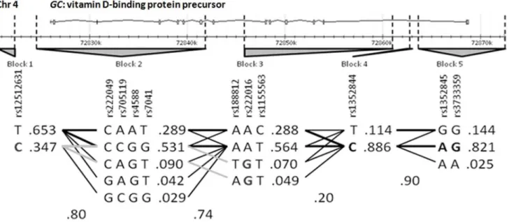
Haplotype Analysis and Association with Melanoma Risk We performed haplotype analyses using the 11 tag-SNPs inGC (the gene on which we found an associated SNP) selected previously from HapMap data. These SNPs were organized into five independent blocks according to the Haploview program for the use of data from a European subset of samples (HapMap_-CEU). Block 1 included rs12512631 in a 39downstream region of GC. Block 2 is represented by one SNP located 39downstream and three SNPs within the 39end of the gene, two of them corresponding to exon 9. Blocks 3, 4 and 5 contained six SNPs from various intron regions, including intron 1, and SNP with rs3733359, located on 59UTR. Block 5 represented the most likely promoter region with consensus sequences for TF binding sites. Data analysis was done according to the block structures detailed in Figure 1. The case-control analysis of the haplotype distribution revealed a statistical association with melanoma susceptibility on two SNPs, rs12512631 (p-value 961023), and rs222016 (p-value 0.031), representing Blocks 1 and 3, respectively (Table S2). These data were coherent with the associations described above.
Functional Implication
To assess for possible functional implications of polymorphisms, we carried out a prioritization of SNPs based on functional properties using the Pupasuite 3.1 software. Results showed non-synonymous changes of aminoacid on rs4588 (K436T) and rs7041 (E416D). TRANSFAC revealed three SNPs located in consensus sequence affecting the TF binding sites; rs4588 FOXJ2, rs7041 HNF-1 and rs222014 E2F-1 (located within intron 3 and grouped on block 3 with tag-SNP rs222016). None of the other SNPs seemed to have any additional functionality.
Discussion
GWAS on vitamin D levels, with further validation on CM patients, has pointed again to the GC gene as a candidate for melanoma susceptibility when vitamin D levels are taken into consideration [16,21].
Our study suggests that the 39downstream region of the GC gene, marked through the SNP rs12512631, is associated with CM risk. Furthermore, evidence of association was suggested by the tag-SNP rs222016, which marks a large disequilibrium block
within the gene, including intron 1. SNP rs222016 is in LD with SNP rs222014, which could have functional implications through the disruption of a transcription factor element (E2F-1), but we cannot discard other mechanisms such as alternative splicing signalling. Previous studies have shown an association between tag-SNP rs12512631 and susceptibility to prostate and colorectal cancer, which supports our results [28,29].
Table 4.Genotypic distribution among cases and controls in a Spanish population.
Controls (N = 314) n (%) Cases (N = 530) n (%)
Gene SNP
Major
homozygotes Heterozygotes Minor homozygotes
Major
homozygotes Heterozygotes Minor
homozygotes OR (95% CI) p-value
GC rs12512631 154 (49.04) 115 (36.62) 38 (12.10) 199 (37.55) 261 (49.25) 61 (11.51) 1.29 (1.04–1.60) 0.0190 rs222049 240 (76.43) 50 (15.92) 1 (0.32) 441 (83.21) 74 13.96) 2 (0.38) 0.83 (0.57–1.20) 0.3150
rs705119 102 (32.48) 136 (43.31) 58 (18.47) 152 (28.68) 276 (52.08) 90 (16.98) 1.06 (0.86–1.31) 0.5649
rs4588 154 (49.04) 128 (40.76) 30 (9.55) 248 (46.79) 229 (43.21) 42 (7.92) 1.00 (0.81–1.25) 0.9910
rs7041 101 (32.17) 140 (44.59) 60 (19.11) 145 (27.36) 271 (51.13) 100 (18.87) 1.11 (0.90–1.36) 0.3333
rs188812 261 (83.12) 44 (14.01) 2 (0.64) 425 (80.19) 97 (18.30) 1 (0.19) 1.25 (0.86–1.80) 0.2422
rs222016 236 (75.16) 58 (18.47) 5 (1.59) 368 (69.43) 127 (23.96) 8 (1.51) 1.30 (0.95–1.77) 0.1020
rs1155563 150 (47.77) 121 (38.54) 30 (9.55) 252 (47.55) 217 (40.99) 45 (8.61) 0.99 (0.79–1.25) 0.9633
rs1352844 221 (70.38) 51 (16.24) 3 (0.96) 416 (78.49) 93 (17.55) 8 (1.51) 1.02 (0.73–1.42) 0.9137
rs1352845 204 (64.97) 70 (22.29) 4 (1.27) 365 (68.87) 138 (26.04) 8 (1.51) 1.09 (0.81–1.48) 0.5675
rs3733359 254 (80.89) 18 (5.73) 0 (0) 483 (91.13) 38 (7.17) 0 (0) 1.10 (0.62–1.97) 0.7400
VDR rs11574143 238 (75.80) 55 (17.52) 1 (0.32) 383 (72.26) 88 (16.60) 1 (0.19) 0.98 (0.68–1.41) 0.9143
rs739837 74 (23.57) 160 (50.96) 66 (21.02) 121 (22.83) 258 (48.68) 115 (21.70) 0.97 (0.79–1.19) 0.7731
rs731236 109 (34.71) 141 (44.90) 44 (14.01) 186 (35.09) 248 (46.86) 64 (12.09) 0.95 (0.76–1.18) 0.6325
rs2228570 140 (44.59) 130 (41.40) 39 (12.42) 217 (40.94) 225 (42.52) 58 (11.01) 1.02 (0.82–1.27) 0.8502
rs4334089 138 (43.95) 121 (38.54) 25 (7.96) 261 (49.25) 192 (36.23) 36 (6.79) 0.86 (0.68–1.08) 0.1898
rs4237855 102 (32.48) 76 (24.20) 91 (28.98) 165 (31.13) 129 (24.34) 140 (26.42) 0.98 (0.81–1.17) 0.7977
rs7299460 131 (41.72) 137 (43.63) 32 (10.20) 236 (44.53) 224 (42.26) 47 (8.87) 0.90 (0.73–1.13) 0.3680
rs4760658 136 (43.31) 134 (42.68) 27 (8.60) 225 (42.45) 212 (40) 67 (12.64) 1.13 (0.91–1.39) 0.2795
rs4516035 106 (33.76) 149 (47.45) 45 (14.33) 183 (34.53) 228 (43.02) 94 (17.74) 1.06 (0.86–1.29) 0.5980
Heterozygotes and minor homozygotes individuals count for cases and controls and their percentages are calculated among the total of samples including fails. OR (CI 95%) means Odds Ratio and its 95% confidence interval in parentheses. OR and p values are calculated via unconditional logistic regression considering differences between cases and controls genotypes.
Bold denotes statistically significant results considering as significant p values lower than 0.05. doi:10.1371/journal.pone.0059607.t004
Table 5.Genotypic analyses of SNPs with Cutaneous Melanoma risk association.
Non-adjusted Adjusted* Enlarged sample
SNP Statistical model OR (CI 95%) p-value OR (CI 95%) p-value OR (CI 95%) p-value
rs12512631 Genotypic 1.29 (1.04–1.60) 0.019 1.32 (1.01–1.72) 0.041 1.11 (0.96–1.28) 0.153 Dominant 1.63 (1.23–2.17) 761024 1.71 (1.20–2.45) 3
61023 1.28 (1.05–1.56) 0.012
Recessive 0.94 (0.61–1.45) 0.774 0.94 (0.55–1.60) 0.809 0.90 (0.68–1.19) 0.458
rs222016 Genotypic 1.30 (0.95–1.77) 0.102 1.23 (0.83–1.82) 0.312 1.11 (0.92–1.35) 0.277
Dominant 1.37 (0.98–1.93) 0.068 1.30 (0.85–1.98) 0.222 1.22 (0.97–1.52) 0.083
Recessive 0.95 (0.31–2.93) 0.929 0.65 (0.14–3.09) 0.586 0.65 (0.35–1.20) 0.168
Bold denotes statistically significant results.
OR (CI 95%) means Odds Ratio and its 95% confidence interval.
*Adjusted for eye colour, hair colour, skin colour, lentigines, number of naevi and childhood sunburn. The enlarged sample makes a total of 684 controls and 1045 cases.
We did not observe any evidence of association between CM andVDRvariants overall, in accordance with our previous results [22]. Some other controversial results are reported in the literature. A review by Ko¨stner indicates only one variant in VDRassociated with CM risk, Fok1 (rs2228570) [33], but a meta-analysis of various studies revealed that the only variant that presents solid evidence is Bsm1 (not considered in our study) and remarked on the need to adjust by phenotype or environmental characteristics [34]. Gapska et al. found an association only when analyzing the haplotypes, but not the variants themselves [14]. More recent studies have not detected an association with CM risk on the VDR variants unless adjusting by vitamin D levels [15,35,36]. This last situation might explain why we have not found a significant association; vitamin D levels were not taken into account.
The strength of this study is the homogeneity of the Spanish population sample and the ability to control for established risk factors for CM through a structured questionnaire. We recognize, however, some potential for misclassification of phenotypic characteristics due to the subjective nature of the phenotypic attributes considered. Controls participated on a volunteer basis, which may have introduced some selection bias.
We show a comprehensive study of genetic variation on the vitamin D pathway genes VDR (vitamin D receptor) and GC (vitamin D transporter), and examine their putative role in CM susceptibility. We observed statistically significant results for CM susceptibility with two variants in theGCgene, but none in the VDR gene. One of these associations remained significant after correction for multiple testing, SNP rs12512631. This association suggests that GC may also play a role in modulating the susceptibility to CM.
We encourage replication of these findings in independent studies since the GC gene may well be a new marker for CM predisposition.
Supporting Information
Table S1 SNPs onGC and VDRgenes considered in this
study.GCrefers to Vitamin D binding protein gene;VDRrefers to Vitamin D receptor gene. Bold in sequence context denotes nucleotide change. Location is described considering as the first Exon 1 of consensus sequence. DWST means downstream, UTR means untranscribed region and UPST means upstream. (DOCX)
Table S2 Case-control study based on haplotypes in the
GCgene conducted in Spanish population. Bold denotes
statistically significant results. Italic Haplotypes on each block mark the risk haplotype. The Marker number indicates the order of the tag-SNP on the gene. LD Block means linkage disequilibrium block. Frequencies are calculated for the associa-tion alleles of each haplotype among 530 cases and 314 controls. (DOCX)
Acknowledgments
We would like to give thank to all the patients and volunteers who gave their consent for the study. We also thank the medical staff of Gregorio Maran˜o´n, La Paz and Ramo´n y Cajal Hospitals for collecting the samples and the Biobank of the Instituto Valenciano de Oncologı´a for providing the material for the genetic analysis. Genotyping with Kaspar was performed at the Unidad Central de Investigacio´n Me´dica of the Facultad de Medicina of the University of Valencia.
Author Contributions
Conceived and designed the experiments: MPC MIV AL EN GR. Performed the experiments: MPC MIV GR. Analyzed the data: MPC MIV GR. Contributed reagents/materials/analysis tools: MMG MF CGF DP GC AL EN. Wrote the paper: MPC MIV EN GR.
References
1. Jerant AF, Johnson JT, Sheridan CD, Caffrey TJ (2000) Early detection and treatment of skin cancer. Am Fam Physician 62: 357–368, 375–356, 381–352.
2. Holick MF (2003) Vitamin D deficiency: what a pain it is. Mayo Clin Proc 78: 1457–1459.
3. Holick MF (2003) Evolution and function of vitamin D. Recent Results Cancer Res 164: 3–28.
4. Field S, Newton-Bishop JA (2011) Melanoma and vitamin D. Mol Oncol 5: 197– 214.
5. Lurie G, Wilkens LR, Thompson PJ, Carney ME, Palmieri RT, et al. (2011) Vitamin D receptor rs2228570 polymorphism and invasive ovarian carcinoma risk: pooled analysis in five studies within the Ovarian Cancer Association Consortium. Int J Cancer 128: 936–943.
6. Alimirah F, Peng X, Murillo G, Mehta RG (2011) Functional significance of vitamin D receptor FokI polymorphism in human breast cancer cells. PLoS One 6: e16024.
7. Touvier M, Chan DS, Lau R, Aune D, Vieira R, et al. (2011) Meta-analyses of vitamin D intake, 25-hydroxyvitamin D status, vitamin D receptor polymorph-isms, and colorectal cancer risk. Cancer Epidemiol Biomarkers Prev 20: 1003– 1016.
8. Smedby KE, Eloranta S, Duvefelt K, Melbye M, Humphreys K, et al. (2011) Vitamin D receptor genotypes, ultraviolet radiation exposure, and risk of non-Hodgkin lymphoma. Am J Epidemiol 173: 48–54.
9. Karami S, Brennan P, Navratilova M, Mates D, Zaridze D, et al. (2010) Vitamin d pathway genes, diet, and risk of renal cell carcinoma. Int J Endocrinol 2010: 879362.
10. Bektas-Kayhan K, Unur M, Yaylim-Eraltan I, Ergen HA, Toptas B, et al. (2010) Association of vitamin D receptor Taq I polymorphism and susceptibility to oral squamous cell carcinoma. In Vivo 24: 755–759.
11. Chang CK, Mulholland HG, Cantwell MM, Anderson LA, Johnston BT, et al. (2011) Vitamin D Receptor Gene Variants and Esophageal Adenocarcinoma Risk: A Population-Based Case-Control Study. J Gastrointest Cancer. 12. Srinivasan M, Parwani AV, Hershberger PA, Lenzner DE, Weissfeld JL (2010)
Nuclear vitamin D receptor expression is associated with improved survival in non-small cell lung cancer. J Steroid Biochem Mol Biol 123: 30–36. 13. Kidd LC, Paltoo DN, Wang S, Chen W, Akereyeni F, et al. (2005) Sequence
variation within the 5’ regulatory regions of the vitamin D binding protein and receptor genes and prostate cancer risk. Prostate 64: 272–282.
14. Gapska P, Scott RJ, Serrano-Fernandez P, Mirecka A, Rassoud I, et al. (2009) Vitamin D receptor variants and the malignant melanoma risk: a population-based study. Cancer Epidemiol 33: 103–107.
15. Orlow I, Roy P, Reiner AS, Yoo S, Patel H, et al. (2011) Vitamin D receptor polymorphisms in patients with cutaneous melanoma. Int J Cancer. 16. Ahn J, Yu K, Stolzenberg-Solomon R, Simon KC, McCullough ML, et al.
(2010) Genome-wide association study of circulating vitamin D levels. Hum Mol Genet 19: 2739–2745.
17. Pani MA, Regulla K, Segni M, Hofmann S, Hufner M, et al. (2002) A polymorphism within the vitamin D-binding protein gene is associated with Graves’ disease but not with Hashimoto’s thyroiditis. J Clin Endocrinol Metab 87: 2564–2567.
18. Disanto G, Ramagopalan SV, Para AE, Handunnetthi L (2010) The emerging role of vitamin D binding protein in multiple sclerosis. J Neurol 258: 353–358. 19. Chishimba L, Thickett DR, Stockley RA, Wood AM (2010) The vitamin D axis
in the lung: a key role for vitamin D-binding protein. Thorax 65: 456–462.
20. Wang TJ, Zhang F, Richards JB, Kestenbaum B, van Meurs JB, et al. (2010) Common genetic determinants of vitamin D insufficiency: a genome-wide association study. Lancet 376: 180–188.
21. Davies JR, Chang YM, Snowden H, Chan M, Leake S, et al. (2011) The determinants of serum vitamin D levels in participants in a melanoma case-control study living in a temperate climate. Cancer Causes Control 22: 1471– 1482.
22. Barroso E, Fernandez LP, Milne RL, Pita G, Sendagorta E, et al. (2008) Genetic analysis of the vitamin D receptor gene in two epithelial cancers: melanoma and breast cancer case-control studies. BMC Cancer 8: 385.
23. Laayouni H, Calafell F, Bertranpetit J (2010) A genome-wide survey does not show the genetic distinctiveness of Basques. Hum Genet 127: 455–458. 24. (2003) The International HapMap Project. Nature 426: 789–796.
25. Barrett JC, Fry B, Maller J, Daly MJ (2005) Haploview: analysis and visualization of LD and haplotype maps. Bioinformatics 21: 263–265. 26. Newton Bishop JA, Bishop DT (2005) The genetics of susceptibility to cutaneous
melanoma. Drugs Today (Barc) 41: 193–203.
27. Kvaskoff M, Whiteman DC, Zhao ZZ, Montgomery GW, Martin NG, et al. Polymorphisms in nevus-associated genes MTAP, PLA2G6, and IRF4 and the risk of invasive cutaneous melanoma. Twin Res Hum Genet 14: 422–432. 28. Ahn J, Albanes D, Berndt SI, Peters U, Chatterjee N, et al. (2009) Vitamin
D-related genes, serum vitamin D concentrations and prostate cancer risk. Carcinogenesis 30: 769–776.
29. Poynter JN, Jacobs ET, Figueiredo JC, Lee WH, Conti DV, et al. (2010) Genetic variation in the vitamin D receptor (VDR) and the vitamin D-binding protein (GC) and risk for colorectal cancer: results from the Colon Cancer Family Registry. Cancer Epidemiol Biomarkers Prev 19: 525–536.
30. Hibler EA, Hu C, Jurutka PW, Martinez ME, Jacobs ET (2012) Polymorphic Variation in the GC and CASR Genes and Associations with Vitamin D Metabolite Concentration and Metachronous Colorectal Neoplasia. Cancer Epidemiol Biomarkers Prev 21: 368–375.
31. Shen LH, Zhang XM, Su DJ, Yao SP, Yu BQ, et al. (2010) Association of vitamin D binding protein variants with susceptibility to chronic obstructive pulmonary disease. J Int Med Res 38: 1093–1098.
32. Abbas S, Linseisen J, Slanger T, Kropp S, Mutschelknauss EJ, et al. (2008) The Gc2 allele of the vitamin D binding protein is associated with a decreased postmenopausal breast cancer risk, independent of the vitamin D status. Cancer Epidemiol Biomarkers Prev 17: 1339–1343.
33. Kostner K, Denzer N, Muller CS, Klein R, Tilgen W, et al. (2009) The relevance of vitamin D receptor (VDR) gene polymorphisms for cancer: a review of the literature. Anticancer Res 29: 3511–3536.
34. Mocellin S, Nitti D (2008) Vitamin D receptor polymorphisms and the risk of cutaneous melanoma: a systematic review and meta-analysis. Cancer 113: 2398– 2407.
35. Newton-Bishop JA, Beswick S, Randerson-Moor J, Chang YM, Affleck P, et al. (2009) Serum 25-hydroxyvitamin D3 levels are associated with breslow thickness at presentation and survival from melanoma. J Clin Oncol 27: 5439–5444. 36. Randerson-Moor JA, Taylor JC, Elliott F, Chang YM, Beswick S, et al. (2009)