Cop
yright
© ABE&M t
odos os dir
eit
os r
eser
vados
.
Arq Bras Endocrinol Metab. 2014;58/5
Cop
yright
© ABE&M t
odos os dir
eit
os r
eser
vados
.
Recommendations of the Brazilian
Society of Endocrinology and
Metabology (SBEM) for the diagnosis
and treatment of hypovitaminosis D
Recomendações da Sociedade Brasileira de Endocrinologia e Metabologia (SBEM) para o diagnóstico e tratamento da hipovitaminose D
Sergio Setsuo Maeda1, Victoria Z. C. Borba2, Marília Brasilio Rodrigues
Camargo1, Dalisbor Marcelo Weber Silva3, João Lindolfo Cunha
Borges4, Francisco Bandeira5, Marise Lazaretti-Castro1
ABSTRACT
Objective: The objective is to present an update on the diagnosis and treatment of hypovitami-nosis D, based on the most recent scientiic evidence. Materials and methods: The Department of Bone and Mineral Metabolism of the Brazilian Society of Endocrinology and Metabology (SBEM) was invited to generate a document following the rules of the Brazilian Medical As-sociation (AMB) Guidelines Program. Data search was performed using PubMed, Lilacs and SciELO and the evidence was classiied in recommendation levels, according to the scientiic strength and study type. Conclusion: A scientiic update regarding hypovitaminosis D was pre-sented to serve as the basis for the diagnosis and treatment of this condition in Brazil. Arq Bras Endocrinol Metab. 2014;58(5):411-33
Keywords
Vitamin D; cholecalciferol; PTH; osteoporosis; deiciency; insuficiency; diagnosis; treatment
RESUMO
Objetivo: Apresentar uma atualização sobre o diagnóstico e tratamento da hipovitaminose D baseada nas mais recentes evidências cientíicas. Materiais e métodos: O Departamento de Metabolismo Ósseo e Mineral da Sociedade Brasileira de Endocrinologia e Metabologia (SBEM) foi convidado a conceber um documento seguindo as normas do Programa Diretrizes da Associação Médica Brasileira (AMB). A busca dos dados foi realizada por meio do PubMed, Lilacs e SciELO e foi feita uma classiicação das evidências em níveis de recomendação, de acordo com a força cientíica por tipo de estudo. Conclusão: Foi apresentada uma atualização cientíica a respeito da hipovitaminose D que servirá de base para o diagnóstico e tratamento dessa condição no Brasil. Arq Bras Endocrinol Metab. 2014;58(5):411-33
Descritores
Vitamina D; colecalciferol; PTH; osteoporose; deiciência; insuiciência; diagnóstico; tratamento
1 Endocrinology Division,
Federal University of São Paulo (Unifesp), Escola Paulista de Medicina, São Paulo, SP, Brazil
2 Department of Medicine,
Paraná Federal University (UFPR), Curitiba, PR, Brazil
3 Department of Medicine, Univille
Medical School, Joinville, SC, Brazil
4 Endocrinology Division,
Brasília Catholic University (UCB), Brasília, DF, Brazil
5 Endocrinology Division, Agamenon
Magalhães Hospital, University of Pernambuco (UPE), Escola de Medicina, Recife, PE, Brazil
Correspondence to: Sergio Setsuo Maeda
Rua Conselheiro Furtado, 847, ap. 93 01511-001 – São Paulo, SP, Brazil ssetsuo@terra.com.br Recebido em Mar /31/2014 Aceito em Jun/18/2014
DOI: 10.1590/0004-2730000003388
INTRODUCTION
H
ypovitaminosis D is highly prevalent and represents a public health problem in the entire world. Studies show an elevated prevalence of this di sease in several geographic regions, including Brazil. It can affect more than 90% of individuals, depending on the population studied (1).
Vitamin D is essential in functions related to bone metabolism, but it seems to be related in the patho
Cop
yright
© ABE&M t
odos os dir
eit
os r
eser
vados
.
412
The correct diagnosis of this condition and the identiication of improvement or worsening factors can help the elaboration of more eficient strategies for the treatment of risk populations, such as the elderly and postmenopausal women.
This document represents the efforts of the Depart ment of Bone Metabolism of the Brazilian Society of Endocrinology and Metabology (SBEM) for the devel opment of recommendations based on evidence availa ble in the scientiic literature regarding the diagnosis and treatment of this condition. The objective of this document is to respond daily questions and to be a guideline for endocrinologists and clinicians in the Bra zilian context.
MATERIALS AND METHODS
The elaboration of this guideline was motivated by the SBEM within its Practical Guidelines program. The model applied followed the Guidelines Program of the Brazilian Medical Association (AMB) and the Federal Council of Medicine (CFM). After the selection of collaborators, with a signiicant role and relevant publi cations in the area, clinical questions to be approached were elaborated.
The publication search was performed using Med LinePubMed and SciELOLilacs. We used the Oxford Classiication, which evaluates the study design and considers the best available evidence for each question, to categorize the recommendation level or evidence strength of each article (4,5).
The levels of recommendation and evidence strength were reported as:
A: experimental or observational studies with bet ter consistency.
B: experimental or observational studies with less consistency.
C: case reports (noncontroled studies).
D: opinion lacking critical evaluation, based on guidelines, physiological studies or animal mo dels.
DEFINITION AND PHYSIOLOGY
1. What is vitamin D: a nutrient or a prohormone?
Although it is deined as a vitamin, conceptually it is a prohormone. In conjunction with the parathyroid hormone (PTH), it acts as an important regulator of calcium homeostasis and bone metabolism.
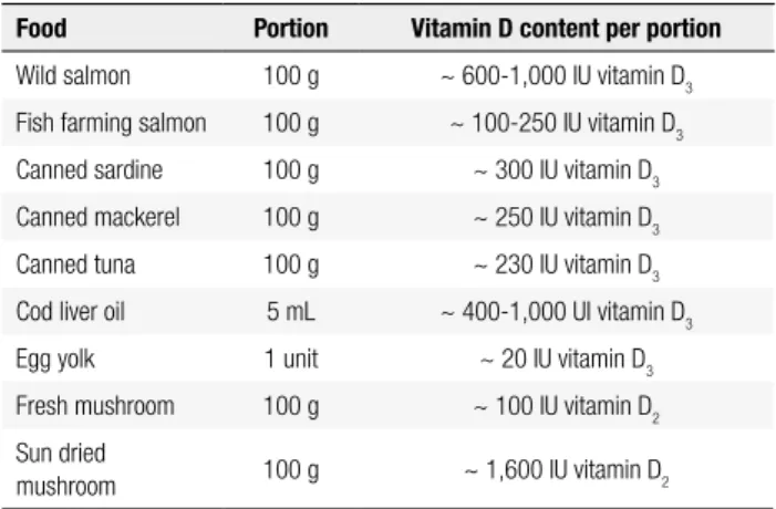
It can be obtained from food sources, such as cod liver oil and from other fatrich ish (salmon, tuna, mackerel), or from endogenous cutaneous synthesis, which represents the most important source of this “vi tamin” for the majority of human beings (2,3,6,7) (A). Table 1 shows some food sources of vitamin D (3).
Vitamin D can be found in the form of ergocalci ferol or vitamin D2 and cholecalciferol or vitamin D3 (8). Vitamin D2 can be obtained from some yeast and plants, being produced for commercial use, through ir radiation of the ergosterol present in some mushrooms (8)(D).
Figure 1. Photobiosynthesis of vitamin D.
7-dehidrocholesterol Cholecalciferol or vitamin D or D
3
Ergocalciferol or
vitamin D2 25 hydroxilase
25(OH) vitamin D
1 a hydroxilase
1,25 (OH)2D3
24, 25(OH)2 D3 Receptor
Skin Ultra-Violet Rays
In the skin, the precursor is the 7dehydrocholeste rol (7DHC) (8,9). During sun exposure, UVB pho tons (ultraviolet B, 290315 nm) penetrate the epi dermis and produce a photochemical fragmentation to originate precholecalciferol. This intermediate is converted to vitamin D (or cholecalciferol) through a temperaturedependent isomerization (Figure 1).
Table 1. Vitamin D food sources
Food Portion Vitamin D content per portion
Wild salmon 100 g ~ 600-1,000 IU vitamin D3
Fish farming salmon 100 g ~ 100-250 IU vitamin D3
Canned sardine 100 g ~ 300 IU vitamin D3
Canned mackerel 100 g ~ 250 IU vitamin D3
Canned tuna 100 g ~ 230 IU vitamin D3
Cod liver oil 5 mL ~ 400-1,000 UI vitamin D3
Egg yolk 1 unit ~ 20 IU vitamin D3
Fresh mushroom 100 g ~ 100 IU vitamin D2
Sun dried
mushroom 100 g ~ 1,600 IU vitamin D2
Cop
yright
© ABE&M t
odos os dir
eit
os r
eser
vados
.
Arq Bras Endocrinol Metab. 2014;58/5
Cholecalciferol is transported to the liver by DBP (vitamin D binding protein). In the liver, there is the hydroxylation of carbon 25 (CYP27B1), forming the 25hydroxyvitamin D (25(OH)D), through a process which is not strictly regulated, since it happens without control, and depends on the combination of cutaneous and diet stocks of vitamin D (8).
After the liver step, 25(OH)D is transported to the kidneys by DBP, where it is converted to calcitriol or 1,25dihydroxyvitamin D [1,25(OH)2D] (Figure 1). This is the most active metabolite and it is responsible for stimulating intestinal calcium and phosphate absorp tion. The kidney hydroxylation is stimulated by PTH and suppressed by phosphate and FGF23. Calcitriol production is strictly controlled by negative feedback, in a way to inluence its own synthesis by the decrease of 1ahydroxylase. It is responsible for accelerating its inactivation through the conversion of 25(OH)D to 24,25(OH)2D. This mechanism relects a direct action of 1,25(OH)2D in the kidneys, however there is still an inhibitory action on PTH production in the parathy roids (8,9). The 1ahydroxylase can also be found in other cells and tissues, such as the skin, prostate, breast, intestine, lungs, pancreatic β cells, monocytes and para thyroid cells. The 1,25(OH)2D molecule can also be locally synthesized by these cells and tissues (8,9) (D).
The vitamin D receptor (VDR) belongs to the super family of nuclear receptors regulating the transcription factors of the steroid hormones, retinoic acid, thyroid hormones and vitamin D. After binding 1,25(OH)2D to VDR, it interacts with the retinoic acid receptor, form ing a heterodimeric complex (RXRVDR), which then binds speciic sequences of DNA, known as Vitamin D Responsive Elements (VDRE) (10,11). The main target organs for 1,25(OH)2D are the intestine, bones, para thyroid glands and kidneys. However, its receptors have been found in several other tissues (10,11) (D).
SBEM recommendation: food sources are scarce in vitamin D and humans depend mainly on the skin production catalysed by UVB sunlight exposure (Evi dence A).
2. What are the effects on bone metabolism?
SBEM recommendation: active vitamin D mo dulates PTH synthesis, increases intestinal calcium absorption, and improves bone mass and muscular function (Evidence A).
The best known and studied actions of vitamin D are related to bone metabolism, where it plays a cru cial role. It participates in intestinal calcium absorption, muscle function, modulation of PTH secretion and bone cell function.
Parathyroid cells express the 1ahydroxylase en zyme, and can synthetize the active form of vitamin D (1,25(OH)2D) inside the cells using the 25(OH)D serum pool (12) (B). In hypovitaminosis D, due to a minor intracellular synthesis, there is a secondary hy perparathyroidism, which is associated to an increase in bone resorption (2,1316) (B), besides the fact that the circulating levels of 1,25(OH)2D are, generally, normal. There is an inverse correlation between PTH and 25(OH)D, described in children (17) and the el derly (2). Several cut off values for 25(OH)D for the PTH normalization have been published, the majority, being around 28 and 40 ng/mL (70 to 100 nmol/L) (2,1823) (C). Other causes of secondary hyperpara thyroidism also have to be investigated, such as chronic kidney disease (creatinine clearance below 60 mL/ min), Paget’s disease, hungry bone syndrome and the calcium and vitamin D malabsorption syndromes (24).
Intestinal calcium absorption depends on the active vitamin D action in the duodenum, through a transcellu lar saturable process, whose stimulus leads to the synthe sis of proteins such as calbindinD9K (CaBP9k) and the epithelial apical channel TRPV6 (13,14) (D). However, there is evidence that the nonsaturable transport, which happens with part of calcium absorption in the human ileum is also vitamin D sensitive (25). According to He aney and cols., individuals with 35 ng/mL of 25(OH)D presented higher absorption than those with 25 ng/mL (26) (B). Increase of calcium absorption with increasing dose of vitamin D3 or serum 25(OH)D was recently ob served, but there is no evidence of what the minimum value of 25(OH)D to ensure calcium absorption from the intestine in the range of 1652 ng/mL evaluated in the study (27).
Population studies correlated positively vitamin D concentration with bone mass, mainly of the hip, but with 25(OH)D cut off points varying from 12 to 36 ng/mL (3090 nmol/L) (2830) (C).
Cop
yright
© ABE&M t
odos os dir
eit
os r
eser
vados
.
414
of 800 IU of cholecalciferol for 12 weeks decreased in 49% the number of falls (32) (B). Cholecalciferol use is associated with the prevention of falls among the elderly with hypovitaminosis D, but not among those presenting normal serum values (33) (B).
In a metaanalysis of the main osteoporosis in tervention studies; BischoffFerrari and cols. indi cated again 25(OH)D serum concentration above 30 ng/mL (75 nmol/L) to be the most beneicial for health in general (A). Bone health, here represented by a better bone mineral density (BMD), decreased risk of fall and femural and nonvertebral osteopo rotic fractures, seems to be beneited by 25(OH)D concentrations equal to or higher than 30 ng/mL (75 nmol/L), concentrations around 36 ng/mL (90 nmol/L) being suggested as the most advanta geous (3436). The same 25(OH)D values seem to beneit the muscle strength of lower limbs, which was evaluated by the TUG (Time Up and Go) test, where the individual is evaluated based on the time he needs to walk a distance equivalent to eight steps. Indivi duals presenting 25(OH)D in the range of 36 to 40 ng/mL (90 and 100 nmol/L) seem to perform with higher speed. Evidence also suggests that higher 25(OH)D values are associated with a lower risk for colorectal cancer and periodontal disease (36) (A).
Regarding the bone tissue, evidence suggests that 1,25(OH)2D stimulates mineralization, through an indirect process that happens with the increase in in testinal absorption of the minerals which are incorpo rated into the bone matrix. Physiological concentra tions of calcitriol promote calcium mobilization to the bones, while the administration of large doses pro motes excessive bone remodeling. Osteoblasts present 1,25(OH)2D receptor. This hormone modulates the gene expression of alkaline phosphatase and osteo calcin. Therefore, in the process of bone remodeling, 1,25(OH)2D is important for bone formation and re absorption (37).
Priemel and cols. evaluated 675 bone biopsies and correlated the histomorphometry indings with serum 25(OH)D concentration. The presence of bone mi neralization defects was only found in individuals with concentrations below 30 ng/mL (75 nmol/L) (38)
(B).
The role of vitamin D in nonbone related end points, such as mortality, cardiovascular risk, cancer and autoimmune diseases is still controversial (39,40).
DIAGNOSIS
3. How to deine hypovitaminosis D?
SBEM recommendation: the analysis of the 25 hydroxyvitamin D metabolite (25(OH)D) should be used for the evaluation of the vitamin D status of an individual (Evidence A).
There is a consensus that 25(OH)D (calcidiol) is the most abundant metabolite and the best indicator for the evaluation of vitamin D status (A), the indivi duals being classiied as: deicient, insuficient of suf icient in vitamin D (3,6,41,42). On the other hand, there is no consensus regarding the cut off value for the deinition of “vitamin D suficiency” (6,43,44).
The values discussed in the medical literature, based on populational studies, with emphasis on calcium homeostasis and bone health, vary from 20 to 32 ng/mL (50 to 80 nmol/L) (26,4347). Sev eral specialists agree that for correction of second ary hyperparathyroidism, reduction of the risk of fall and fractures and maximum calcium reabsorption, the best 25(OH)D cut off value is 30 ng/mL (75 nmol/L) (6,41,46). Thus, serum concentrations be low 20 ng/mL (50 nmol/L) are classiied as dei ciency; those ranging from 20 to 29 ng/mL (50 to 74 nmol/L) as insuficiency and between 30 and 100 ng/mL (75 and 250 nmol/L) as suficiency. Therefore, many consider 25(OH)D serum con centrations below 30 ng/mL as hypovitaminosis D (3,4143,48,49). These values were recognized by the Endocrine Society guideline, although they differ from the ones accepted (20 ng/mL) by the Institute of Medicine (IOM) (50) (B). In general population, there is no evidence of beneit in the measurement of 25(OH)D due to the high cost, but according to the Endocrine Society, to maximize bone health; supple mentation is recommended for children up to 1 year with at least 400 IU/day; between 1 and 70 years, at least 600 IU/day while over 70 years old, 800 IU/ day (41).
Cop
yright
© ABE&M t
odos os dir
eit
os r
eser
vados
.
Arq Bras Endocrinol Metab. 2014;58/5
4. What are the methodological implications for the plasma determinations of 25(OH)D?
SBEM recommendation: the methods based on chromatography are considered gold standard for the laboratory evaluation of 25(OH)D, although, automated immunometric assays can be used in the clinical practice taking into account the good correla tion with the method of excellence, besides the prac ticality and lower cost. However, the clinician should be aware of the possible mistakes caused by several interfering conditions, possibly leading to diagnostic classiication errors (Evidence B).
Circulating 25(OH)D level is the best method to evaluate the individual vitamin D status. Neverthe less, there are controversies regarding the best method for 25(OH)D determination. Some factors should be considered when the levels of this vitamin are evalu ated, such as the lack of a precise physiological regula tory control (feedback), the variability of methods and standards, the inclusion of contaminant metabolites in the analysis, among others. Radioimmunoassays (RIA) used in the past underestimated the levels of 25(OH)D when the dominant levels were 25(OH)D2. RIA have been replaced by automated chemiluminescent immu noassays, resulting in higher concentrations and by im munoenzymatic assays which measure total 25(OH)D, a combination of vitamin D2 (25(OH)D2) and vitamin D3 (25(OH)D3 (51) (B).
The methods that do not employ direct immune detection are high performance liquid chromatogra phy (HPLC) coupled to mass spectrometry (LCMS), which can distinguish individual levels of 25(OH) D2 and 25(OH)D3. These are considered the gold standard for analysis and currently used as reference (52) (B). Both 1,25(OH)2D and 25(OH)D circu late predominantly bound to proteins and their con centration can be determined. However, to evaluate the vitamin D status, 25(OH)D total serum level is used, including both D3 and D2 forms. The results can be reported in nanograms per milliliter (ng/mL) or nanomol per liter (nmol/L). For conversion, you just need to multiply the value obtained in ng/mL by 2.5, to obtain the value in nmol/L. Automated meth ods allow the use in clinical routines, they are fast and report 25(OH)D2 and 25(OH)D3 together, while
LCMS methods can distinguish between 25(OH)D2
and 25(OH)D3, being useful, then, for the evaluation
of the effectiveness of D2 supplementation, versus endogenous D3 production. These chromatographic methods, although more precise, are more laborious and expensive (53) (B).
SBEM recommendation: for conversion of 25(OH) D concentration from ng/mL to nmol/L, the multi plication factor 2.5 is applied.
The accuracy of the measurements varies widely between laboratories and between different assays, and even when testing identical samples, this varia tion can achieve 17 ng/mL (53). The immunoassay requires the development of selective antibodies for 25(OH)D2 and 25(OH)D3, which preferentially do not show cross reaction with any other metabolite. Matrix effects can still occur, caused by endogenous components that modify the binding of the antibody to the material to be analyzed. Metabolites with lower physio logical potential end up being included in the quantiication, such as the 3epimer of the 25(OH) D, which can correspond to up to 5% of the to tal 25(OH)D. As its molecular weight is identical to 25(OH)D, these are not separated by LCMS. Finally, 24,25 dihydroxyvitamin D (24,25(OH)2D), consid ered an inactive metabolite, can correspond to up to 20% of the 25(OH)D measured, whereas some assays show 100% crossreaction (51,54).
The use of a standard cut off value to evaluate vita min D status is problematic if applied to all laboratories and all methods, considering there are still differences on vitamin D extraction from its binding protein, cross reaction between 25(OH)D2, 25(OH)D3 and other metabolites, besides the lack of standardization (52,53) and for this reason, quality control tools, such as DE QAS (International Vitamin D External Quality Assess ment Scheme) were created, as an attempt to decrease the variation in data analysis (55).
The most used methods nowadays are competitive assays, based on speciic antibodies and nonradioactive markers, the improvement in the comparison between results obtained from different methodologies being necessary. Whatever the method employed is a precise deinition of the normality range is fundamental (56). It is also important to highlight that the intraindivi dual variability can vary from 12.1 to 40.3% (57).
Cop
yright
© ABE&M t
odos os dir
eit
os r
eser
vados
.
416
light exposure. Polymorphisms in CYP27B1, which codes for 1ahydroxylase, showed strong correlation with variations in 25(OH)D level. The vitamin D bind ing protein (DBP) is the main transporter for vitamin D metabolites, its phenotype helping predict 25(OH)D serum concentrations. Certain polymorphic forms can be more eficient for vitamin D binding, activation and metabolism, interfering with circulating levels. Genetic polymorphisms greatly contribute to the heterogeneity of clinical manifestations of hypovitaminosis D, espe cially among ethnic groups (51,58) (B).
EPIDEMIOLOGY
5. Which are the risk populations for hypovitaminosis D? What is the prevalence in Brazil?
SBEM recommendation: the analysis of 25(OH) D concentration is not recommended for the gene ral population. It is recommended for speciic groups belonging to populations at risk or for those with re levant clinical condition in which deiciency is suspec ted (Evidence A).
The Department of Bone and Mineral Metabolism from SBEM agrees with the guidelines published by the Endocrine Society, which does not recommend the 25(OH)D test for the general population con sidering the cost of this evaluation. The laboratory test is recommended for individuals under risk for hy povitaminosis D or for those with a relevant clinical condition. The candidates to be tested are the ones presenting the following conditions: rickets or osteo malacia, osteoporosis, history of falls and fractures in the elderly, obesity, pregnant and lactating women,
patients with malabsorption syndromes (cystic i brosis, inlammatory bowel disease, Chron’s disease, bariatric surgery), renal or liver insuficiency, hyper parathyroidism, medications interfering in vitamin D metabolism (anticonvulsants, glucocorticoids, anti fungal, antiretroviral, cholestyramine, orlistat), gran ulomatous diseases and lymphomas (41,59) (A). It is also useful for the evaluation of hypothesis of vitamin D intoxication.
Besides that, it is important to highlight that all the conditions that limit sunlight exposure can potentially cause hypovitaminosis D and can be added to the list of individuals in photoprotection regimen (60) (D) and religious garment users (veil, burqa, cassock and others) (6163) (C).
Hypovitaminosis D is a world health problem and Brazil is part of this scenario, also presenting an elevated prevalence of hypovitaminosis D in the population (B). Table 2 presents some of the main Brazilian and inter national studies that included Brazil, published in the last decade. In general, in several regions of the country, the values indicate suboptimum vitamin D concentra tions, verifying high prevalence of hypovitaminosis D in several age groups. The majority of the studies included mainly the elderly and postmenopausal women which are the populations at risk for osteoporosis (C). Ho wever, three studies involving adolescents showed high prevalence of hypovitaminosis D in this age group of the Brazilian population (6466). The factors that seem to favor the presence of higher serum concentrations in our population are: younger age (64,6769), commu nity life (70), the practice of outdoors physical activity (64,71), vitamin D oral supplementation (72), season of the year (spring, summer) (6873), residence in sun ny beach areas (74,75) and in lower latitudes (76).
Table 2. Prevalence of hypovitaminosis D in Brazil
Author, year
n Population Age
(years) Average ±
SD
Local, Latitude
25(OH)D (ng/ mL) Average
± SD
25(OH)D (ng/mL) Cut off
value
Prevalence (%)
Vitamin D intake or oral supplementation
Saraiva, 2005 (73) and 2007 (70)
420
177
243
Elderly, > 65 years
Institutionalized 125 W and 52 M
From the community 168 W and 75 M
76 ± 9
79 ± 6
São Paulo, SP 23ºS
14.4 ± 9.2
19.6 ± 11.2
< 10 < 20 < 40 < 10 < 20 < 40
41 71 99 16 42 96
7% with oral supplementation: daily dose 125-1.000 IU 4% with supplementation
10% with supplementation
Cop
yright
© ABE&M t
odos os dir
eit
os r
eser
vados
.
Arq Bras Endocrinol Metab. 2014;58/5 Lips, 2006
(78)
151 Post-menopausal women with osteoprosis, > 41 years, in ambulatorial
follow up
67 Vitória, ES
20ºS
32.4 < 20
< 30
15 42
Values not mentioned
Maeda, 2007 (68)
121 Volunteers,
17-33 years, 72 W and 49 M
24 ± 2 São Paulo, SP 23ºS
31.2 ± 13.2 < 28.8 < 40 50 75 Without supplementation Silva, 2008 (72) 180 Endocrinology ambulatorial patients, 14-91 years, 165 W and 15 M
58 Belo
Horizonte, MG 19ºS
39.6 ± 16.8 < 14 < 32
0.8 42
27% with oral supplementation: varied doses Peters, 2008 (64) 136 Adolescents, 16-20 years, 72 W and 64 M
18 Indaiatuba, SP 23ºS
29.2 ± 0.8 ≤ 10
≤ 30
0 62
Average daily intake 140 IU
Russo, 2009 (77)
251 Post-menopausal volunteers with low bone mass, 50-85
years
67 ± 6 Rio de
Janeiro, RJ 22ºS
26 ± 10.4 <10 < 20 < 30 < 40 2 27 67 92 Without supplementation Kuchuk, 2009 (79) 1.486 Post-menopausal women with osteoporosis, 50-85 years - Latitude 15°S-23ºS
28 ± 7.6 < 10 < 20 < 30
0.5 12.5
66
Values not mentioned
Lopes, 2009 (80)
415 Post-menopausal women with and without fractures
Without fracture: 72.1 ± 4.4
with fracture: 74.6 ± 5.8
São Paulo, SP 23ºS
Without fracture: 20.7
± 10.7 with fracture
16.9 ± 8.2
< 30 Without fracture: 82.3% with fracture:
93.65%
Supplementation users were excluded
Maeda, 2010 (71)
99 Physical exercise practitioners, 2 hours
a week of outdoors physical activity, 52 W and 47 M
67 ± 6 São Paulo, SP 23ºS
31.6 ± 12.4 < 10 < 20
3 19
7% with oral supplementation: 200-400 IU Unger, 2010 (67) 603 Volunteers: employees and students from USP, 18-80 years, 485 W
and 118 M
47 ± 13 São Paulo, SP 23ºS Median post winter: 21.4 Post summer: increase of 10.6
< 30 77 Parameters not evaluated
Bandeira, 2010 (74)
93 Healthy
post-menopausal women
65 ± 7 Recife, PE 8ºS
28.8 ± 14.8 < 20 24 Parameters not evaluated
Neves, 2012 (75)
91 The elderly with SAH, > 60 years, 81 W and 10 M
69 ± 7 João Pessoa, PB 7ºS
Adequate: 44.8 ± 12.5
Non-adequate: 24 ± 3.5 < 20 < 30 4 33 Without supplementation Santos, 2013 (66)
234 Adolescents 7-18 years
13.0 ± 1.9 Curitiba, PR 25º
Varied from 20.8 to 22 in
the studied tertiles < 30 < 20 90,6 63,7 Without supplementation Oliveira, 2013 (65) 160 Adolescents, 15-17 years, 71 W and 89 M
16 Juiz de Fora, MG 21ºS
- ≤ 10
≤ 30
1,3 70,6
Average daily intake 88 IU Author,
year
n Population Age
(years) Average ± SD Local, Latitude 25(OH)D (ng/ mL) Average ± SD 25(OH)D (ng/mL) Cut off value Prevalence (%)
Vitamin D intake or oral supplementation
Cop
yright
© ABE&M t
odos os dir
eit
os r
eser
vados
.
418 Maeda, 2013 (69)
591 Volunteers,
17-100 years, 388 W and 203 M
Nursing homes: 76.2 ± 9.0 Community: 79.6 ± 5.3
Exercise: 67.6 ± 5.4 young: 23.9
± 2.8
São Paulo, SP 23ºS
Nursing homes: 15.0
± 11.9 community: 19.8 ± 11.0 Exercise: 31.5
± 12.4 young: 34.5 ±
14.0
< 10 < 20 < 30
19 47 73
6% with oral supplementation: 200-400 IU
Arantes, 2013 (76)
1.933 Post-menopausal women with low bone
mass, 60-85 years
67 ± 5 Latitude
8°S-33°S
27.2 ± 8.4 ≤ 30 68,3 Parameters not evaluated
Martini, 2013 (81)
636 Adolescents, adults and the elderly
- São Paulo, SP
23ºS
M: 16.7 W: 19.2
< 20 - Intake varied from 108 to 140 IU/d
Cabral, 2013 (82)
284 Men, skin phototype evaluated
69.4 ± 6.5 Recife, PE 8ºS
28.0 ± 13.6 < 20 < 30
31,5 66,7
2,5% took supplements
M: men; W: women; IU: international units.
TREATMENT
6. How to treat hypovitaminosis D in patients who are at high risk for the deiciency?
SBEM recommendation: generalized vitamin D su pplementation is not indicated for the entire popu lation. The beneits of the vitamin D treatment are more evident in populations presenting high risk for the deiciency (Evidence A).
Current evidence does not support the concept of general population supplementation (41) (A). As the adequacy of vitamin D concentration in our popula tion is closely related to the cutaneous production, secon dary to sunlight exposure, individuals with low exposure represent the main population of deicient indivi duals. Therefore, a simple interview can bring important information on the probability of vitamin D deiciency in a speciic individual.
The complementation of the daily needs, as well as the treatment of the deiciency should be performed for individuals with hypovitaminosis D risk (see Epidemi ology section) or those to whom sunlight exposure is prohibited, due to skin cancer, transplants or systemic lupus erythematosus (A).
The most available vitamin D form for treatment and supplementation is cholecalciferol or vitamin D3 and this is the metabolite that has been shown to be the most effective one. Ergocalciferol or vitamin D2 can also be used as a supplement, however the studies show that, as its halflife is a little shorter than the one of D3, it should be used preferentially daily (83). Be sides that, some laboratory methods that test 25(OH) D recog nize only 25(OH)D3, what can bring problems for the control of plasma levels when vitamin D2 is used for supplementation. Therefore, although supplemen tation and treatment can be done with both vitamin D metabolites, preference should be given to vitamin D3, due to the advantages on the maintenance of more stable concentrations.
The treatment doses vary according to the degree of deiciency and the target to be achieved. Apparent ly, 25(OH)D concentrations higher than 12 ng/mL would be suficient to avoid rickets and osteomalacia, as well as to normalize intestinal absorption of calcium (84,85). However, to reduce fractures, concentrations above 24 ng/mL are necessary (86), while to avoid the development of secondary hyperparathyroidism, concentrations above 30 ng/mL are desirable (69). Therefore, especially during osteoporosis treatment, it Author,
year
n Population Age
(years) Average ±
SD
Local, Latitude
25(OH)D (ng/ mL) Average
± SD
25(OH)D (ng/mL) Cut off
value
Prevalence (%)
Cop
yright
© ABE&M t
odos os dir
eit
os r
eser
vados
.
Arq Bras Endocrinol Metab. 2014;58/5
is recommended that plasma 25(OH)D is kept above 30 ng/mL.
As a practical rule, one can predict that, for every 100 IU supplemented, an increase of 0.7 to 1.0 ng/mL is gained in the concentration of 25(OH)D (41). How ever, other studies show that this doseresponse curve is not linear.
In a study developed in Brazil, with an institutiona lized population, showing high prevalence of hypo vitaminosis D (40.4% with 25(OH)D < 20 ng/mL), supplementation with 7,000 IU/day, produced an average elevation of 7.5 ng/mL in 25(OH)D con centration after three months (87) and this elevation achieved a plateau around six weeks. However, as it has already been recognized by other authors, this in crease was more evident among those individuals with lower initial values (< 20 ng/mL), in whom the ave rage increase was 10.3 ng/mL after three months of treatment, while those showing 25(OH)D concentra tion above 20 ng/mL, increased on average only 5.18 ng/mL. Besides that, 45% of the individuals still kept insuficient (30 ng/mL) and 10% still kept deicient (< 20 ng/mL) at the end of three months of supplemen tation. This demonstrates that, for values lower than 20 ng/mL, doses higher than 1,000 IU/day will be necessary if the target to be achieved is 30 ng/mL (B).
In a similar institutionalized population, Moreira Pfrimer and cols. demonstrated, in a randomized, dou ble blind prospective placebo controlled study, that an average dose of 3,700 IU/day of vitamin D3 for six months was able to take the treated group to average concentrations of 34.6 (variation from 20.9 to 48.4) ng/mL, while the placebo group kept in 20.7 (varia tion from 9.4 to 41.2) ng/mL (p < 0.0001). There was a signiicant increase in calcemia for the treated group, but no patient developed hypercalcemia (88) (A).
Those institutionalized and bedridden are a popula tion with elevated risk for deiciency. Mocanu and cols. evaluated the effect of the fortiication of the bread roll with 320 mg of calcium and 5,000 IU of vitamin D on an institutionalized population for 12 months. It was possible to verify an effective increase in 25(OH) D (initial average 11.4 ng/mL and inal average 50.0 ng/ mL), with 92% of the individuals achieving concentra tions higher than 30 ng/mL. No individual developed hypercalcemia ou hypercalciuria. PTH concentrations were reduced during treatment and there was a signii cant increase in BMD of the lumbar spine and proxi mal femur (89). However, when reevaluated, this same
population, three years after removal of supplementa tion, it was veriied that the beneits gained with vita min D supplementation had been lost (90).
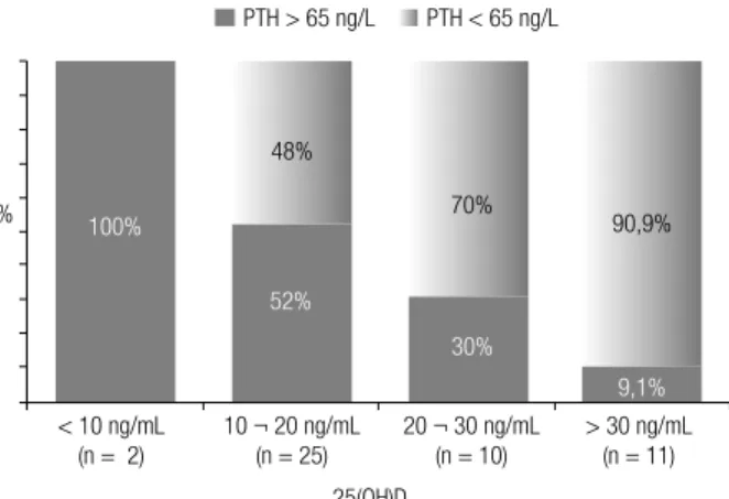
In a population of postmenopausal women under going treatment for osteoporosis, followed in a speciic outpatient clinic, Camargo (91) demonstrated that weekly doses higher than 7,000 IU (> 1,000 IU/day) are necessary to achieve vitamin D suficiency (25(OH) D > 30 ng/mL), which is in accordance with the propo sition of the Endocrine Society for the elderly at risk (Table 3). According to this Brazilian study, 73% of the patients, followed for at least three months in am bulatory directed to the treatment of osteoporosis were below the desired target concentration (> 30 ng/mL) (Figure 2). In the same study, a positive correlation was found between 25(OH)D concentrations and the fe mur BMD and a negative correlation with PTH (91).
Table 3. Vitamin D daily maintenance doses recommended for the general population and the population at risk for the deiciency
Age groups General population
(IU)
Population at risk (IU)
0 – 12 months 400 400 – 1,000
1 – 8 years 400 600 – 1,000
9 – 18 years 600 600 – 1,000
19 – 70 years 600 1,500 – 2,000
> 70 years 800 1,500 – 2,000
Pregnant women 14 – 18 years
600 600 – 1,000
Pregnant women > 18 years
600 1,500 – 2,000
Lactating 14 – 18 years 600 600 – 1,000
Lactating > 18 years 600 1,500 – 2,000
Adapted from the nutritional tables of the Institute of Medicine and the Endocrine Society.
Figure 2. Percentage of non-adequacy of vitamin D according to the plasma concentrations of 25-hydroxyvitamin D (25(OH)D) in a population of individuals presenting osteoporosis at a medical school teaching ward (91).
100 90 80 70 60 50 40 30 20 10 0
ng/mL (n) < 10 (19) 5,2
36,4
73,3
< 20 (132) < 30 (266)
25(OH) D
Cop
yright
© ABE&M t
odos os dir
eit
os r
eser
vados
.
420
In children and adolescents, the doses, apparently, do not vary much from the ones in the adults, with the exception of the irst year of life (Table 3). Winzenberg and cols., in a metaanalysis involving six studies that evaluated vitamin D supplementation in healthy chil dren, were able to group 343 participants that received placebo and 541 participants that received vitamin D and the analysis suggested a beneit of the supplemen tation on the in lumbar spine bone mineral density and total body bone mineral content of those children that were previously deicient (92). Therefore, as already observed in other studies, the positive endpoints of the supplementation are always much more evident when the populations studied were initially deicient (A).
Vieth in a study done in Canada, tested two vitamin D3 doses, compared to placebo. The irst dose was 1,400 IU/week (or 200 IU/day), the same quantity recom mended at current nutritional tables from the Ministry of Health in Brazil. The second dose was 14,000 IU/ week, both groups being followed for 12 months. The group that received 1,400 IU/week did not present signiicant increase in 25(OH)D concentration, while the group receiving 14,000 IU obtained an increment of 15 to 30 ng/mL at the end of 12 months (93). Ta king from that and similar studies, an alteration on the vitamin D daily recommendation tables was proposed in several countries.
In 2011, the Institute of Medicine, in the USA, an organ that regulates the reference tables for daily recom mended intake (DRI) for the general population, increased the daily recommendation to 600 IU for in dividuals between the ages of 1 and 70, and to 800 IU for those older than 70 (Table 3) (50,94). However, the Brazilian nutritional table remains with the daily recommended intake of 200 IU, although several na tional studies have demonstrated that the typical meals of the Brazilian population are not a relevant vitamin D source (64,95), that we depend on cutaneous synthesis to obtain suficiency and that the deiciency is present in all age and populational groups, especially among the elderly (70,74,76,80) (C).
Generally speaking, when 25(OH)D is lower than the target concentration (below 20 ng/mL), an at tack dose is necessary to replenish the body stocks. The most used scheme currently is to administer 50,000 IU/week (or 7,000 IU/day) of vitamin D for six to eight weeks (41). In case the desired concentration of 25(OH)D is not achieved, a new cycle can be pro posed. As there can be some individual variation in the
response to treatment, the reevaluation of the plasma values after each cycle is ideal, especially in the cases of more serious deiciencies, up to achieving the desired concentration. After this period, the maintenance dose should be deined and it varies according to the age group and concurrent conditions (Table 3). For the adults, maintenance doses vary between 400 and 2,000 IU, depending on the sunlight exposure and skin tone. For the elderly, the recommended doses vary from 1,000 to 2,000 IU/day or 7,000 to 14,000 IU/week. Obese individuals, presenting malabsorption condi tions or in use of anticonvulsants might need doses that are twice or three times higer (41) (A).
SBEM recommendation: doses recommended for the maintenance of vitamin D suficiency are indi cated in table 3, varying for the different age groups. Howe ver, for deiciency correction, higher doses (at tack doses) are necessary (Evidence A).
Due to the fact that it is a liposoluble substance, vita min D is absorbed with fats and follows the enterohepatic circulation, meaning it is normally secreted through the gallbladder and reabsorbed in the small intestine. Thus, especially in cases of malabsorption, doses which are much higher than usual can be necessary in order to normalize 25(OH)D concentrations. Besides that, for supplementa tion studies and in the daily practice it is possible to no tice some individual variation in blood concentrations of 25(OH)D reached in response to a same dose of vitamin D, suggesting individuals might present different compe tencies in intestinal absorption or its metabolization (96). It seems there is no difference considering vitamin D ab sorption in relation to fasting or meal type (97).
SBEM recommendation: the risk of intoxication with the deined doses in this document is almost null (Evidence A). Special care must be taken when pharmaceutical compounding is used (Evidence D) and with patients that have diseases with anomalous production of 1,25 dihydroxyvitamin D by the tis sues, such as in granulomatous diseases and some lymphomas (Evidence B).
Cop
yright
© ABE&M t
odos os dir
eit
os r
eser
vados
.
Arq Bras Endocrinol Metab. 2014;58/5
dificult to achieve with these routine doses (99). In rare clinical situations, such as in some cases of granu lomatous diseases (sarcoidosis, tuberculosis and chronic fungal infections) and some lymphoma, activated mac rophages can locally produce 1,25(OH)2D in excess and induce hypercalcemia and hypercalciuria (B). Children with Williams syndrome are more predisposed to hy percalcemia. Therefore, under these conditions, supple mentation should be more criterious and follow fre quent monitoring of plasmatic and urinary calcium (41).
7. What are the differences between vitamin D2 and D3?
SBEM recommendation: vitamin D deiciency tre atment and supplementation can be done either with vitamin D3 (cholecalciferol) or with vitamin D2 (er gocalciferol), although the irst metabolite presents some advantages over the second, due its commercial avaiability, for being more evaluated in clinical stu dies, for allowing more posologic formulations, pro moting more effective increases and for being identi ied by all laboratory methods (Evidence B).
Vitamin D sources are: sunlight exposure, diet and supplementation. The main difference between vitamin D2 (ergocalciferol) and vitamin D3 (cholecalciferol) is the source. In summary, vitamin D2 is the vitamin D from the plant sources, while the one from animal sources is in the form of vitamin D3. The D2 and D3 sources differ only due to the presence of an additional double bond and a methyl group incorporated to the long side chain of the biological form called D2 (100). The two forms present equivalent biological power and are activated in equally eficient ways by hydroxylases in humans. However, there is controversy on the bioequivalence of these formulations for supplementation. A metaanalysis evaluating only controlled and randomized studies that use vitamin D2 and D3 showed that vitamin D3 increased 25(OH)D levels more signiicantly when compared to vitamin D2 (p = 0.001), the single or in bolus dose of vitamin D3 being better than D2 (p = 0.0002). However, this advantage was lost in daily supplementation (101)
(A). Previous studies had already shown a small superi ority of vitamin D3 when administered in single dose, to maintain 25(OH)D le vels for longer time (102).
When the two formulations were compared in dai ly use for 25 weeks, it was observed that those u sing vitamin D2 presented 25(OH)D average concentra tions lower when compared to those that received D3, although, without altering PTH levels (103) (A).
A recent study did not demonstrate difference in effec tiveness when higher 25(OH)D levels were found and also in sustained serum concentration of 1,25(OH)2D3, after 11 weeks of supplementation with 1,000 IU of vitamin D2 or D3 per day (104) (A).
The same was observed in the treatment of children with rickets and controls, where there was a similar in crease in 25(OH)D and 1,25(OH)D levels with both formulations (105) (A). It is possible to conclude that both forms are equivalent in relation to daily supple mentation and that vitamin D3 presents superiority in relation to the maintenance of the 25(OH)D levels for single dose supplementation.
8. What is the difference between vitamin D and calcitriol?
SBEM recommendation: vitamin D active forms, such as calcitriol or alfacalcidol, should not be used when the objective is supplementation, or in the tre atment of vitamin D deiciency, because of their hi gher risk of side effects (Evidence A).
Cop
yright
© ABE&M t
odos os dir
eit
os r
eser
vados
.
422
control of calcemia and calciuria, because hypercalce mia can frequently occur (14,41) (A).
9. How to do supplementation in special cases?
a) In chronic kidney disease
The patient with chronic renal disease presents higher risk for vitamin D deiciency. In renal disease, PTH con centrations also correlate with circulating 25(OH)D levels (A). Therefore, it is believed that vitamin D dei ciency contributes to the development of secondary hy perparathyroidism in chronic renal patients, independent on the renal calcitriol production. Nowadays it is known that several tissues, such as macrophages and osteoblasts have the capacity of producing active vitamin D (cal citriol) and that this synthesis depends on the subs trate, therefore, it is not strictly regulated as the renal synthe sis. Because of that, the treatment of the deiciency and the adequacy of the circulating levels of 25(OH)D is always recommended each time plasma concentrations are lower than 30 ng/mL (106109). According to the opinion of the committee responsible for writing the treatment guidelines for osteomineral disease in chronic renal patients in Brazil, 25(OH)D quantiication is rec ommended at the end of each cycle of attack doses, until the target concentration is achieved, and from then on, every six months (110) (D).
b) In the treatment of osteoporosis
A good part of vitamin D beneits on the risk of fractures observed in the literature has been associated with the concomitant use of calcium. Therefore, the adequacy of calcium intake, either through diet, or through the use of calcium salts, is part of any protocol for osteo porosis treatment. Recommended vitamin D doses are those capable of taking and maintaining plasma con centration to 30 ng/mL or above, avoiding, this way, the secondary hyperparathyroidism and the increase in bone resorption (111113) (A). The nonadequacy of vitamin D concentration is considered one of the po tential failures in medicinal treatment of osteoporosis (signiicant BMD loss and fractures).
SBEM recommendation: for patients with osteopo rosis and increased risk of fractures, it is recommen ded that the 25(OH)D concentrations are maintained above 30 ng/mL for full beneits, for the prevention of secondary hyperparathyroidism, decreased risk of fall and improvement of BMD. To this end, daily doses be tween 1,000 and 2,000 IU are necessary (Evidence A).
c) In obesity and post-bariatric surgery
Obese patients present lower vitamin D concentration when compared to nonobese and are considered a population at risk for the deiciency (41) (A). Nowa days, bariatric surgery is a very used alternative to in duce weight loss in these individuals, possibly further aggravating this deiciency. Therefore, it is advisable to correct this condition prior to surgery. Santos and cols. demonstrated that women submitted to bariat ric surgery at least three years before, presented lower 25(OH)D values when compared to normal paired controls, 77.1% of them presenting vitamin D insuf iciency/deiciency and 41.7% presenting secondary hyperparathyroidism (Figure 3) (114). 25(OH)D con centrations correlated inversely with PTH (r = 0,57, p < 0,05) and directly with bone remodeling markers (CTX and osteocalcin) which, together, can justify the increased risk for fracture observed in this population by other researchers (C). Depending on the surgical technique used and the degree of disabsorption pro moted, some individuals might have a lot of dificulty normalizing 25(OH)D and PTH concentrations, being necessary to follow these parameters and to consider a new attack dose or even higher maintenance doses that should be individually adjusted (D). In some cases, the orientation for frequent sunlight exposure and use of parenteral vitamin D are resources that can be neces sary (115117).
Figure 3. Prevalence of secondary hyperparathyroidism in a population of women submitted to bariatric surgery at least three years prior to the study enrollment. Different ranges of circulating 25(OH)D (114).
100%
< 10 ng/mL (n = 2)
> 30 ng/mL (n = 11)
25(OH)D
25(OH)D: 25-hydroxyvitamin D. Signiicance level of the chi-square test (p < 0.025). 10 ¬ 20 ng/mL
(n = 25)
20 ¬ 30 ng/mL (n = 10)
52%
30%
9,1%
90,9% 70%
48%
PTH > 65 ng/L PTH < 65 ng/L
Cop
yright
© ABE&M t
odos os dir
eit
os r
eser
vados
.
Arq Bras Endocrinol Metab. 2014;58/5
SBEM recommendation: obese people are a popula tion at risk for deiciency and after bariatric surgery it is further aggravated, leading to a secondary hy perparathyroidism and an increased risk for fractures (Evidence A). 25(OH)D test in this situation is very useful for the titration of the daily vitamin D doses, which can be as high as ten times the routine ones (Evidence D).
d) Pregnancy
It is a critical period, because women are oriented to avoid sunlight exposure. Vitamin D deiciency in pregnant women was associated to low birth weight of the newborn, besides some late endpoints, such as low bone mass and cardiovascular risk markers in school age children. In a recent metaanalysis, Agha jafari and cols. analyzed 31 studies, including 18,869 individuals and they concluded that the serum le vels of 25(OH)D are related to gestational diabetes, pre eclampsia, low birth weight newborn and bacterial vaginosis (118). In another metaanalysis, vitamin D supplementation showed positive effect on the low birth weight reduction (119) (A). The doses recom mended for supplementation in this period of life can be found in table 3.
When vitamin D deiciency is suspected, the treat ment with higher doses is still indicated, but daily doses are preferable. The 25(OH)D concentration in the newborn shows high correlation with the one found in the mother. The placenta presents the 1ahydroxylase enzyme and therefore, has the capacity of converting 25(OH)D to calcitriol. Apparently, this production is not strict controlled as that what happens in renal tu bules, and depends only on the amount of substrate (41) (C). For this reason, the recommendation during pregnancy is to avoid weekly or monthly doses.
SBEM recommendation: for pregnant women pre senting deiciency risk, treatment brings beneits for the mother (Evidence B) and the newborn (Evidence A). Daily doses of vitamin D are recommended dur ing pregnancy and weekly or monthly higher doses should be avoided, because the placental production of calcitriol is substratedependant (Evidence C).
10. When should active vitamin D analogues be used?
SBEM recommendation: vitamin D analogues can be used to suppress PTH concentrations in patients with hyperparathyroidism secondary to chronic kidney disea se (CKD), the indication and the dose depending on the CKD stage and the PTH level (Evidence A). There is no superiority of the analogues currently avai lable in Brazil in comparison with cholecalciferol and calcitriol for the prevention of fractures (Evidence B). The analogues are not recommended for the preven tion of cancer (Evidence B).
Active vitamin D analogues are synthetic sub stances that directly bind the vitamin D receptor (VDR). They present different selectivity to the para thyroid cells, depending on its chemical structure, calcitriol (1,25(OH)2D3) shows smaller selectivity, promoting more side effects such as hypercalcemia, hyperphosphatemia and vascular calciications. More selective compounds such as paricalcitol (19nor 1a,25(OH)2D2), maxacalcitol (22oxa1a,25(OH)2D3) and doxercalciferol (1a(OH)D2) and eldecalcitol (1a,25(OH)22β(3hydroxypropyloxy)D3) and doxer calciferol (1a(OH)D2) and eldecalcitol (1a,25(OH)2 2β(3hydroxypropyloxy)D3) promote less adverse ef fects. Doxercalciferol and the alfacalcidol require liver 25hydroxylation to become active (120) (A).
a) Use in secondary hyperparathyroidism
The analogues are classically used to suppress PTH levels in patients with secondary hyperparathyroidism (SHPT) and chronic kidney disease (CKD). In CKD there is an increase in PTH levels, secondary to an alter ation of the regulation of the ibroblast growth factor (FGF23) in the PTHvitamin D axis and the decrease of calcitriol production due to CKD itself. The suppres sion of PTH levels in patients in stages 34 of CKD is more than 40% in 90% of the patients (121,122) (A).
Cop
yright
© ABE&M t
odos os dir
eit
os r
eser
vados
.
424
nine times the upper limit of normality. There is no consensus on the doses of doxercalciferol and paricalci tol, some studies calculated the dose in relation to the initial PTH value dividing it by 80 to 120, to minimize the excessive suppression of PTH or hypercalcemia and hyperphosphatemia (106) (B).
The use of vitamin D analogues minimizes bone loss in CKD due to the suppression of PTH levels and pre vents bone remodeling reduction, due to the effect on the differentiation of normal osteoblasts and inhibition of osteoclastogenesis. However, the concern with the excessive suppression, which would lead to adynamic bone disease persists (122) (B).
Several studies have demonstrated beneit on the survival of patients undergoing dialysis treated with cal citriol or vitamin D analogues. Besides that, there is a smaller risk of progression for terminal renal disease and an increase in the survival in patients with CKD stages 34 (123,124) (B). There is doubt in relation to the beneit of vitamin D active analogues, compared to placebo, in relation to fractures, quality of life, hos pitalizations, muscle function and fall in these patients (125,126) (A).
The most selective analogues, compared to calci triol, demonstrate lower mortality, lower number of hospitalizations and lower duration of each hospitali zation per year (124) (B). Doxercalciferol, similar to paracalcitol, presents higher beneit in relation to sur vival, when compared to calcitriol. A doseresponse was observed in the beneit when the levels of PTH were adjusted according to vitamin D (127) (B).
The use of analogues to avoid the evolution of ne phropaty in diabetic patients is discussed, although the results are still conlicting, using microalbuminu ria and albuminuria as markers. Results from VITAL study are awaited for a more deinitive observation (120,128,129) (B).
b) Fracture prevention
The relationship between vitamin D levels with falls and fractures has been described, also as a signiicant linear predictor of major osteoporotic fractures in ten years (130) (B). A recent metaanalysis demonstrated a modest decrease in the prevention of fractures with the use of 1,25(OH)2D3 (calcitriol) and 1ahydroxy vitamin D3 (alfacalcidol), similar to that obtained using doses higher than 700 IU/day of vitamin D3 (35) (A). Eldecalcitol (1a,25dihydroxy2β[3hydroxypro piloxy] vitamin D3 is a new analogue of vitamin D
active form, recently approved for the treatment of osteoporosis in Japan. Eldecalcitol presents a strong inhibitory effect on bone resorption and signiicantly increases bone mineral density. This drug showed a 26% decrease in the incidence of new vertebral frac tures in three years and 71% decrease in the risk of wrist fracture, in comparison to alfacalcidol, but showed no bene it against hip fracture. An increase in serum cal cium was observed, although hypercalcemia was only seen in 0.4% of patients (131) (A). Edelcalcitol com pared to alfacalcidol promoted better quality of life and decreased severity of vertebral fractures (125,126) (A).
NON-BONE ACTIONS OF VITAMIN D
11. What is the evidence for extra-skeletal effects of vitamin D?
SBEM recommendation: although observational studies show an association between low vitamin D concentration and alterations in several extraskeletal systems, in the moment it is still not possible to pro ve a causeeffect relationship (Evidence B).
Traditionally, vitamin D was associated only with cal cium metabolism functions. The possibility of existence of extraskeletal effects occurred after the disco very of vitamin D receptor (VDR) in tissues not involved with calcium metabolism (e. g. skin, placenta, breast, pros tate, and colon cancer cells) and the identiication of the enzyme 1ahydroxylase in extrarenal tissues. The question to be discussed is the real biological meaning of the presence of VDR and 1ahydroxylase in different tissues (132).
Nagpal and cols. (133) reported that 1,25(OH)2D3 through its transcriptional activity was able to directly or indirectly regulate at least 200 genes. These genes are involved in the control of proliferation, apoptosis and angiogenesis in several tissues. The etiological connec tion between vitamin D deiciency and speciic extra skeletal diseases still needs to be identiied in humans. Findings using animal models, regarding the beneicial effects of 1,25(OH)2D3 suggest mechanisms that in volve similar human signaling pathways (134,135) (B). The main nonskeletal effects studied in the literature will be described here.
a) Vitamin D and cardiovascular disease
Cop
yright
© ABE&M t
odos os dir
eit
os r
eser
vados
.
Arq Bras Endocrinol Metab. 2014;58/5
vational studies that demonstrate a strong association between vitamin D deiciency and mortality due to cardiovascular disease, the mechanism being unclear in the literature (136) (B). Potential hypothesis include the action in the regulation of genes involving renin production, proliferation of cardiac and vascular mus cle cells, negative regulation of reactive C protein and other involvement in other proinlammatory processes. Fiscella and Franker (137) showed that black indivi duals, presenting calcitriol levels in the lower quartile presented 40% increase in the risk of death from coro nary arterial disease (CAD) or stroke (B).
Serious vitamin D deiciency in patients with stabi lized CVD is related to 50% more deaths from stroke and three to ive times more sudden death (138). On the other hand, another study, demonstrated that very high levels are associated with an increase in the risk of ischemic myocardial disease (139). An increased risk of systemic arterial hypertension and metabolic syndrome has been demonstrated when suboptimum vitamin D levels are detected (140). Observational and cohort studies led to the potential vitamin D use as an antihy pertensive agent. Some studies demonstrated decreased levels of systolic blood pressure upon supplementation. However, larger studies were not able to prove these positive effects. Two prospective studies did not show reduction in cardiovascular mortality upon vitamin D supplementation (131). A metaanalysis presented 8% reduction in the mortality from all causes with mo dest doses (141). Nonetheless, a recent metaanalysis in cluding 51 studies, concluded that supplementation did not have a signiicant effect in the mortality (RR 0.96), CAD incidence (RR 1.02) and stroke (RR 1.05) (136). So far, there is no strong evidence for the screening of vitamin D deiciency in patients under risk for CVD, as well as patients with previous CVD. Good prospective studies are necessary for a better understanding of the eficacy of supplementation in the risk reduction of car diovascular disease (132134) (B).
b) Vitamin D and diabetes
Epidemiological and observational studies demonstrate a potential involvement of vitamin D in the patho genesis of the inlammatory process and in the preven tion and control of both diabetes mellitus type 1 and type 2 (DM1 and DM2). Studies performed in animals and humans suggest that vitamin D can be a potential modiier of these diseases (142) (A).
Animal studies demonstrate that immunomodula tory and antiinlammatory actions of vitamin D reduce autoimmune insulinitis in DM1. It seems to suppress the antigen capacity of macrophages, inhibit the matu ration of dendritic cells, modulate the development of CD4 lymphocytes and inhibit the production of cyto kines such as interferon (IFN) and interleukin2 (IL2). These cytokines are known for activate macrophages and cytotoxic T cells, that lead to the destruction of pancreatic cells (143).
In DM2, vitamin D acts reducing insulin resistance and increasing its secretion, through the modulation of the immune and inlammatory process. DM2 is as sociated to an increase in the levels of tumor necrosis factors a and β, Creactive protein (CRP), plasminogen activating factor and interleukin6 (142).
Epidemiological studies demonstrate that children with vitamin D deiciency present 2.4 times higher risk of developing DM1. In the EURODIAB study there was as a reduction in the risk of developing DM1 in 33% among supplemented children (144) (B). The same way the maternal supplementation demonstrates a protective effect to the newborn. A metaanalysis con cluded that, childhood supplementation seems to be protective against the development of DM1 (130). In adults with the disease, a reduction in the insulin dose was seen with the calcitriol supplementation (145) (B).
In rats with vitamin D deiciency, after supplemen tation there was improved insulin secretion (146). Two large studies evidenced that the combined use of cal cium and vitamin D reduced the risk of DM2. In a doseresponse analysis, DM2 risk was reduced in 4% at each increment of 4 ng/mL in the concentration of 25(OH)D (147) (B). In a metaanalysis, it was con cluded that the insuficiency of calcium and vitamin D can negatively inluence glycemia and that supplemen tation of both can be beneicial for the optimization of glucose metabolism (146147) (B).
There is evidence suggesting that vitamin D has a role in the prevention and treatment of DM1 and DM2, through its action on the immune system, insu lin secretion and resistance. However, further studies, using larger populations, are necessary to better eluci date the mechanisms of action and the doses necessary to present the best beneits (132134,148) (A).
c) Vitamin D and cancer
Cop
yright
© ABE&M t
odos os dir
eit
os r
eser
vados
.
426
types of cancer, as well as skin color seems to be related to an increase in the prevalence of colorectal, breast and prostate cancer (149) (B). The risk of development and death due to neoplasia is more elevated in places of higher latitude and this can be related to lower sunligh exposure. Women presenting vitamin D insuficiency show a higher risk of developing colorectal cancer when compared to women presenting suficient vitamin D levels, although no beneit was observed in the use of vitamin D for prevention (150,151).
In tissues where 25(OH)D is available, there is paracrine production of 1,25(OH)2D3, which through binding to its receptor, VDR, regulates transcription of target genes, that act in the differentiation of nor mal and tumor cells. Epidemiological and preclinical studies suggest the action of vitamin D in cancer pre vention and treatment. Polymorphisms in the VDR gene are associated with an increased risk for the de velopment of neoplasias (150). Local production of 1,25(OH)2D3 does not have any function in the con trol of calcium metabolism, but it presents autocrine and paracrine effects. In vitro, it is possible to observe a decrease in 1ahydroxylase (CYP27B1) and in vitamin D receptor (VDR) as the tumor progresses, associated to an increase in 24hydroxylase (CYP24A1), which is inactivating. In vitro and in vivo studies demonstrate the direct or indirect effect of 1,25(OH)2D3 and its analogues on proliferation, differentiation, apoptosis, angiogenesis, invasion and inlammation of malignant cells. Microarray studies show that 1,25(OH)2D3 in luences the transcription of a great number of genes, mainly related to apoptosis control (149,152) (B).
Low vitamin D levels make the tissues more sensi tive to procarcinogenic events. Vitamin D analogues are not able to eradicate tumor cells, however they can be used as adjuvants in cancer treatment. It is believed that high doses of these substances are necessary for a real beneit to be observed, although it increases ad verse effects (153) (B).
A doubleblind placebo controlled study deter mined that the use of 1,25(OH)2D3 in preleukemia showed promising results at the beginning, although it determined an increase in calcemia during the blastic crisis (154). In prostate cancer, the administration of 2,000 IU/day resulted in PSA levels reduction, though the presence of severe hypercalcemia determined the end of the study (134).
Some nonhypercalcemic calcitriol analogues are as sociated with good prognosis in patients that present
elevated VDR expression. However, the use of calcitriol and analogues for the treatment of cancer patients is so far uncertain. Most of the clinical studies were con ducted in patients with prostate cancer and patients with advanced cancer that do not respond to traditional therapies. Laboratory evidence indicates that calcitriol generates a biological response that results in the inhi bition of the neoplastic progress. However, large scale clinical studies are necessary to conirm the beneits of vitamin D use in neoplasias (155,156) (B).
d) Vitamin D and autoimmune disease
Vitamin D action on the immune system seems to be mediated by B and T lymphocytes. VDR is present in these cells. The molecule 1,25(OH)2D3 inhibits the pro liferation of T cells, suppresses the synthesis and prolif eration of immunoglobulins, prevents the formation of IFNγ (interferonγ) and IL2 (interleukin2); besides increasing the activity of suppressor T cells (TH2). In humans, there is epidemiological evidence of the impor tance of vitamin D in the immune system (157) (C).
Sunlight or vitamin D are environmental factors in the etiology of multiple sclerosis and can have a role together with class II MHC inherited factors. Epide miological studies suggest that adults with elevated vi tamin D levels present lower risk of developing multiple sclerosis. Women with high vitamin D intake have 42% less chance to develop this disease (134,158) (B).
Preliminary studies suggest that activated vitamin D can be an eficacious treatment for rheumatoid arthri tis. Treated mice showed decreased activity of the cell responsible for this disease. In humans, epidemiological studies also conirmed a negative association between the levels of vitamin D and the prevalence of the dis ease. Other autoimmune diseases that are being asso ciated with vitamin D are: autoimmune encephalitis, systemic lupus erythematosus, intestinal inlammatory disease and autoimmune thyroiditis. Additional studies are necessary for the conirmation of data, and the use of vitamin D for the prevention and treatment of auto immune diseases (155,156).
e) Vitamin D and innate immunity
Recent studies suggest that vitamin D can modulate in nate immunity. Hypovitaminosis D can present a nega tive impact in infectious diseases. It has been observed that 1,25(OH)2D3 has an antimicrobial activity, includ