Universidade Nova de Lisboa
Instituto de Higiene e Medicina Tropical
Differential expression and functional characterization of
cattle
tick genes in response to pathogen infection (
Babesia
bigemina
)
Sandra Isabel da Conceição Antunes
DISSERTAÇÃO PARA A OBTENÇÃO DO GRAU DE DOUTOR EM CIÊNCIAS BIOMÉDICAS, ESPECIALIDADE DE PARASITOLOGIA
MÉDICA
Universidade Nova de Lisboa
Instituto de Higiene e Medicina Tropical
Thesis:
“Differential expression and functional chara
cterization ofcattle tick genes inresponse to pathogen infection (Babesia bigemina)”
Author: Sandra Isabel da Conceição Antunes
Supervisor: Investigadora DoutoraAna Isabel Amaro Gonçalves Domingos, (IHMT, UNL)
Co-supervisor: ProfessorDoutor Virgílio Estólio do Rosário (IHMT,UNL)
Professor Doutor José de la Fuente (IREC, UCLM)
Dissertation presented to obtain a PhD degree in Biomedical Sciences, speciality of Medical Parasitology.
Universidade Nova de Lisboa
Instituto de Higiene e Medicina Tropical
Tese: Expressão diferenciada e caracterização funcional de genes de carraças em
resposta a infecção poragentes patogénicos(Babesia bigemina)
Autor: Sandra Isabel da Conceição Antunes
Orientador: Investigadora Doutora Ana Isabel Amaro Gonçalves Domingos (IHMT, UNL)
Co-orientador: ProfessorDoutorVirgílio Estólio do Rosário (IHMT,UNL)
Professor Doutor José de la Fuente (IREC, UCLM)
Dissertação apresentada para cumprimento dos requisitos necessários à obtenção do grau deDoutor em Ciências Biomédicas, especialidade em Parasitologia Médica.
The results obtained during the development of this PhD project incited the
following original research articles:
Antunes S, Galindo RC, Almazán C, Rudenko N, Golovchenko M, Grubhoffer L, Shkap V, do Rosário V, de la Fuente J, Domingos A. 2012. “Functional genomics studies of Rhipicephalus (Boophilus) annulatus ticks in response to infection with the cattle protozoan parasite, Babesia bigemina.” Int J Parasitol: 42(2):187-95
Antunes S, Merino O, Mosqueda J, Moreno-Cid JA, Bell-Sakyi L, Fragkoudis R, Weisheit S, Pérez de la Lastra JM, Alberdi P, Domingos A, de la Fuente J. 2014. “Characterization of tick proteins involved in tick-pathogen interactions as potential antigens for the control of tick infestation and pathogen infection.” Parasit Vectors: 7: 42
Dedico esta tese á minha Mãe…
i
Acknowlegments
This thesis would not have been realized without the support and collaboration from a great many people over the years and I would like to take this opportunity to thank them all. First and foremost, I would like to express my deep gratitude to Ana Domingos and Jose de la Fuente my research supervisors, for their patient guidance, enthusiastic encouragement, useful critiques that pushed me further and also for their friendship. Also I want to express my appreciation to Prof. Virgílio do Rosário for his help; whenever necessary he was always present.
ii
Lorena, Uriel, Sarahy and Orlando you were not forgot! Thanks for the smiles and rides along the city! In my second time around in Mexico, now in Querétaro, I had the pleasure of meeting Doctor Juan Mosqueda and Gabriela and to work again with Octávio. Doctor Juan, I’m grateful for all the advices and support you gave me in this last part of the project. Octávio again thank you so much for sharing the work and your good vibes! Finally, aos meus colegas e amigos portugueses! Esta viagem começou no INETI no grupo da UTPAM onde conheci a Ana Domingos, a Bé, o Tiago, Ana Armada, Dr. Carlos, o Fernando, a Ângela, a Maria, a Raquel e a Cristina. Este grupo maravilhoso aceitou-me de braços abertos e apesar de já não existir deixa muitas saudades. No IHMT tenho de agradecer a todos, os quais me ajudaram directa ou indirectamente a terminar este projecto de doutoramento: Renato, Cátia, Tiago, Pedro, Isabel, Ana Afonso, Manuela, Prof. Paulo Almeida, Prof. Silvana Belo, obrigado pelo interesse no meu trabalho e pela amizade. À Fábia, Joana e Lara o meu sincero obrigado por partilharem comigo algum do trabalho!
Quero deixar um agradecimento especial a três pessoas que de facto foram muito importantes para mim neste percurso: Cristina, muito obrigado por seres minha amiga apesar de eu ser relaxada e esquecida por sorrires mesmo when you´re feeling down. Raquel és uma pessoa extraordinária com muita força e determinação e adorei estar grávida contigo. Ana, o apoio que me dá em todas as alturas e em todos os campos tem sido fundamental, obrigado por partilhar comigo os bons e maus momentos.
Não posso deixar de agradecer à minha família por todo o apoio: Aos meus pais, Alberto e Susete, pelos sacrifícios que fizeram e ainda fazem para me darem esta oportunidade. Ao meu irmão Hugo, pelo apoio sempre discreto. À minha irmã Zélia e ao meu cunhado Zé por acreditarem e me incentivarem a lutar pelo que quero. Claro aos meus sobrinhos adorados Sara e Pedro! Maria Inês, não me esqueci de ti, nem dos teus pais e avós! À Carina (que já é da família!) obrigado porque sim… Não podia sonhar com uma família melhor! Comparo muitas vezes a minha família a um circo onde há espaço para todos, alegria todos dias e força para enfrentar os desafios da vida!
iv
Expressão diferenciada e caracterização funcional de genes de carraças em resposta a infecção poragentes patogénicos(Babesia bigemina)
Sandra Isabel da Conceição Antunes
Palavras-chave: Carraça, Vacina, Babesia, RNA de interferência, TROSPA
O conceito “One health” reconhece a necessidade do trabalho conjunto de veterinários,
profissionais de saúde e cientistas, dada a interface dinâmica entre pessoas, animais e ambiente. Este conceito é muito importante em zoonoses, tais como doenças associadas a carraças (DAC´s) que dependem de animais como reservatório. Os protozoários do género
Babesia são agentes patogénicos transmitidos por carraças que causam a doença denominada babesiose num variado número de animais incluindo o Homem. Particularmente a B. bovis e
B. bigemina são transmitidas por carraças, relacionadas com gado, Rhipicephalus (Boophilus) annulatus e R. microplus sendo estas consideradas os ectoparasitas de maior importância,
com largo impacto económico na produção animal. O uso tradicional de acaricidas no controlo de carraças apresenta diversas desvantagens incluindo a seleção de carraças acaricido-resistentes e contaminação ambiental com resíduos químicos. As vacinas destacam-se como uma alternativa ao uso de acaricidas. O objetivo destas vacinas é a proteção contra DAC´s através do controlo das infestações pelos vectores e redução de transmissão de doença. As proteínas envolvidas nas interações carraça-agente patogénico podem ser bons candidatos para essas novas vacinas mas a sua identificação e validação continuam a ser obstáculos. Os objetivos do presente estudo foram, primeiro, a identificação de genes de R. annulatus diferenciadamente expressos em resposta à infeção por B. bigemina, segundo, a validação da influência destes genes no processo de infeção e finalmente a caracterização dos antigénios identificados, a fim de selecionar os melhores candidatos, para o desenvolvimento de uma potencial nova vacina. A fim de alcançar os objectivos propostos, clones de uma biblioteca de hibridização subtrativa por supressão (SSH) foram sequenciados e analisados. Os genes diferenciadamente expressos com prováveis funções relacionadas com a interface carraça- agente patogénico, foram selecionadas para validação dos resultados de SSH por real time RT-PCR. A análise funcional conduzida por RNA de interferência mostra que, nas condições do presente estudo, o silenciamento dos genes que codificam para as proteínas, sérica amilóide A e TROSPA levam á redução de níveis de infecção em R. annulatus e em R. microplus em comparação com o grupo controlo. Em R. microplus é demonstrada a
vi
Differential expression and functional characterization ofcattle tick genes in response to pathogen infection (Babesia bigemina)
Sandra Isabel da Conceição Antunes
Keywords: Tick, Vaccine, Babesia, RNA interference, TROSPA
The “One Health” approach recognizes the need for veterinarians, human health
professionals, and environmental scientists to work together given the dynamic interface among people, animals, and the environment. This approach is increasingly important for zoonotic diseases, such as tick borne diseases (TBD´s) which rely on animals as reservoirs.
Babesia spp. are tick-borne pathogens that cause a disease called babesiosis in a wide range of animals and also humans. Particularity, B. bovis and B. bigemina are transmitted by cattle ticks, Rhipicephalus (Boophilus) annulatus and R. microplus being considered the most important cattle ectoparasites with major economic impact on cattle production. The
traditional use of chemicals to control ticks has serious drawbacks, including the selection of acaricide-resistant ticks and contamination of the environment with chemical residues. Research on alternatives to the use of acaricides is strongly represented by tick vaccines considered a more cost-effective and environmentally safe strategy. The ultimate goal of tick vaccines is to protect against TBD´s through the control of vector infestations and reducing pathogen infection and transmission. Tick proteins involved in tick-pathogen interactions may provide good candidate protective antigens for these new vaccines but their identification and validation are still limiting steps. The objectives of this study were first to identify R. annulatus genes differentially expressed in response to infection with B. bigemina, second to validate the influence of these genes in the infection in both R. annulatus and R. microplus ticks and finally to characterize identified antigens to select the best candidates for future vaccine development. In order to achieve these goals, suppression-subtractive hybridization (SSH) library clones were sequenced and analyzed. After molecular function gene ontology assignment differentially expressed genes with putative functions in tick-pathogen interactions were selected for validation of SSH results by real-time RT-PCR. Functional analysis by RNA interference showed that under the conditions of the present study, knockdown of trospa and serum amyloid A significantly reduced B. bigemina infection levels in R. annulatus and R. microplus when compared to controls. In R. microplus also calreticulin showed infection reduction. TROSPA and CRT were selected, recombinant proteins were obtained using Escherichia coli expression system and poly/monoclonal
antibodies were generated. Their specificity against tick recombinant proteins was confirmed by Western blotting and against native proteins in tick tissues using immunofluorescence. Capillary-fed ticks ingested antibodies added to the blood meal and the effect of these antibodies on tick weight and/or oviposition was shown. No significant effect was observed on pathogen acquisition. The results highlighted the advantages and disadvantages of in vitro
tick capillary feeding for the characterization of candidate tick protective antigens. Several studies have characterized the tick-pathogen interface at the molecular level. However, to our knowledge this is the first report of functional genomics studies in ticks infected with B. bigemina. The results reported here increased our understanding of the role of tick genes in
viii
µg Microgram
µl Microliter
µm Micrometer
ºC Degree Celsius
64TRP 64 tick recombinant protein
Abs Absorbance
A.D. Anno Domini
AKR Akirin
AMA-1 Apical membrane antigen 1
Amp Ampicillin
AP Alkaline Phosphatase
B.C. Before Christ
Bbo-MIC1 Recombinant microneme protein from Babesia bovis
bp Base pair
BSA Bovine serum albumin
cDNA Complementary DNA
CRT Calreticulin
dATP Deoxyadenosine triphosphate
dCTP Deoxycytidine triphosphate
dNTPs Deoxynucleotide triphosphates
dGTP Deoxyguanosine triphosphate
dTTP Deoxythymidine triphosphate
DAPI 4,6-diamidino-2-phenylindole
DDT Dichlorodiphenyltrichloroethane
DMEM Dulbecco’s Modified Eagle Medium
DMSO Dimethyl sulfoxide
DTT Dithiothreitol
DNA Desoxyribonucleic acid
dsRNA Double-stranded ribonucleic acid EDTA Ethylene diamine tetra acetic acid
e.g. exempli gratia
ix
ELISA Enzyme-linked immunoabsorbent assay
ER Endoplasmatic reticulum
EST Expressed sequence tag
FBS Fetal bovine serum
FPLC Fast protein liquid chromatography
G Gauge
g G-force
GO Gene ontology
HAT Hypoxanthine-aminopterin-thymidine
HAZV Hazara vírus
HGPRT Hypoxanthine guanidine phosphoribosyltransferase
His Histidine
i.e. id est
IFA Indirect fluorescent antibody assay
IgGs Imunoglobulin G
IPTG Isopropyl-β-D-thiogalactopyranoside
Kb Kilo base
kDa Kilo Dalton
kHz Kilo Hertz
KTPI Kunitz-type protease inhibitor
LB Luria Bertani
M Molar
mA Milliampere
Ml Millilitre
min Minute
mM Millimolar
Mab´s Monoclonal antibodies
mRNA Messenger RNA
nm Nanometer
OD Optical density
ospA Outer surface protein A
x
OTEs Off-target effects
PBS Phosphate buffered saline
PCR Polymerase chain reaction
PEG Polyethylene glycol
PVC Packed cell volume
PVDF Polyvinylidene fluoride
RAS-3 Rhipicephalus appendiculatus serpin 3 RAS-4 Rhipicephalus appendiculatus serpin 4
RBC Red blood cells
rCRT Recombinant calreticulin
rDNA Ribossomal DNA
RIM36 Immuno-dominant protein of Rhipicephalus appendiculatus
rTROSPA Recombinant TROSPA
RNA Ribonucleic acid
RNAi RNA interference
RPM Rotations per minute
RT-PCR Reverse transcriptase polymerase chain reaction
siRNAs Small interfering RNAs
TAE Tris acetate buffer
TBD Tick borne diseases
TBE Tick borne encephalitis
TBE Tris/Borate/EDTA
TBS Tris buffered saline
Tris-HCl Tris hydrochloride
TROSPA Tick receptor for outer surface protein A
TTBS Tween-Tris buffer saline
RNAseq RNA sequencing
s Second
s.s. sensu stricto
s.l. sensu lato
SDS Sodium dodecyl sulfate
xi
spp. Species (plural)
SSH Suppression-Subtractive Hybridization S.O.C. Super-optimal catabolite repression médium
SUB Subolesin
V Volt
v/v Volume per volume
w/v Weight per volume
xiii
Acknowlegments ... i
Resumo ... iii
Abstract ... v
Abbreviations ... vii
Table of contents ... xii
Index of figures ... xvii
Index of tables ... xx
Introduction ... xxii
1.1TICKS ... 23 1.2HISTORICAL OVERVIEW ... 23 1.3.CHARACTERIZATION, IDENTIFICATION, MORPHOLOGY ... 24
1.3.1. Classification ... 24
1.3.2. Life cycle ... 25
1.3.3. Tick-host interactions ... 26
1.3.4. Overview of tick anatomy and physiology... 28
1.3.5. From Boophilus to Rhipicephalus ... 31
1.4.TICK CONTROL ... 32
1.4.1. Biological tick control ... 32
1.4.2. Genetic tick control ... 33
1.4.3. Chemical tick control- Acaricides ... 34
1.4.4. Immunological tick control - Vaccines... 35
1.5.TICK-BORNE DISEASES ... 37
1.5.1. Viral diseases ... 38
1.5.2. Bacterial diseases ... 39
1.5.3. Protozoan diseases ... 40
1.6.THE GENUS BABESIA ... 41
1.6.1. Bovine babesiosis ... 42
1.6.2. Symptoms of bovine babesiosis ... 43
1.6.3. Diagnosis of bovine babesiosis ... 44
1.6.5. Babesia bigemina life cycle ... 45 1.6.5.1. Events on the vertebrate host ... 46 1.6.5.2. Events in the tick ... 47
1.6.6. Hosts immunity to Babesia sp. ... 48 1.6.6.1. In the vertebrate host ... 48 1.6.6.2. In the tick ... 49
xiv
1.6.4.1. Chemotherapy ... 51 1.6.4.2. Vaccines ... 52
1.7.TICK AND TICK BORNE DISEASES CONTROL ... 53
1.7.1. Tick antigens ... 54
1.7.2. Antigens identification ... 56
1.8.PREMISES AND AIMS OF THIS PHD PROJECT ... 60
Material and methods ... 62
2.1IDENTIFICATION OF DIFFERENTIALLY EXPRESSED GENES IN RHIPICEPHALUS ANNULATUS FEMALE TICKS ... 63
2.1.1 Tick total DNA, RNA and protein extraction ... 63 Agarose gel electrophoresis: ... 64
2.1.2 Detection of Babesia bigemina in ticks by polymerase chain reaction (PCR) ... 64 Cloning of PCR products: ... 65 Transformation:... 65 Selection of transformants: ... 65 Colony screening by PCR: ... 65 Plasmid DNA purification: ... 66
2.1.3 Complementary DNA (cDNA) library construction and SSH ... 66 cDNA library cloning: ... 66 Plasmid purification for sequencing: ... 67 Sequencing and analysis of clones: ... 67
2.1.4 Confirmation of differential expression by Real time PCR ... 68
2.2 IN VIVO GENE SILENCING IN TICKS BY RNA INTERFERENCE ... 71
2.2.2 dsRNA synthesis ... 71
2.2.3 dsRNA injection in ticks ... 72 Treatment of ticks after injection: ... 72 Analysis of tick phenotype after RNAi: ... 73
2.2.3 Gene knockdown assessment by real time reverse transcriptase PCR and infection
quantification ... 73
2.3EXPRESSION AND PURIFICATION OF RECOMBINANT PROTEINS INVOLVED IN TICK-PATHOGEN
INTERACTIONS ... 74
2.3.1 Amplification of selected genes ... 74 Calreticulin: ... 74 TROSPA: ... 74
xv
SDS-PAGE (sodium dodecyl sulphate polyacrylamide gel electrophoresis): ... 77 Western-Blotting: ... 77 Purification of recombinant proteins: ... 78 Determination of protein concentration: ... 79
2.4PRODUCTION AND CHARACTERIZATION OF ANTIBODIES AGAINST TICK PROTEINS INVOLVED IN TICK -PATHOGEN INTERACTIONS ... 80
2.4.1 Poly and Monoclonal antibodies production ... 80 Immunization: ... 80 Antibody titer monitoring of mouse serum using ELISA: ... 80 Generation of hybridomas: ... 81 Screening for antibody producing hybridomas: ... 82 Hybridomas culture and storage:... 81
2.4.2 Immunolocalization assays ... 83 Western blotting: ... 83 Indirect immunofluorescence: ... 83
2.4.3 Tick capillary feeding ... 85 Infected and uninfected bovine blood: ... 85 Antibodies: ... 85 Artificial feeding: ... 86 PCR to determine pathogen infection levels in ticks: ... 86 Evaluation of tick mRNA levels of genes encoding for antigens: ... 87
Results ... 88
3.1.IDENTIFICATION OF DIFFERENTIALLY EXPRESSED GENES IN RHIPICEPHALUS ANNULATUS FEMALE TICKS ... 89 3.2DIFFERENTIAL GENE EXPRESSION INBABESIA BIGEMINA-INFECTED RHIPICEPHALUS ANNULATUS TICKS 92 3.3.SEQUENCE ANALYSIS OF TICK GENES DIFFERENTIALLY EXPRESSED IN RESPONSE TO BABESIA BIGEMINA
INFECTION ... 93 3.4FUNCTIONAL ANALYSIS OF TICK GENES DIFFERENTIALLY EXPRESSED IN RESPONSE TO B. BIGEMINA
INFECTION ... 95 3.5AMPLIFICATION OF CALRETICULIN AND TROSPA CDNA FRAGMENT ... 98 3.6EXPRESSION OF RECOMBINANT PROTEINS CALRETICULIN AND TROSPA ... 100 3.7PRODUCTION OF MONOCLONAL ANTIBODIES ... 103 3.8IMUNOLOCALIZATION ... 106
3.8.1 Western-blot analysis ... 106
3.8.2. Immunofluorescence assays ... 108
3.9CAPILLARY FEEDING ASSAYS ... 113
3.9.1. Effect of antibodies against tick proteins on tick weight, oviposition and pathogen infection. 113
3.9.2. Effect of antibodies and pathogen infection on the mRNA levels of genes encoding for tick
xvi
Discussion ... 117
4.1.ANTIGENS IDENTIFICATION ... 118 4.2.TROSPA AND CRT EXPRESSION AND IMMUNOLOCALIZATION ... 122 4.3.CHARACTERIZATION OF TICK PROTEINS INVOLVED IN TICK-PATHOGEN INTERACTIONS AS POTENTIAL ANTIGENS FOR THE CONTROL OF TICK INFESTATION AND PATHOGEN INFECTION. ... 125 4.4.GENERAL CONCLUSIONS ... 128
References ... 130
Appendix ... 174
xviii
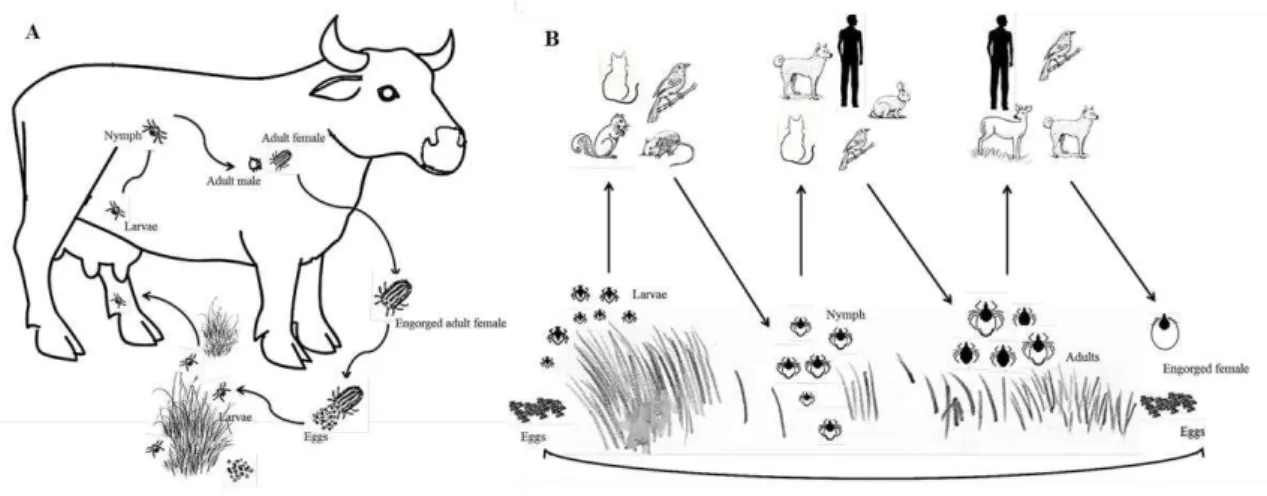
Figure 1: Drawing of hyena-like animal showing tick infestation... 23 Figure 2: Tick examples. ... 25 Figure 3: Characteristic Ixodid life cycles. ... 27 Figure 4: Dissection of an engorged Rhipicephalus annulatus female tick. ... 29 Figure 5: Dissected salivary glands from Rhipicephalus annulatus engorged female. ... 30 Figure 6: Dissected ovary from an engorged Rhipicephalus annulatus female tick. ... 31 Figure 7: The life cycle of Babesia bigemina in cattle and the ixodid tick vector Rhipicephalus (Boophilus)
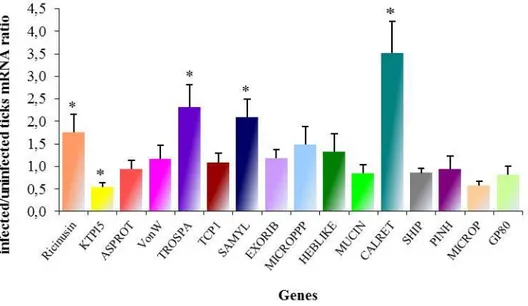
microplus. ... 46 Figure 8: Example of the total RNA extracted using TRI-Reagent SIGMA®. ... 89 Figure 9: Detection of Babesia bigemina in Rhipicephalus annulatus ticks by PCR. ... 90 Figure 10: Cloning of SSH library sequences. ... 91 Figure 11: Functional grouping of tick genes differentially expressed in B. bigemina-infected R. annulatus,
based on Gene Ontology (GO) molecular function assignments ... 92 Figure 12: Differential gene expression in B. bigemina infected R. annulatus ticks. ... 93 Figure 13: TROSPA multiple amino acid sequence alignment. ... 94 Figure 14: Analysis of ricinusin ortholog sequences. ... 95 Figure 15: Amplification of fragments of interest for dsRNA synthesis. ... 96 Figure 16: Synthesis of dsRNA. ... 96 Figure 17: Amplification of fragments of interest by RT-PCR (A) and PCR (B). ... 99 Figure 19: Expression of rCRT. ... 101 Figure 18: Confirmation of correct fragment size cloning in pET101/D-TOPO® plasmid. ... 101 Figure 20: rCRT purification probed by WB with an anti-His antibody. ... 102 Figure 21: Purified recombinant calreticulin. ... 102 Figure 22: Purified recombinant TROSPA... 103 Figure 23: Serum titer for each mouse after 4 immunizations with recombinant proteins. ... 104 Figure 24: Screening of supernatants from hybridoma cultures by WB against rTROSPA. ... 105 Figure 25: Screening of supernatants from hybridoma cultures by WB against rCRT. ... 105 Figure 26: SDS-PAGE gel with R. annulatus protein extracts. ... 106 Figure 27: WB analysis of specificity of rTROSPA and rCRT immunized mice serum and 2D7 and 3C8
supernatants against R. annulatus protein extract. ... 107 Figure 28: WB analysis of specificity of rCRT 3C8 supernatant against different R. annulatus protein
extracts. ... 107 Figure 29: WB analysis of specificity of rTROSPA immunized mice serum and 2D7 supernatant against
different R. annulatus protein extracts. ... 108
xix
Figure 34: Capillary feeding in R. microplus females. ... 113
Figure 35: Effect of antibodies on tick oviposition. ... 115 Figure 36: Effect of antibodies on B. bigemina infection. ... 115 Figure 37: Fold change in expression of genes in the presence of B. bigemina infection. ... 116 Figure 38: Sequencing of obtained fragment from CRT coding region amplification. ... 178 Figure 39: Alignment of available calreticulin protein sequences, ... 178 Figure 40: Negative control of immunofluorescence analysis of cattle tick tissues. ... 179 Figure 41: Microscopic view of a histological specimen of Rhipicephalus annulatus tick tissues stained
xxi
Table 1: Main features that distinguish Ixodid and Argasid ticks. ... 26 Table 2: Ticks identified in Portugal mainland: associated hosts and vector role. ... 37 Table 3: Sequences of primers used for real-time PCR or real time reverse transcription (RT)-PCR. ... 69 Table 4: Sequences of primers used for double-stranded RNA synthesis. ... 72 Table 5: Primer sequences used for coding region amplification ... 75 Table 6. Babesia bigemina infection levels after gene knockdown by RNA interference (RNAi) in
Rhipicephalus (Boophilus) annulatus ticks. ... 97 Table 7: Babesia bigemina infection levels after gene knockdown by RNA interference (RNAi) in
Rhipicephalus (Boophilus) microplus ticks. ... 97 Table 8: Female tick weight after gene knockdown by RNA intereference (RNAi) in Rhipicephalus
(Boophilus) annulatus and Rhipicephalus microplus ticks. ... 98 Table 9: Sequence identity between different available CRT nucleotide sequences ... 100 Table 10: Hybridomas screened by ELISA. ... 104 Table 11: Parameters associated to the feeding process of partially fed Rhipicephalus (Boophilus)
23
1.1 Ticks
Ticks have evolved to become one of the most important groups of arthropod vectors of pathogens. Found in most terrestrial regions of the earth. Ticks are a highly specialized group of obligate, bloodsucking, nonpermanent ectoparasitic arthropods that feed on mammals, birds, and reptiles (Schwan, 2011). These haematophagous ectoparasites inflict a direct negative impact on the vertebrate hosts while they feed and most importantly they may also act as vectors, as well as reservoirs, of multiple pathogens (Jongejan and Uilenberg, 2004). While considered zoophilic, many species are associated to the transmission of important ethological agents to humans (Silva et al., 2006). To qualify as a vector, a tick must (1) feed on infectious vertebrates (2) acquire the pathogen during the blood meal (3) maintain the pathogen through one or more life stages (4) transmit the pathogen to previously unexposed hosts while feeding again (Kahl et al., 2002, Jongejan and Uilenberg, 2004, Estrada-Pena et al., 2013).
1.2 Historical overview
Fossil records suggest that ticks originated 65–146 million years ago in the Cretaceous period from the Mesozoic Era (Klompen and Grimaldi, 2001, Nava et al., 2009).
Figure 1: Drawing of hyena-like animal showing tick infestation.
Discovered in the Egyptian tomb of Antef, dating from the time of Thutmose III (eighteenth dynasty), about 1500 B.C.
24
A reference to - what could be - 'tick fever' was found on a papyrus scroll dating back to the 16th Century B.C. (Krantz, 1978, Heyman et al., 2010), and an animal draw-ing datdraw-ing back to the Queen Hatshepsut III era (15th Century B.C) (Figure 1) shows what were considered to be three ticks attached to a hyena's ear (Arthur, 1965, Heyman et al., 2010).The Greek philosopher Aristotle (384-322 B.C.) wrote in his “Historia Animalium” “The tick is generated from couch-grass” and that “cattle suffer both from lice and from ticks. Sheep and goats breed ticks, but do not breed lice” (Aristotle, 350 B.C.). In an early encyclopedia “Historia Naturalis”, published A.D.77–79 by Pliny the Elder, ticks are described: ‘a tick simply filled to bursting point with its victim's blood and then died because it had no anus’ (Heyman et al., 2010, Nijhof, 2010). It is obvious that the hostile nature of ticks was long ago recognized.
1.3 Characterization, identification, morphology
1.3.1 Classification
25
Prostriata is regarded as the most primitive line and consist of the Ixodes genus only, which can copulate both on and off the host in contrast to Metastriata adults which mate only on the host (Barker and Murrell, 2004). Examples of hard and soft ticks are represented in figure 2.
Figure 2: Tick examples.
(A) dorsal (left) and ventral (right) view of a Rhipicehalus annulatus female, representative of a hard tick species (original from the author). (B) dorsal (left) and ventral (right) view of an Ornithodoros savignyi Audouin, 1827,female, representative of a soft tick species (original and authorized from Ard Nijhof).
1.3.2 Life cycle
Ticks undergo four stages, namely egg, larvae, nymph and adult (Oliver, 1989, Sonenshine, 1991). Ixodid have only one nymph instar, in contrast to the several nymphal instars of argasid ticks (Oliver, 1989). Ixodid ticks need several days to feed, and once the female is engorged she drops from the host to deposit thousands of eggs and dies. Argasid ticks feed more than once and intermittently and these parasites don´t remain attached to their hosts. These ticks may feed several times during their lifetime and on a number of different hosts laying few hundred eggs in batches. Argasid ticks have a remarkable longevity living for many years and may endure long period of starvation (Sonenshine, 1991). Longevity in fact is remarkable in argasid ticks: there are records of
Ornithodoros canestrinii Clifford, Kohls & Sonenshine, 1964, survival in laboratory of
10 years without food and impressively an unfed adult of O. lahorensis Neumann, 1908,
for 18 years (Hoogstraal, 1985, Oliver, 1989). The most distinctive features between soft and hard ticks are summarized in table 1.
26
Table 1: Main features that distinguish Ixodid and Argasid ticks (adapted from University, 2013)
Features Hard Ticks Soft Ticks
Scutum (dorsal shield) Present Not presente
Capitulum anterior (mouthparts)
Visible from dorsal view
Not visible from the dorsal view
Nymphal stages One Several
Adult feeding time Days 30-60 min
Female blood meals One Several
Oviposition One Several
Total eggs laid 3.000-8.000 400-500
In Ixodid ticks larval, nymphal and adult feeding usually requires 3-7, 4-8 and 7-9 days respectively, during which time active growth of gut and cuticle occurs in order to accommodate the blood meal, most of which will be acquired in the final 24 hours of engorgement. Male Ixodids do not feed as females. They feed intermittently, small quantities of blood and enough for their reproductive organs to mature. Male ticks in the genus Ixodes have active reproductive organs as soon as they moult from the nymphal stage and do not need to feed. In nature, tick life cycles are regulated by seasons. The length of life cycles is quite variable in result to several constrains such as photoperiods, temperature, humidity and availability of suitable hosts. Rhipicephalus microplus
Canestrini, 1887, ticks can ensure three or four generations a year: one generation a year is probably the usual pattern for most of the ticks in the subtropics and warmer regions. But in colder regions, ticks can take one to three years to complete their life cycle protracting it until better conditions arise (Oliver, 1989, Sonenshine, 1991).
1.3.3 Tick-host interactions
27
nymphs reattach feeding again until repletion. After nymphs drop from the host and after some days (or longer) adults hatch and seek a new host to complete the life cycle. One-host ticks attach to a One-host as larvae, and then feed and mature to the adult stage on the same host (Schwan, 2011) (Figure 3A).
Figure 3: Characteristic Ixodid life cycles.
A) The one host life cycle. B) Three-host life cyle (original from the author).
There is some flexibility in this feeding behavior and under certain conditions ticks can use one or two hosts (e.g. Hyalomma scupense, Schulze, 1919) or use two instead of three (e.g. Amblyomma rotundatum, Koch 1844) (Oliver, 1989). Some ticks accept a wide variety of host species, other might be more selective and some others are extremely demanding and attach to only one host species. In fact, with the exception of fish, ticks parasite all vertebrates (mammals, birds, reptiles and amphibians). Nevertheless, mammals are the dominant host for most of Ixodid ticks.
28
with long intervals of immobility which can be interrupted when water uptake can be established or to seek for a host. When on the host, there is no longer danger of drying out or starving, but they can be removed by host’s grooming or feeding might be reduced by host immunity. Most ticks have adaptations in their behavior (e.g. attachment site) and physiology of feeding thus reducing host reactions. Host-acquired immunity might be expressed in various ways ranging from rejection of the parasite to increasing feeding time, inadequate engorgement leading to decreased egg viability or infertility (Parizi et al., 2012, Willadsen and Jongejan, 1999).
1.3.4Overview of tick anatomy and physiology
Externally ticks have bodies which are divided into two primary parts: the anterior capitulum (or gnathosoma) containing the head and mouthparts; and the posterior idiosoma which contains the legs, digestive tract, and reproductive organs. The idiosoma is distinguished to podosoma with chelicerae, palps, hypostome and four pairs of legs in adults, and opisthosoma with anal aperture. The hypostome is modified specifically for blood-feeding and is essential for the attachment to the host. Chemosensillas, mechanosensillas and eyes or photoreceptors are located on the capitulum. Ticks also have a special sensory organ - Haller’s organ – on the tarsus of the first pair of legs that provides information about host location, host odors, or detection of pheromones. Cuticle covers whole tick body and serves as an exoskeleton (like in other arthropods). A layer of cuticle called procuticle, specifically its outer part, becomes sclerotized in certain parts and forms sclerites. The biggest sclerite, scutum, covers the anterior part of the body and protects the dorsal side of it. The major components of the cuticle are proteins and chitin; lipids represent a minor part (Sonenshine, 1991).
29
Figure 4: Dissection of an engorged Rhipicephalus annulatus female tick.
After removal of part of the cuticle midgut, ovary, malpighian tubules and tracheal system are effortlessly observed.
(original from the author).
The midgut is the most prominent organ in the tick body. According to Coons and Alberti (1999), the midgut of ticks is divided into an anterior and a post-ventricular region, lined by a simple pseudo-stratified epithelium composed of cells that have distinct classifications and functions. In general, six different cell types have been described in the anterior midgut of many tick species, namely, replacement (or stem cells), digestive, secretory, undifferentiated, endocrine, and vitelogenic cells (Remedio et al., 2013). During feeding, it occupies almost the whole body cavity of the tick. The midgut branches serve as storage place. Contrary to insect, the digestion in ticks is an intracellular process (with the exception of the intraluminal digestion of erythrocytes) (Sonenshine, 1991, Coons and Alberti, 1999). Ultra structurally, the cells of the midgut of Ixodidae ticks in general are complex, having different organelles and many cytoplasmic inclusions thus reflecting the multifunctional activities of the midgut (Caperucci et al., 2010).
30
secretion of protein factors and water transport (Sonenshine, 1991). Before feeding salivary glands are essential in water balance regulation, during attachment and feeding are responsible for cement proteins secretion as well as other molecules transported by saliva (Sonenshine, 1991). During feeding salivary glands enlarg several times and once females fully engorge undergo degeneration and remodeling processes that are likely under hormonal regulation (Sauer et al., 2000, L'Amoreaux et al., 2003).
Figure 5: Dissected salivary glands from Rhipicephalus annulatus engorged female.
(original from the author).
Female reproductive system consists of a single U-shaped ovary: the paired oviducts, a single uterus, the vagina subdivided into cervical and vestibular regions and the seminal receptacle (organ not present in Argasid and postriate females whereas uterus is enlarged distally where it joins the vagina). Ovary is located in the posterior region of the body. In the unfed females the ovary is rather thin and small but in fed females it´s a large organ with a tube-like structure of luminal epithelium and developing oocytes connected with an epithelium by a short hollow stalk called funiculus (Figure 6) (Sonenshine, 1991). The tick oocytes remain stationary during formation and yolk deposition takes place in situ. The whole ovary with egg cells is expanded into the
31
Figure 6: Dissected ovary from an engorged Rhipicephalus annulatus female tick.
(original from the author).
1.3.5 From Boophilus to Rhipicephalus
Progress in the phylogeny and evolution of ticks, in particular of hard ticks, culminates into the adjustment of nomenclature of ticks.
The genus Boophilus is no longer valid and has recently became part of the Rhipicephalus genera since there is considerable evidence from morphology and nucleotide sequences that Rhipicephalus is paraphyletic with respect to Boophilus
(Murrell et al., 2000, Beati & Keirans, 2001, Murrell and Barker, 2003). To preserve the name, since it is very well known, the genus Boophilus has become a subgenus of the genus Rhipicephalus. In fact, many times these ticks are still referred as only Boophilus
disregarding the current nomenclature. In this dissertation Boophilus may be used when referring R. annulatus and/or R. microplus in order to simplify the allusion to these ticks. There are five valid species of ticks in subgenus Boophilus: Rhipicephalus (Boophilus) annulatus Say, 1821, R. decoloratus Koch, 1844, R. geigyi Aeschlimann & Morel, 1965, R. kohlsi Hoogstraal & Kaiser, 1960 and R. microplus Canestrini, 1888. These ticks, commonly known as cattle ticks, present a characteristic morphology and one-host life-cycle: preferably cattle (except for R. kohlsi with a predilection for small ruminants),
taking about three weeks to complete their cycles on the host from unfed larvae to engorged female (Jongejan and Uilenberg, 2004).
As Rhipicephalus (Boophilus) ticks are one-host ticks, they may become very
32
considerable direct damage. Although these ticks have short mouthparts, damage to hides is considerable as the preferred feeding sites are often of good leather potential (Jongejan and Uilenberg, 2004). In addition to R. microplus, species of most importance to farm animals are R. annulatus which is widespread in tropical and sub-tropical countries and
R. decoloratus which occurs in Africa. These species are involved in the transmission of several pathogens which escalate their importance in animal industry. These cattle ticks, are associated with the transmission of pathogens that cause babesiosis (Babesia bovis
Babes, 1888 and Babesia bigemina Smith & Kilborne, 1893) and anaplasmosis
(Anaplasma marginale Theiler, 1910) (Peter et al., 2009) which are regarded as very
important diseases in cattle, leading to large economic losses (Bock et al., 2004, Shkap et al., 2007a, Suarez and Noh, 2011).
1.4 Tick control
Tick control is essential to reduce impact on livestock productivity and also to contract tick-borne diseases prevalence. Tick control is mainly based on the application of acaricides, despite their disadvantages and limitations, although other control methods such as vaccination or biocontrol agents are available (Willadsen, 2006).
1.4.1 Biological tick control
Ticks have relatively few natural enemies, but the use of predators, parasites, and pathogens has been examined towards tick control. Most predators are non-specific, opportunistic feeders and probably have little impact on ticks. Sub-Saharan African birds such as the yellow and red-billed oxpeckers (Buphagus africanus Linnaeus, 1766 and Buphagus erythrorhynchus Stanley, 1814) can eat 14 grams of ticks per day. Other
33
Ticks can harbor a wide range of endosymbiotic bacteria including Rickettsia da Rocha-Lima, 1916, Francisella Dorofe'ev 1947, Coxiella Philip, 1948, and Arsenophonus
Werren, 2005, amongst others (Alberdi et al., 2012).Tick control strategies could be planned based on interference with their endosymbionts for the control of the vectors and the pathogens they hold. Since these endosymbionts are essential for the subsistence of the arthropod host, their elimination would be deleterious for their survival (Ghosh et al., 2007).
The application of entomopathogenic fungi is a promising approach for controlling ticks. Of the approximately twenty species of entomopathogenic fungi which have been reported to attack and kill ticks, only a handful have been extensively studied, in particular Metarhizium anisopliae Sorokin, 1883and Beauveria bassiana (Bals.-Criv.)
Vuill. (Kaaya et al., 1996, Pirali-Kheirabadi et al., 2007, Quinelato et al., 2012, Monteiro et al., 2013). Entomopathogenic fungi for the control of ticks have been applied in field trials both on- and off-host with reasonable success and commercial products have been developed (Samish et al., 2004, Stafford and Allan, 2010). Biocontrol agents usually favor both human and environmental safety, especially in comparison to the use of acaricides, but few products have been used in spite of their potential. The inability to successfully adopt biocontrol strategies includes factors like environmental stability (e.g.
UV resistance, temperature tolerance), ability to initiate infection at low humidity and potential unspecific damage to non-target invertebrates (Domingos et al., 2013).
1.4.2 Genetic tick control
The genetic resistance of cattle to ticks and tick-borne diseases is complex, but, facing other control methods and their problems, breeds resistance has become an important parameter in some regions. This resistance trait is most commonly manifested in indigenous zebu (Bos indicus Linnaeus, 1758) breeds. The resistance status can be
34
Genetic control of a different order is the suggested release of sterilized ticks into the environment, similar to sterile insect techniques developed for the control of insect pests. Ticks can be sterilized through hybridization (Hilburn et al., 1991), treatment with chemicals (Hayes and Oliver, 1981) or by RNA interference (RNAi) (de la Fuente et al., 2006c, Merino et al., 2011a). Practical difficulties such as the mass rearing of sufficient amounts of sterile ticks and expected difficulties in obtaining the public and political support for the mass release of ticks may pose too high hurdles to make the field application of this technique possible.
1.4.3 Chemical tick control - Acaricides
35
worldwide; products combining active components are available in an attempt to exploit a diverse number of mechanisms of action, to reduce the emergence of insecticide resistance (Veiga et al., 2012).
The introduction of a new product in the market is time-consuming and has a huge economic burden (Graf et al., 2004). The public awareness of the detrimental effects of pesticides on the environment and increasing concerns about resistance of insecticidal compounds, demands the necessity of finding new approaches in tick control.
1.4.4 Immunological tick control - Vaccines
Major alternatives to conventional acaricide treatments have been developed in recent years and anti-tick vaccines are among the most important developments. William Trager demonstrated in 1939 that injection of guinea pigs with extracts of whole larvae, salivary glands or digestive tracts of female Dermacentor variabilis Say, 1821 ticks conferred partial protection against subsequent tick challenge. This partial resistance could be transferred through serum from immune animals to susceptible ones (Trager, 1939, Allen and Humphreys, 1979, Johnston et al., 1986). Immunization with heterogeneous preparations such as tissue extracts results in a polyclonal and multifactorial immune response by the experimental animal. This kind of immunization masks the antigen(s) that triggered the effective response so it became necessary to simplify mixtures injected in the hosts. Increasingly simpler midgut protein combinations were tested until an antigen named Bm86, which conferred significant protection of cattle
against R. microplus infestations, was discovered (Willadsen et al., 1989). This protein of
36
TickGARD of a dairy herd resulted in a 56% reduction of tick numbers in the field over a single generation, a 72% reduction in tick reproductive performance and an increase in cattle live weight gain of 18.6 kg over a 6 month period compared to an unvaccinated control group (Jonsson et al., 2000). In Cuba, within a different context, vaccine based on the same antigen was marketed as Gavac (de la Fuente et al., 2007a, Merino et al., 2013). A retrospective analysis of the field application of Gavac from 1995 to 2003 during which almost 600,000 dairy cattle were vaccinated showed that the number of acaricide treatments was reduced by 87% with an overall reduction of 82% in the country’s acaricide consumption for tick control. The incidence of mortality due to bovine babesiosis was reduced as well (Valle et al., 2004). Despite the effectiveness of these commercial Bm86-based vaccines for the control of cattle tick infestations, they show strain-to strain variation in efficacy being effective mainly against Rhipicephalus tick
species (de la Fuente and Kocan, 2003, Willadsen, 2006, Guerrero et al., 2012b) and since Bm86 protein was found in the midgut of ticks and therefore designated ‘concealed’ antigen, i.e. not exposed to the host’s immune system, regular booster is required to
maintain a strong antibody response which poses a problem to convince farmers to adopt this control approach (Willadsen, 2004, Kiss et al., 2012).
These anti-tick vaccines use recombinant proteins as antigens to immunize animals and are an attractive alternative for the control of tick infestations, since they present several advantages, such as prevention or reduction of pathogens transmission (de la Fuente et al., 1998, Almazan et al., 2005, de la Fuente et al., 2007a, de la Fuente et al., 2011, Merino et al., 2011b) environmental safety, low cost production (Odongo et al., 2007, Kiss et al., 2012), prevention of drug-resistant ticks selection (Parizi et al., 2012) and inclusion of multiple antigens that could target several tick species (de la Fuente et al., 2000, de Vos et al., 2001, Willadsen, 2004, de la Fuente and Kocan, 2006, Willadsen, 2008, Parizi et al., 2012).
37
immunization (ELI) and other technologies allowing for a rapid, systematic and comprehensive approach to tick vaccine discovery, this constraint is being surpassed (de la Fuente and Kocan, 2006, Domingos et al., 2013). Although the availability of these powerful techniques the identification and characterization of effective antigens remains a significant challenge.
1.5 Tick-borne diseases
In history, Theobald Smith and Frederick L. Kilbourne (1889 to 1893) demonstrated how a disease was spread from cattle to cattle by ticks serving as the vector of transmission. Moreover, they were able to identify the pathogen of Texas fever, an intra-erythrocytic protozoan which Smith named Pyrosoma bigeminum now renamed
genus Babesia Babes, 1888 (Assadian and Stanek, 2002). Some years later it was
established that ticks are able to transmit disease to humans. First reports are even prior to the discovery of Smith and Kilborne and associate the bite of soft ticks in Angola and Mozambique to disease (Livingstone, 1857, Schwan et al., 2012) but only in the beginning of the twenty century route of transmission of the ethological agent of Rocky Mountain Fever was proven to be a tick, the hard tick Dermacentor andersoni Stiles,
1908 (Ricketts, 1991, Amsden et al., 2005). Throughout the last century many different associations tick-pathogens were discovered, including bacteria, viruses, protozoan parasites (Jongejan and Uilenberg, 2004, de la Fuente et al., 2008a), nematodes (de la Fuente et al., 2008a, Brianti et al., 2012) and one Trypanosoma species (Morzaria et al., 1986, Latif et al., 2004); the importance of these arthropods by themselves and as vectors is now very well documented and extensively reviewed (Estrada-Pena and Jongejan, 1999, Jongejan and Uilenberg, 2004, de la Fuente et al., 2008a, Colwell et al., 2011, Dantas-Torres et al., 2012). Table 2 shows ticks identified in Portugal mainland and their associated hosts and pathogens.
38
Argas vespertilionis (Latreille, 1802) Bats Issyk-Kul Fever virus; borreliosis 1,2,3
Ornithodoros maritimus (Vermeil and Marguet, 1967) Birds Soldado virus 1,2,3
Ornithodoros erraticus (Lucas 1849) Pigs; Humans
African swine fever virus; Qalyub virus; Borrelia microti, Borrelia hispanica, Borrelia crocidurae; Babesia meri 1,2,3,5
Ixodes acuminatus( Neumann 1901)
Wild carnivores and rodents (occasionally
birds) 1,2,6
Ixodes arboricola (Schulze & Schlottke, 1930) Birds Rickettsia spp. 6
Ixodes bivari (Santos Dias 1990) Rabbit 1,2
Ixodes canisuga (Johnston, 1849) Dogs, cats, wild carnivores 1,2
Ixodes frontalis (Panzer, 1798) Wild birds Candidatus Neoehrlichia mikurensis 1,2,6,7
Ixodes hexagonus (Leach 1815) Domestic and wild carnivores; Ungulates Anaplasma phagocytophilum; B. burgdorferi s.l. 1,2,3,5
Ixodes ricinus (Linnaeus,1758) Domestic and wild mammals; Humans
Anaplasma phagocytophilum; Borrelia burgdorferi s.l., Rickettsia helvetica; R. monacensis; Babesia microti; B.divergens 1,2,3,5
Ixodes simplex (Neumann, 1906) Bats 1,2
Ixodes ventalloi (Gil Collado, 1936)
Domestic and wild mammals (carnivores),
birds, rodents Anaplasma phagocytophilum; Rickettsia helvetica 1,2
Ixodes vespertilionis (Koch, 1844) Bats Issyk-Kul Fever virus 1,2,3
Dermacentor marginatus (Sulzer, 1776)
Domestic and wild mammals (ungulates, carnivores); Humans
Rickettsia slovaca; Borrelia lusitaniae; Babesia canis; Coxiella
burnetti 1,2,3,5
Dermacentor reticulatus (Fabricius, 1794)
Domestic and wild mammals, ungulates and carnivores
Babesia canis; B. caballi; Rickettsia slovaca; Francisella
tularensis 1,2,3,5
Haemaphysalis hispanica (Gil Collado, 1938) Lagomorphs 1,2
Haemaphysalis inermis (Birula, 1895) Ungulates 1,2
Haemaphysalis punctata (Canestrini & Fanzago, 1878) Ungulates, birds Palma virus; Crimean-Congo Hemorrhagic Fever virus 1,2,3,8
Rhipicephalus bursa (Canestrini & Fanzago, 1878) Domestic dog, ungulates; Humans
Babesia ovis; B. bigemina; Anaplasma marginale; A. ovis;
Theileria ovis 1,2,3,5
Rhipicephalus pusillus (Gil Collado, 1936)
Domestic and wild mammals (carnivores,
lagomorphs ungulates) Rickettsia sibirica 1,2
Rhipicephalus sanguineus (Latreille, 1806)
Wild and domestic mammals (carnivores, insectivores, ungulates and rodents)
Anaplasma platys; Babesia canis; B. vogeli;B. gibsoni; Ehrlichia canis; Rickettsia conorii; R. massiliae;Hepatozoon canis 1,2,3,5
Rhipicephalus annulatus (Say, 1821)
Ungulates, carnivores, domestic birds, and
lagomorphs Babesia bigemina; B. bovis; Anaplasma marginale 1,3,5,9
Hyalomma lusitanicum (Koch 1844 )
Domestic and wild mammals (ungulates,
insectivores, carnivores) B. burgdorferi s.l 1,2,9
Hyalomma marginatum (Kosh, 1844) Ungulates; Birds
Theileria annulata; Rickettsia aeschlimannii; Borrelia lusitaniae;
Dhori viruses; Crimean-Congo Hemorrhagic Fever virus 1,2,3,5 Most Common associated Host Most commom associated pathogens
Ticks in Portugal Ref
References [1] (Caeiro, 1999); [2] (Santos-Silva et al., 2011); [3] (de la Fuente et al., 2008a); [4] (Lisboa et al., 2009); [5] (Jongejan and Uilenberg, 2004); [6] (Norte et al., 2012); [7] (Movila et al., 2013); [8] (Dilcher et al., 2012); [9] (Estrada-Pena and Santos-Silva, 2005)
1.5.1 Viral diseases
39
compromises the central nervous system tissues, transmitted mainly by I. ricinus and I. persulcatus Schulze, 1930(Dumpis et al., 1999).
1.5.2 Bacterial diseases
Ticks also transmit several important bacterial diseases. They are traditionally gathered into two groups: tick-borne rickettsioses (Rickettsia spp.) and other bacterial
diseases (Sonenshine, 1991). From the first group diseases like Mountain Spotted Fever (Rickettsia rickettsia), Boutenneuse Fever (R. conorii), Anaplasmosis (Anaplasma spp. Theiler, 1910), and Ehrlichiosis (Ehrlichia spp.Moshkovski, 1945) are good examples. Rickettsia species parasite generally the endothelial cells lining of the vascular system,
Ehrlichia invades monocytes, Cowdria neutrophils, Anaplasma erythrocytes. Anaplasma marginale is the primary cause of anaplasmosis in livestock. It is considered to be one of the most important diseases of cattle and sheep (Kocan et al., 2010). During feeding, the gut infections remain constant (Kocan et al., 1992) and the gut cells serve as a reservoir for repeated infection of the salivary glands. This pattern is significantly different from piroplasms (addressed in the next section) where the gut cells are cleared of the infection as the parasites move to the salivary glands. Infection could be transmitted transstadially, but the transovarial transmission of anaplasmosis does not appear to occur (Kocan et al., 1985). Other tick-borne bacterial diseases include for example Lyme disease (caused by the coil-shaped Gram-negative spirochete Borrelia Swellengrebel, 1907: B. burgdorferi s.s., B. afzelii, and B. garinii among others), and Tularemia (caused by the gram-negative
coccobacillus Francisella tularensis). Lyme disease (borreliosis) is an emerging disease
and the most frequent and important human tick-borne disease (Schuijt et al., 2011, Embers and Narasimhan, 2013). These bacteria are transmitted by ticks from the genus
Ixodes: I. ricinus Linnaeus, 1758 or I. persulcatus in Europe and I. scapularis Say, 1821
40
Borrelia surface protein OspA (Pal et al., 2000, Pal et al., 2004). Borrelia spp. spirochetes at first multiply within the midgut fluid after ingestion with the blood meal, then stop and remain attached to the digestive epithelium as the infected tick digests the blood meal and molts to the next stage (transstadial transmission). During the next tick feeding, the spirochetes are activated, multiply and penetrate into the hemolymph (expression of OspC instead of OspA (Schwan and Piesman, 2002). Within 48 hours after the attachment, the spirochetes can be found adjacent to the salivary glands (Zhu, 1998).
1.5.3 Protozoan diseases
Tick-borne protozoan parasites of the phylum Apicomplexa are important parasites causing disasterous effects and substantial financial losses regarding livestock. In addition, these organisms could infect humans (Babesia spp. transmitted by Ixodes
spp. ticks) (Skrabalo and Deanovic, 1957, Garnham et al., 1969), pets and other animals (Hepatozoon spp. Miller, 1908) (Patton, 1908, Jongejan and Uilenberg, 2004). Apicomplexan parasites of economic importance that are transmitted by ticks comprise
Babesia and Theileria spp. Due to the existence of pear-shaped intraerythrocytic stages;
they have been referred to as piroplasms (Florin-Christensen and Schnittger, 2009, Schnittger et al., 2012). A unique morphological characteristic shared by piroplasmids with other apicomplexan protozoans is the presence of an apical complex: a complex cellular apparatus consisting of conoid, rhoptries, micronemes (Babesia) or microspheres
(Theileria), and other subcellular organelles (Kakoma and Mehlhorn, 1994). These two
piroplasms are phylogenetic close (Allsopp et al., 1994, Florin-Christensen and Schnittger, 2009, Schnittger et al., 2012) and sometimes confusion arises when classifying these genera (Lack et al., 2012). Regardless of this discussion, Babesia spp.
are typically differentiated from Theileria spp. based on life-cycle characteristics,
including distinctions in their biology within tick vectors, the manner by which they are transmitted from vector to vertebrate host, and the location of replication in the vertebrate hosts (e.g., Babesia spp. multiply only in erythrocytes, while Theileria spp. enter lymphocytes and develop into schizonts) (Uilenberg, 2006).
41
pathogen one of the most important protozoan transmitted by ticks (Schnittger et al., 2012).
1.6 The Genus Babesia
It was at the end of the 19th century that the Romanian biologist Victor Babeş discovered micro-organisms in erythrocytes of cattle in Romania and associated them with bovine hemoglobinuria or red water fever (Babes, 1888). Five years later B. bigemina was recognized as the causative agent of Texas Cattle Fever (Smith and
Kilborne, 1893).
Babesiosis is caused by intraerythrocytic parasites of the Apicomplexan genus
Babesia and is a common infection of vertebrate animals worldwide. Currently it is
known that members of the genus Babesia are one of the most ubiquitous and widespread
blood parasites in the world, second only to the trypanosomes. All over the world, there are more than 100 Babesia species
.
(Homer et al., 2000, Hunfeld et al., 2008, Gohil et al.,2013). Ticks from several genera are now known to be vectors and reservoirs of numerous Babesia spp. transmissible to reptiles, birds and mammals (Gohil et al., 2013).
The Babesia species are characterized by transovarial transmission in the vector tick and the limitation of infecting only erythrocytes in the host (Uilenberg, 2006). Completion of a life cycle and therefore the maintenance of Babesia parasites are completely dependent on both the tick and the vertebrate host (Mehlhorn and Schein, 1984, Chauvin et al., 2009) and therefore, the distribution of all the different Babesia
species is primarily ruled by the geographical distribution of their tick vectors. Babesia
organisms can be visualized in a Giemsa stained infected blood smear. They are pyriform (pear shaped) or observed as round or amoeboid forms. Depending on the Babesia
species, a host erythrocyte may be parasitized by single, paired, or multiple organisms. The size of the organisms varies depending on the species being classified as either small (1.0-2.5 μm; B. bovis, B. gibsoni, B. ovis and B. divergens) or large Babesia (2.5-5.0 μm; B. bigemina, B. caballi and B. canis), accordingly to the size of the trophozoites
subunit-42
ribossomal RNA gene (18S rDNA) sequences, showing that small and large Babesia
parasites are divided in two different phylogenetic clusters (Homer et al., 2000).
1.6.1 Bovine babesiosis
The major economic impact of babesiosis is shown in cattle industry. Bovine babesiosis, in which the most important pathogens are B. bigemina, B. bovis and B. divergens (prevalent only in Europe and potentially zoonotic), threaten the health and
safety of millions of bovines in tropical and subtropical regions of the world (Bock et al., 2004). References, to what it may have been a babesial infection, were found in the biblical book of Exodus (Homer et al., 2000).
Since the discovery of this disease a great amount of time and money have been spent to eradicate/control this type of cattle disease since it can induce animal mortality, abortions, reduction of milk/meat production, and sometimes, neurological symptoms (Brown and Palmer, 1999, Suarez and Noh, 2011). If in one hand we have the economic losses related to the disease on the other we will have the high cost of tick control, disease detection, prevention and treatment (de Leon et al., 2010, Mosqueda et al., 2012). Furthermore there is an indirect and underestimated cost of the disease related with the refusal of cattle farmers in endemic areas to improve the production of beef and milk in their herds by introducing pure-breed animals, most of them from tick-free areas, because they will present an acute form of the disease and many will die in the following weeks to their arrival. The consequence is that the quality of cattle in endemic areas remains low, therefore impeding the development of the cattle industry and the well being of producers and their families (Mosqueda et al., 2012).
In almost a century and a quarter since the first report of the disease there is none safe and efficient vaccine available, chemotherapeutic choices are limited and there are few low-cost, reliable and fast detection methods (Mosqueda et al., 2012).
43
1.1 million are bovine, which are susceptible to tick infestation and consequently, to infection with tick-borne diseases including babesiosis (Silva et al., 2010a). From the ticks found to parasitise Portuguese cattle three,Rhipicephalus (Boophilus) annulatus, R. bursa Canestrini & Fanzago, 1878 and Ixodes ricinus are known vectors for babesiosis (Estrada-Pena and Santos-Silva, 2005). Three fairly different studies confirm the presence of bovine babesiosis in Portugal. First a quite small study in 100 bovines report the presence of about 76% of Babesia spp. in blood smears with reference to mixed infections with Anaplasma spp.(29%) and Theileria spp.(5%) (Antunes, 2008). The
second and more relevant study comprising a molecular methodology shows that in 1104 cattle blood samples collected from Central and Southern regions of Portugal and analyzed by PCR–reverse line blotting (RLB), 5.2% were positive for Babesia spp.
Furthermore B. divergens was the most prevalent with 4.2% and several mixed infections
were detected (Silva et al., 2010a). In 2013 a methodologically similar study in approximately 1400 bovines confirmed the presence of babesial infections, with 7, 9% of the studied population but remarkably most of the infections (7,8%) are B. bigemina
(Gomes et al., 2013). Despite some differences these studies demonstrate the presence and dispersal of bovine babesiosis in Portugal.
1.6.2 Symptoms of bovine babesiosis
Despite being closely related, B. bovis and B. bigemina cause remarkably different
symptons in cattle. The clinical signs associated with B. bovis infections are fever,
44
disease can develop very rapidly with sudden and severe anaemia, jaundice and death (Bock et al., 2004).
1.6.3 Diagnosis of bovine babesiosis
Diagnosis of clinical cases of babesiosis can be achieved by microscopy, immunological or using molecular detection methods (Mosqueda et al., 2012). From the first group blood smears stained with Giemsa or acridine orange can behighlighted. Thin blood films from B. bovis are prepared from capillary blood, as blood of general
circulation may contain up to 20 times fewer parasites due to sequestration of infected erythrocytes in capillaries of brain and other organs (Bose et al., 1995). In B. bigemina
infections, parasitized cells are evenly distributed throughout blood circulation. For low levels of parasitemia, especially in cases where B. bovis is involved, diagnosis is based on thick smears of infected blood stained with Giemsa (Morzaria et al., 1992, Mosqueda et al., 2012). The advantage of the thick smear is that a large amount of erythrocytes is analyzed in a reduced amount of space; therefore the probability of finding infected cells is ten times higher than in a thin smear. These techniques are inexpensive and reasonably portable although accuracy of diagnosis relies on the training and skills of the microscopist.
45
other techniques (Mosqueda et al., 2012). Immunological methods to detect Babesia spp. parasites have the disadvantage of relying on the presence of specific antibodies against those parasites, which may take days or weeks to develop in an infected animal or they are present for months after the infection has disappeared, making their usefulness very limited in acute disease cases, vaccinated or cleared-by-treatment animals.
The different molecular diagnosis methods rely on the presence and amplification of pathogen DNA (PCR based assays) so they can distinguish active infections. With the evolution of more sensitive PCR based techniques, several methods for the detection and differentiation of bovine babesiosis infections have been described, including nested PCR (Figueroa et al., 1993), reverse line blot (RLB) hybridization (Gubbels et al., 1999), LAMP (Loop-Mediated Isothermal PCR) (Iseki et al., 2007) and real time PCR (Buling et al., 2007, Criado-Fornelio et al., 2009). Despite the advantages of these techniques regarding sensitivity, due to one factor or another (costs, contaminations, validation), none of these methods is used globally.
1.6.5 Babesia bigemina life cycle
The complex life cycle of Babesia spp. takes place in two hosts, vector and
46
Figure 7: The life cycle of Babesia bigemina in cattle and the ixodid tick vector Rhipicephalus (Boophilus) microplus.
(adapted from Bock et al, 2004).
1.6.5.1 Events in the vertebrate host
47
2004). The vacuole membrane gradually disintegrates, and the parasite is left with the defining piroplasm feature of a single membrane, in contrast to Plasmodium species, which invade by a similar mechanism but retain the host membrane in addition to its own (Rudzinska et al., 1976, Homer et al., 2000). Once inside the erythrocyte, it transforms into a trophozoite, by binary fision, from which two merozoites develop by a process of merogony. The merozoites lyse the cell and go on to infect additional erythrocytes. Four parasites can be formed at the same time giving rise to a Maltese cross form which is characteristic in Babesia sp. infection. Rapid reproduction destroys the host cell and leads
to hemoglobinuria in the host. Multiplication is asynchronous and different divisional stages can be seen in the bloodstream at the same time (Chauvin et al., 2009). Some trophozoites develop into a diploid ovoid type of merozoite, called a gamont precursor. These gamont precursors do not develop further until they are taken up by the tick in the blood meal (Mackenstedt et al., 1995, Chauvin et al., 2009) later on, when in the gut of the tick, even prior to leaving the erythrocytes these precursors develop into gametocytes (Homer et al., 2000).
1.6.5.2 Events in the tick