The
SCA1
(Spinocerebellar ataxia type 1) and
MJD
(Machado-Joseph
disease) CAG repeats in normal individuals: segregation analysis and allele
frequencies
Cláudia Emília Vieira Wiezel, Maria do Carmo Tomitão Canas and Aguinaldo Luiz Simões
Departamento de Genética da Faculdade de Medicina de Ribeirão Preto, Universidade de São Paulo,
Ribeirão Preto, SP, Brazil.
Abstract
Spinocerebellar ataxia type 1 (SCA1) and Machado-Joseph disease (MJD/SCA3) are autosomal dominant neurodegenerative diseases caused by expansions of a CAG trinucleotide repeat in theSCA1 and MJD genes. These expanded sequences are unstable upon transmission, leading to an intergeneration increase in the number of repeats (dynamic mutation). The transmission of the CAG repeat was studied in normal mother-father-child trios, referred for paternity testing (SCA1, n = 367; MJD, n = 879). No segregation distortion was detected. The CAG allele frequencies were determined in 330 unrelated individuals (fathers from couples tested for paternity). The allele frequency distributions did not differ from those previously reported for European populations. The estimated values for the statistic parameters indicating diversity at theSCA1 locus did not differ much from those reported previously for other STRs in the Brazilian population, while those for theMJD locus were close to or higher than the maximum values of previous reports. This shows thatSCA1 and MJD are highly informative loci for applications in genetic and population studies and for forensic analysis.
Key words:segregation distortion, Spinocerebellar ataxia type 1, Machado-Joseph disease. Received: April 2, 2002; accepted: April 7, 2003.
Introduction
Spinocerebellar ataxia type 1 (SCA1) and Machado-Joseph disease (MJD/SCA3) are autosomal dominant neurodegenerative diseases caused by expansions of a CAG repeat in theSCA1andMJDgenes, respectively. Seg-regation distortion favoring the transmission of mutated or normal alleles during meiosis has been reported for both genes. (Ikeuchiet al., 1996; Riesset al., 1997; Takiyamaet al., 1997; Iughetti et al., 1998). Rubinsztein and Leggo (1997) added to these observations, reporting the preferen-tial transmission of alleles with smaller CAG repeats by normal females.More recently, however, Mac Millanet al.
(1999) did not find any evidence of segregation distortion upon transmission of CAG repeat alleles by normal indi-viduals.
Normal variation in size of the CAG repeats ofSCA1
and MJD genes has been reported in a few surveys (Rubinszteinet al., 1995; Watkinset al., 1995; Limprasert
et al., 1996; Richards et al., 1996; Jodice et al., 1997;
Limprasertet al., 1997; Takanoet al., 1998), as well as in comparative studies carried out in affected and control groups (Dürret al.,1996; Goldfarbet al.,1996; Maruyama
et al., 1996; Matsumura et al., 1996; Hsieh et al.,1997; Soonget al.,1997; Zhouet al.,1997; Lokkegaardet al.,
1998).
In spite of being highly polymorphic, triplet repeats in the normal range are consistently transmitted unchanged from parents to children (Limprasertet al., 1994; Richards and Sutherland, 1994), and the repeat size variation in the
DRPLA(dentatorubral-pallidoluysian atrophy) gene has re-cently been shown to be reliable for use in forensic tests, es-pecially paternity tests (Pelottiet al., 1998).
There are no frequency estimates of the SCA1 and
MJDalleles in the Brazilian population, and segregation analysis has produced conflicting results (Rubinsztein and Leggo, 1997; MacMillanet al.,1999). We investigated al-lele segregation in mother-child and father-child pairs, and estimated the frequencies of SCA1 andMJDalleles in a sample of clinically normal individuals from the northeast-ern region of the State of São Paulo, Brazil. We also evalu-ated their applications in paternity investigations, by comparisons with other markers. The absence of segrega-www.sbg.org.br
Send corresponding to Aguinaldo Luiz Simões. Departamento de Genética, Faculdade de Medicina, USP, Avenida Bandeirantes 3900, 14049-900 Ribeirão Preto, SP, Brazil. E-mail: alsimoes@ fmrp.usp.br.
tion distortion herein reported corroborates the data pre-sented by MacMillanet al.(1999).
Material and Methods
The study sample comprised individuals seeking pa-ternity investigation at the Clínica Civil of the Ribeirão Preto University Hospital, University of São Paulo, during the years 1996 and 1997. All individuals were white (skin color was determined visually), originating from Ribeirão Preto and nearby cities. They formed mother-child-alleged father trios, routinely submitted to the investigation of a set of six or more STRs.
For calculating theSCA1andMJDallele frequencies, we considered mothers and alleged fathers of 165 trios (Sample I) consecutively submitted to the same set of tests for paternity determination, totaling 330 genetically unre-lated individuals, or 660 chromosomes. For estimating seg-regation distortion, 118 trios with confirmed paternity were selected from this sample. In addition, other 249 trios were examined for theSCA1locus, and 761 trios for theMJD lo-cus. The last two groups (Sample II) were neither consecu-tively collected, nor submitted to the same set of paternity tests, but the individuals were ethnically similar to those in Sample I, and paternity was confirmed with a probability of 99,99%. The difference in sample size between the two groups reflects nothing but the longer time of use of the
MJDlocus in the laboratorial routine.
Genomic DNA was extracted from 300µL of whole
blood, as described by Higuchi (1989). The fragments con-taining the CAG repeats of theSCA1andMJDgenes were amplified by PCR, using the primers and conditions de-scribed by Orret al.(1993) and Kawaguchiet al.(1994), respectively. The amplified products were electrophoresed on a 12% denaturing polyacrylamide gel, followed by sil-ver nitrate staining. The PCR products of about 150
sam-ples were initially genotyped after electrophoresis; the procedure was repeated with samples of apparently equal mobility placed side by side, thus allowing the visualization of an allele ladder and the selection of samples for sequenc-ing, in order to determine the exact number of CAG re-peats. These sequenced samples were used as reference.
Exact tests were performed using the GENEPOP pro-gram (Raymond and Rousset, 1995). For estimating segre-gation distortion, we prepared a program that allows to determine the inherited allele by direct genotype analysis of the trios, and records which one of the alleles, the larger or the smaller, was transmitted. The proportions were com-pared using aχ2test, considering the expected ratio of 50%.
This study was approved by the Research Ethics Committee of the Ribeirão Preto University Hospital, Ribeirão Preto School of Medicine, University of São Paulo (HCRP nr. 5158/98).
Results
The alleles were designated according to the number of CAG repeats. Nineteen differentSCA1alleles were iden-tified, with 19 to 39 repeats. At theMJDlocus, 21 alleles were found, with 14 to 40 repeats.SCA1alleles 20, 24, 38 and 39, as well as theMJDallele 40, were observed only in Sample II, and were not considered in the allele frequency analysis. In the segregation distortion analysis, 367 trios were analyzed for theSCA1locus, including 734 meioses (Table 1), and 879 trios for theMJDlocus, totaling 1758 meioses (Table 2). No segregation distortion for theSCA1
alleles was revealed by the analysis of informative meioses (p = 0.8516 and p = 0.3604, for 257 maternal and 269 pater-nal meioses, respectively; Table 1) no distortion was ob-served for theMJDlocus either (Table 2), analyzed in 745 maternal and 695 paternal informative meioses (p = 0.6339 and p = 0.2713, respectively).
Table 1- Transmission of larger and smallerSCA1alleles by normal individuals.
Meiosis Transmitted allele χ2
Smaller Larger Non-informative Total
Maternal 130 127 110 367 0.035 (p = 0.8516)
Paternal 142 127 98 367 0.8364 (p = 0.3604)
Total 272 254 208 734 0.6158 (p = 0.4326)
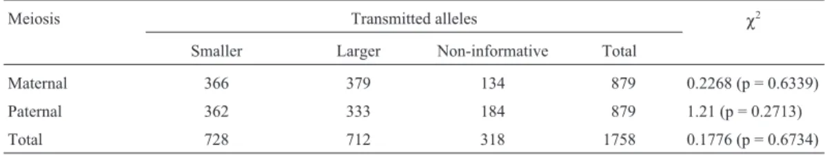
Table 2- Transmission of larger and smallerMJD alleles by normal individuals.
Meiosis Transmitted alleles χ2
Smaller Larger Non-informative Total
Maternal 366 379 134 879 0.2268 (p = 0.6339)
Paternal 362 333 184 879 1.21 (p = 0.2713)
The allele frequencies for theSCA1 and MJD loci were determined in 330 unrelated individuals (660 chromo-somes) of Sample I: 44 genotypes forSCA1and 75 forMJD
were detected; 76 individuals were homozygous at the
SCA1locus and 50, at theMJDlocus.
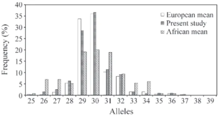
The allele frequencies found were similar to those re-ported in the literature for European populations (Figures 1 and 2). At both loci, the most frequent alleles (29 and 30 for
SCA1and 14 and 23 forMJD) were less frequent in African populations.
The parameters of forensic interest were calculated for both loci: Power of Discrimination (PD): SCA1 = 0.9087 andMJD= 0.9572; Power of Exclusion (PE): SCA1 = 0.4583 and MJD = 0.50; Polymorphism Information Content (PIC): SCA1 = 0.7259 and MJD = 0.8218; Heterozygocity (H):SCA1= 0.7697 andMJD= 0.8485.
Discussion
This study comprises the largest sample of individu-als analyzed forSCA1andMJDloci reported in the litera-ture, corresponding, respectively, to 24 and 28% of the total. It revealed the largest number of different alleles in a single sample hereto described, though no new allele was outside the range already reported in the literature.
We did not observe preferential transmission of nor-malSCA1andMJDalleles: 526 informative meioses (257 maternal and 269 paternal) for theSCA1locus and 1,440 in-formative meioses (745 maternal and 695 paternal) for the
MJDlocus. These results are in accordance with those of Mac Millanet al.(1999), who did not find evidence of seg-regation distortion inMJD,SCA1andDRPLAloci in their study of 377 pairs of twins and their normal parents. How-ever, the preferential transmission of the smaller alleles of theMJDlocus has been reported by Rubinsztein and Leggo (1997) in normal women whose smaller allele was trans-mitted in 166 out of the 290 meioses analyzed, while the men transmitted the smaller allele in 126 out of the 269 meioses.
In the present study, the distributions of the allele fre-quencies at both loci showed a great similarity with the dis-tributions reported in the literature for European populations (Figures 1 and 2). The most prevalent alleles (29 and 30 in theSCA1locus, and 14 and 23 in theMJD lo-cus, respectively) are less frequent in African populations, which gives them a greater heterozygozity. For theMJD lo-cus, we observed the three modes present in European pop-ulations, which correspond to alleles 14, 23, and 27, while, in African populations, up to five modes occur, correspond-ing to alleles 14, 22, 28, 30, and 33. For theSCA1locus, our sample showed a mode around allele 30, similar to Euro-pean populations, while two modes occur in the African populations, corresponding to alleles 26 and 30. The greater heterozygozity in African populations and the pro-file of frequencies with multiple modes may be either
ca-sual or the result of peculiarities of the places of origin of the few samples hereto analyzed.
Since our study is restricted to a sample from a small region of the State of São Paulo, investigations of other se-ries of individuals from different geographical areas of the country are desirable.
For genes with trinucleotide repeats that expand as dynamic mutations, the alleles in the normal range are eas-ily amplified by PCR, but there is a possibility that larger alleles are not amplified, as is the case in myotonic dystro-phy (Gennarelliet al., 1998), resulting in an apparently ho-mozygous genotype. Nevertheless, the CAG expanded repeat has been easily diagnosed by PCR (Brice, 1998). The amplification products corresponding to the normal and expanded alleles can be visualized, and the number of repeats accurately determined after denaturing poly-acrylamide gel electrophoresis and silver staining (Vuillaumeet al., 1998; Maruyamaet al., 1996). We did not detect expanded or intermediate alleles in this study, but, considering the sensitivity of the method used to mea-sure the alleles, the frequency of homozygotes (23.3% for
SCA1and 15.15% forMJD) seems likely to represent the real frequencies in the population.
The estimated values for the statistical parameters of forensic interest (Heterozygozity, Polymorphism Informa-tion Content, Power of Exclusion and Power of
Discrimina-Figure 1- Allele frequencies of theSCA1locus in three population sam-ples. The European and African means were calculated from Watkinset al. (1995); Jodiceet al.(1997); Limprasertet al. (1997); Takanoet al. (1998).
tion) for theSCA1locus did not differ much from the values reported previously for other loci in the Brazilian popula-tion (Pagottoet al., 1999; Leboute, 2000), while those for theMJDlocus were close to or higher than the maximum values reported. This demonstrates that these loci are highly informative markers for general applications in ge-netics and population studies, as well as for paternity tests in forensic investigations, much in the same way as the
DRLPAlocus (Pelottiet al., 1998).
Acknowledgments
We thank Ana Lúcia Pimentel for laboratory techni-cal assistance, Dr. Lewis Joel Greene for comments on ear-lier drafts of this article, to CAPES (Coordenação de Aperfeiçoamento de Pessoal de Nível Superior) and to FAPESP (Fundação de Amparo à Pesquisa do Estado de São Paulo) for financial support.
References
Brice A (1998) Unstable mutations and neurodegenerative disor-ders. J Neurol 245:505-510.
Dürr A, Stevanin G, Cancel G, Duyckaerts C, Abbas N, Didierjean O, Chneiweiss H, Benomar A, Lyon-Caen O, Julien J, Serdaru M, Penet C, Agid Y and Brice A (1996) Spinocerebellar Ataxia 3 and Machado-Joseph Disease: Cli-nical, molecular and neuropathological features. Ann Neurol 39:490-499.
Gennarelli M, Pavoni M, Amicucci P, Novelli G and Dallapiccola B (1998) A single Polymerase Chain Reaction-bases proto-col for Detecting normal and expanded alleles in Myotonic Dystrophy. Diag Mol Pathol 7 (3):135-137.
Goldfarb LG, Vasconcelos O, Platonov FO, Lunkes A, Kipnis V, Kononova S, Chabrashvili T, Vladimirtsev VA, Alexeev VP and Gajdusek DC (1996) Unstable triplet repeat and pheno-typic variabily of Spinocerebellar Ataxia Type 1. Ann Neurol 39:500-506.
Higuchi R (1989) Simple and rapid preparation of samples for PCR. In: Erlich, HA (ed) PCR technology – principles and aplications for DNA amplification. New York, Stockton Press, pp 36.
Hsieh M, Tsai HF, Lu TM, Yang CY, Wu HM and Li SY (1997) Studies of the CAG repeat in the Machado-Joseph disease gene in Taiwan. Hum Genet 100:152-162.
Ikeuchi T, Igarashi S, Takiyama Y, Onodera O, Oyake M, Takano H, Koide R, Tanaka H and Tsuji S (1996) Non- Mendelian transmission in Dentatorubral-Pallidoluysian Atrophy and Machado-Joseph Disease: the mutant allele is preferentially transmitted in male meiosis. Am J Hum Genet 58:730-733. Iughetti P, Otto P, Zatz M, Bueno MRP and Marie SK (1998)
Dif-ferent behavior in the paternallyvs.maternaly inherited mu-tated allele in Brazilian Machado-Joseph (MJD1) families. Am J Med Genet 77:246-248.
Jodice C, Giovannone B, Calabresi V, Bellocchi M and Terrenato L (1997) Population variation analysis at nine loci contain-ing expressed trinucleotide repeats. Ann Hum Genet 61:425-438.
Kawaguchi Y, Okamoto T, Taniwaki M, Aizawa M, Inoue M, Katayama S, Kawakami H, Nakamura S, Nishimura M,
Akiguchi I, Kimura J, Narumiya S and Kakizuka A (1994) CAG expansions in a novel gene from Machado-Joseph dis-ease at chromosome 14q32.1. Nat Genet 8:221-228. Leboute APM (2000) Polimorfismos genéticos em três amostras
da população urbana brasileira. PhD Thesis, Universidade de São Paulo, Ribeirão Preto.
Limprasert P, Nouri N and Keats BJB (1994) Meiotic stability and polymorphism of CAG repeat in normal chromosome at SCA1 locus. Am J Hum Genet (supl): A55:193.
Limprasert P, Nouri N, Heyman RA, Nopparatana C, Kamonsilp M, Deininger PL and Keats BJB (1996) Analysis of the Machado-Joseph gene in human, chimpanzee and monkey populations: a variant nucleotide is associated with the num-ber of CAG repeats. Hum Mol Genet 5(2):207-213. Limprasert P, Nouri N, Nopparatana C, Deininger PL and Keats
BJB (1997) Comparative studies of the CAG repeats in the Spinocerebellar Ataxia Type 1 (SCA1) gene. Am J Med Genet 74:488-493.
Lokkegaard T, Nielsen JE, Hasholt L, Fenger K, Werdelin L, Tranebjaerg L, Lauritzen M, Colding-Jorgensen E, Gronbech-Jensen M, Henriksen AO and Sorensen SA (1998) Machado-Joseph disease in three Scandinavian fami-lies. J Neurol Sci 156:152-157.
Mac Millan JC, Voisey J, Healey SC and Martin NG (1999) Men-delian segregation of normal CAG trinucleotide repeat al-leles at normal CAG trinucleotide repeat alal-leles at three autosomal loci. J Med Genet 36:258-259.
Maruyama H, Kawakami H and Nakamura S (1996) Reevaluation of the exatc CAG repeat length in hereditary cerebellar ataxias using highly denaturing conditions and long PCR Hum Genet 97:591-595.
Matsumura R, Takayanagi T, Murata R, Futamura N, Hirano M and Ueno S (1996) Relationship of (CAG)nC configuration to repeat instability of the Macahdo-Joseph disease gene. Hum. Genet 98:643-645.
Orr HT, Chung M, Banfi S, Kwiatkowski Jr TJ, Servadio A, Beaudet AL, McCall AE, Duvick LA, Ranum LPW and Zoghbi HY (1993) Expansion of an unstable trinucleotide CAG repeat in spinocerebellar ataxia type 1. Nat Genet 4:221-225.
Pagotto RC, Canas MCT, Brito ROAA and Simões AL (1999) Al-lele frequencies of the human von Willebrand factor gene (vWF) in a Brasilian population sample. Int J Legal Med 112:326-328.
Pelotti S, Mantovani V, Esposti PD, D’Apote L, Bragliani M, Maiolini E, Abbondanza A and Pappalardo G (1998) The DRPLA CAG repeats in aan Italian population sample: eval-uation of the polymorphism for forensic applications. J Fo-rensic Sci 43(2):410-412.
Raymond M and Rousset F (1995) GENEPOP (version 1.2): pop-ulation genetics software for exat tests and ecumeniscism. J Hered 86:248-249.
Richards RI and Sutherland GR (1994) Simple repeat DNA is not replicated simply. Nat.Genet 6:114-116.
Riess O, Epplen JT, Amoiridis G, Przuntek H and Schöls L (1997) Transmission distortion of the mutant alleles in spinocerebellar ataxia. Hum Genet 99:282-284.
Rubinsztein DC, Leggo J, Coetzee GA, Irvine R, Bucley M and Ferguson-Smith MA (1995) Sequence variation and size ranges of CAG repeats in the Machado-Joseph disease, spinocerebellar ataxia type 1 e androgen receptor genes. Hum Mol Genet 4(9):1585-1590.
Rubinsztein DC and Leggo J (1997) Non-Mendelian transmission at the Machado-Joseph disease locus in normal females: preferential transmission of the alleles with smaller CAG re-peats. J Med Genet 34:234-236.
Soong B, Cheng C, Liu R and Shan D (1997) Machado-Joseph disease: Clinical, molecular, and metabolic characterization in Chinese Kindreds. Ann Neurol 41:446-452.
Takano H, Cancel G, Ikeuchi T, Lorenzetti D, Mawad R, Stevanin G, Didierjean O, Dürr A Oyake M, Shimohata T, Sasaki R, Koide R, Igarashi S, Hayashi S, Takiyama Y, Nishizawa M, Tanaka H, Zoghbi H, Brice A and Tsuji S (1998) Close asso-ciations between prevalences of dominantly inherited spinocerebellar ataxias with CAG-repeat expansions and frequencies of large normal CAG alleles in japanese and caucasian populations. Am J Hum Genet 63:1060-1066.
Takiyama Y, Sakoe K, Soutome M, Namekawa M, Ogawa T, Nakano I, Igarashi S, Oyake M, Tanaka H, Tsuji S and Nishizawa M (1997) Single sperm analysis of the CAG re-peats in the gene for Machado-Joseph disease (MJD1): evi-dence for non-Mendelian transmission of the MJD1 gene and for the effect of the intragenic CGG/GGG polymor-phism on the intergenerational instability. Hum Mol Genet 6(7):1063-1068.
Vuillaume I, Schraen S, Rousseaux J and Sablonnière B (1998) Simple nonisotopic assays for detection of (CAG)n repeats expansions associated with seven neurodegenerative disor-ders. Diagn Mol Pathol 7(3):174-179.
Watkins WS, Bamshad M and Jorde LB (1995) Population genet-ics of trinucleotide repeat polymorphisms. Hum Mol Genet 4(9):1485-1491.
Zhou YX, Takiyama Y, Igarashi S, Li YF, Zhou BY, Gui DC, Endo K, Tanaka H, Chen ZH, Zhou LS, Fan MZ, Yang BX, Weissenbach J, Wang GX and Tsuji S (1997) Machado-Joseph disease in four Chinese pedigrees: Molecular analy-sis of 15 patients including two juvenile cases and clinical correlations. Neurology 48:482-485.