Arq Neuropsiquiatr 2009;67(4):1133-1142
SPINOCEREBELLAR ATAXIAS
Hélio A.G. Teive
Abstract – Spinocerebellar ataxias (SCAs) constitute a heterogeneous group of neurodegenerative diseases characterized by progressive cerebellar ataxia in association with some or all of the following conditions: ophthalmoplegia, pyramidal signs, movement disorders, pigmentary retinopathy, peripheral neuropathy, cognitive dysfunction and dementia. Objective: To carry out a clinical and genetic review of the main types of SCA. Method: The review was based on a search of the PUBMED and OMIM databases. Results: Thirty types of SCAs are currently known, and 16 genes associated with the disease have been identified. The most common types are SCA type 3, or Machado-Joseph disease, SCA type 10 and SCA types 7, 2, 1 and 6. SCAs are genotypically and phenotypically very heterogeneous. A clinical algorithm can be used to distinguish between the different types of SCAs. Conclusions: Detailed clinical neurological examination of SCA patients can be of great help when assessing them, and the information thus gained can be used in an algorithm to screen patients before molecular tests to investigate the correct etiology of the disease are requested.
KEY WORDS: spinocerebellar ataxias, cerebellar atrophy, genotype, phenotype.
Ataxias espinocerebelares
Resumo – As ataxias espinocerebelares (AECs) compreendem um grupo heterogeneo de enfermidades neurodegenerativas, que se caracterizam pela presença de ataxia cerebelar progressiva, associada de forma variada com oftalmoplegia, sinais piramidais, distúrbios do movimento, retinopatia pigmentar, neuropatia periférica, disfunção cognitiva e demência. Objetivo: Realizar uma revisão clínico-genética dos principais tipos de AECs. Método: A revisão foi realizada através da pesquisa pelo sistema do PUBMED e do OMIM. Resultados: Na atualidade existem cerca de 30 tipos de AECs, com a descoberta de 16 genes. Os tipos mais comuns são a AEC tipo 3, ou doença de Machado-Joseph, a AEC tipo 10, e as AECs tipo 7, 2 1, e 6. As AECs apresentam grande heterogeneidade genotípica e fenotípica. Pode-se utilizar um algoritmo clínico para a pesquisa dos diferentes tipos de AECs. Conclusões: O exame clínico neurológico minucioso nos pacientes com AECs pode auxiliar sobremaneira na avaliação clínica destes pacientes, utilizando-se desta forma de um algoritmo, com os dados clínicos, que pode servir como um instrumento de triagem para a solicitação dos testes de genética molecular, para a correta investigação etiológica.
PALAVRAS-CHAVE: ataxias espinocerebelares, atrofia cerebelar, genótipo, fenótipo.
MD, PhD, Movement Disorders Unit, Neurology Service, Internal Medicine Department, Hospital de Clínicas, Federal University of Paraná, Curitiba PR, Brazil.
Received 9 August 2009. Accepted 11 August 2009.
Dr. Helio A.G. Teive – Rua General Carneiro 1103/102 - 80060-150 Curitiba PR - Brasil. E-mail: [email protected]
Spinocerebellar ataxias (SCAs) constitute a large, com-plex group of heterogeneous autosomal dominant degen-erative diseases characterized by progressive degenera-tion of the cerebellum and its afferent and efferent con-nections. Other nervous system structures are usually af-fected, including the basal ganglia, brainstem nuclei, py-ramidal tracts and posterior column and anterior horn of the spinal cord, as well as the peripheral nerves1-8.
SCAs are clinically characterized by the presence of cerebellar gait and limb ataxia (with dysmetria,
dysdiado-chokinesia, intention tremor, dysarthria and nystagmus), which may be accompanied by extracerebellar signs such as ophthalmoplegia, pyramidal signs, movement disorders (including parkinsonism, dystonia, myoclonia and chorea), dementia, epilepsy, visual disorders (including pigmentary retinopathy), lower motor neuron disease and peripher-al neuropathy1-10.
Type 1 is characterized by cerebellar ataxia with op-tic atrophy, ophthalmoplegia, dementia, amyotrophy and extrapyramidal signs.
Type 2 involves retinal degeneration and can be ac-companied by ophthalmoplegia and extrapyramidal signs. Type 3 can be considered a type of “pure” cerebellar ataxia, while type 4 may present with deafness and myo-clonia in addition to the cerebellar ataxia11.
SCAs have an prevalence of around 1 to 5 cases per 100,000 people12,13.
SCA type 3 is the most common form of the dis-ease worldwide; types 1, 2, 6, 7 and 8 have greatly vary-ing prevalences dependvary-ing on the ethnic background of the population1,2,14-19.
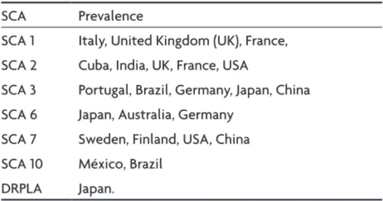
As a general rule, certain types of spinocerebellar atax-ia have a higher incidence in particular countries. Table 1 shows the geographic variation in the prevalence of the main spinocerebellar ataxias in various countries.
In Brazil, Cassa carried out an epidemiologic study in 1996 in which he investigated the prevalence of heredi-tary ataxias – Machado-Joseph disease (MJD) in particular – in the states of Minas Gerais, São Paulo, Goiás and Es-pírito Santo. He found that 426 patients in 33 families had SCA, corresponding to a mean incidence of 6.55 cases per 100,000 people (with variations from 0.78 to 228)20.
In 1997 Lopes-Cendes et al. investigated the frequency of mutations that cause SCAs (SCA1, SCA2, SCA3 and den-tatorubral-pallidoluysian atrophy - DRPLA) in a large se-ries of Brazilian patients. They found that a mutation was present in 59% of their cases and that SCA 3 was present in 44% of these, SCA 2 in 9% and SCA 1 in 6%15.
In 2001 Jardim et al. carried out a study of SCAs in the state of Rio Grande do Sul in which they investigated 66 cases of the disease. The authors concluded that the inci-dence of MJD found was very high and suggested that this might have been due to the presence of a founder effect of Azorean origin. They estimated the incidence of MJD in the region to be 1.8/100,000 and that of other forms of autosomal dominant ataxia to be 0.2/100,00017.
In a southern Brazilian series of 100 families with SCAs, a mutation was identiied in two-thirds of the cases. SCA 3 was the most frequent form of the disease (73.5%), fol-lowed by SCA 10 (11.8%), SCA 2 (7.4%), SCA 7 (4.4%), SCA 1 (2.9%) and SCA 6 (1.5%)16.
The whole degenerative neuropathological process has been studied in depth in transgenic mice and Droso-phila models of SCAs1,3,6-8.
Neuroimaging, particularly magnetic resonance imag-ing, reveals cerebellar atrophy, with or without brainstem involvement (olivopontocerebellar atrophy)1-8.
The onset of SCAs usually occurs between 30 and 50 years of age; however, cases starting before the age of 20 and after the age of 60 have been described1-3.
Current previously unimaginable advances in molecu-lar genetic techniques together with the use of PCR (poly-merase chain reaction) have resulted in various genetic loci and genes being identiied on different chromosomes, allowing a more rational clinical and genetic classiication of the disease1-3,8,21-24.
Table 2 shows the main types of SCAs currently known and gives the genetic loci, mutations and proteins associ-ated with the disease.
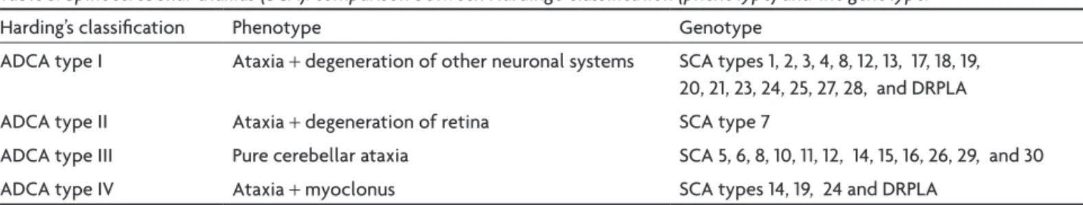
The phenotypical classiication of SCAs described by Harding in 1984 can be compared with the current geno-typical classiication8,11. Table 3 gives a summary of this
comparison.
SCAs-gENETIC ASPECTS
Thirty different types of SCA, known as SCA 1 to SCA 30, have been identiied to date. The particular gene re-sponsible for each type of the disease has been identiied for SCAs types 1-3, 5-8, 10-15, 17, and 27. The other types – SCAs 4, 18-23, 25, 26, 28, 29 and 30 – have been deined by linkage studies, as the genes and mutations associated with them have not yet been identiied1,2,19,21,22.
It should be stressed that SCAs types 9 and 24 remain undeined and that these two type numbers have been reserved for disorders yet to be described in the litera-ture. The mutation in the puratrophin-1(PLEKHG4) gene on chromosome 16q22 is associated with the pure Japanese form of SCA 4 (without sensory axonal neuropathy) and was redeined as SCA 4R. SCA 6 has been identiied as be-ing identical to SCA 15. SCA types 29 and 15 and 22 and 19, may represent different allelic forms of the same gene1,2,19.
SCA types 1, 2, 3, 6, 7 and 17 and dentatorubral pallidol-uysian atrophy (DRPLA) are caused by mutations in the cod-ing region of the disease gene characterized by the pres-ence of an expanded, unstable polymorphic CAG trinucle-otide repeat. The product of the gene is a protein known as ataxin. This protein contains polyglutamine stretches, and these disorders are now known as polyglutamine dis-eases8. The mutant protein ataxin can cause neurodegen-Table 1. Spinocerebellar ataxias (SCA): geographic variation of prevalence.
SCA Prevalence
SCA 1 Italy, United Kingdom (UK), France, SCA 2 Cuba, India, UK, France, USA
SCA 3 Portugal, Brazil, Germany, Japan, China SCA 6 Japan, Australia, Germany
SCA 7 Sweden, Finland, USA, China SCA 10 México, Brazil
DRPLA Japan.
eration by a gain of toxic function that triggers the degen-erative process, forming nuclear inclusions in cerebellar Purkinje cells, with clear involvement of the ubiquitin pro-teasome pathway as well as the system of proteins known as chaperones. In short, neurodegenerative polyglutamine diseases are characterized by expansion of a polyglutamine stretch within the mutant protein that causes the illness. Expression of the mutant protein induces progressive loss of neuronal function and subsequent neurodegenera-tion of a speciic group of neurons for each disease1,8,21-24.
A characteristic that is particular to these types of SCAs is the phenomenon of anticipation, i.e., the increas-ingly early onset and increasincreas-ingly severe nature of the clinical picture in successive generations of affected fam-ilies. The phenomenon of anticipation is related to the number of expanded CAG repeats1-3,5-8.
A second group of SCAs, which includes SCAs types 8, 10 and 12, is caused by a repeat expansion that is locat-ed outside the coding region of the genes associatlocat-ed with the disease and results in deregulation of gene expression
Table 2. Spinocerebellar ataxias (SCA): summary of genetics defects.
SCA Chromossome Gene Mutation Protein
SCA 1 6p22.3 ATAXIN1 CAG Ataxin 1
SCA 2 12q24.13 ATAXIN2 CAG Ataxin 2
SCA 3 14q32.12 ATAXIN3 CAG Ataxin 3
SCA 4 16q24-qter SCA4 (PLEKHG4) ?
-SCA 5 11q13.2 SPTBN2 D/MM Beta-III Spectrin
SCA 6 19p13.13 CACNA1A CAG CACNA1A
SCA 7 3p14.1 ATXN7 CAG Ataxin.7
SCA 8 13q21 KLHLIAS CTG Kelch-like 1
SCA 9 ? - -
-SCA 10 22q13.31 ATXN10 ATTCT Ataxin.10
SCA 11 15q14-q21.3 SCA11 -
-SCA 12 5q32 PPP2R2B CAG PPP2R2B
SCA 13 19q13.33 KCNC3- MM KCNC3
SCA 14 19q13.42 PRKCG MM PRKCG
SCA 15 3p24.2-3ptr ITPR1 PM
-SCA 16 8q23-q24.1 - -
-SCA 17 6q27 TBP CAG TBP
SCA 18 7q31-q32 - -
-SCA 19 1p21-q21 - -
-SCA 20 11 - -
-SCA 21 7p21.3-p15.1 - -
-SCA 22 1p21-q23 - -
-SCA 23 20p13-p12.2 - -
-SCA 24 1p36 - -
-SCA 25 2p21-p15 - -
-SCA 26 19p13.3 - -
-SCA 27 13q33.1 FGF14 MM FGF14
SCA 28 18p11.22-q11.2 - -
-SCA 29 3p26 - -
-SCA 30 4q34.3-q35.1 - -
-DRPLA 12p13.31 ATN1 CAG Atrophin 1
EA 1 12q13 KCNA1 PM K Channel
EA 2 19p13 CACNA-1A PM Ca Channel
EA 3 1q42
EA 4
EA 5 2q22-q23 CACNB4 PM Cav2.1
EA 6 5p SLCIA3 PM EAATI
Others EA
(duenas). SCA 8 is associated with a CTG expansion; SCA 10 with a pentanucleotide (ATTCT) repeat; and SCA 12 is caused by deregulation of the activity of protein phos-phatase 2 (PP2), an enzyme that has an important func-tion in Purkinje cells1-3.
A third group of SCAs – types 5, 11, 13, 14, 15 and 27 – are caused by different mechanisms involving changes in the amino acid composition of the following proteins: βIII spectrin, tau tubulin kinase (TTBK2), potassium channels (KCNC3), protein kinase C (PRKCG), inositol 1,4,5-triphos-phate receptor, type 1 (ITPR1) and ibroblast growth fac-tor 14 (FGF14), respectively1-3.
Table 4 gives a summary of the normal and expanded nucleotide repeat sequences found in the main types of spinocerebellar ataxias.
SCAs – ThE mOST fREquENT TyPES IN SOuThERN BRAzIL
SCA 3 (Machado-Joseph disease)
This form of autosomal dominant ataxia, which is known as Machado-Joseph disease (MJD), has been described as the most common form of SCA identiied in different mo-lecular genetic studies throughout the world1-3,5-8,14-20,25.
The disease is characterized by a CAG triplet
expan-sion that has been mapped to chromosome 14q32.12 and has from 56 to 86 repeats1-3,8,16,17,25.
Neuropathological features include neuronal loss with reactive gliosis in the following structures: the substantia nigra; the dentate nucleus of the cerebellum; the red nucle-us; the pontine nuclei and the nuclei of other motor crani-al nerves; Clarke’s column; cells in the anterior horn of the spinal cord; and the spinocerebellar tracts. The globus pal-lidus can also be affected. Involvement of the olivary nu-clei and the cerebellar and cerebral cortex is uncommon1-5,7.
Studies using neuroimaging show the presence of pontocerebellar atrophy, usually without involvement of the olives. However, a study by Murata et al. published in 1998 in which the authors used magnetic resonance im-aging also showed the presence of pontocerebellar atro-phy and atroatro-phy of the globi pallidi and frontal and tem-poral lobes2,3,26.
MJD sometimes presents with cerebellar ataxia in as-sociation with pyramidal signs; peripheral amyotrophy; nystagmus, ophthalmoparesis and bulging eyes; fascicula-tions of the face, tongue and occasionally the limbs; and dystonia and parkinsonism1-5,27-29.
In 1980 Lima and Coutinho proposed the following diagnostic criteria for MJD: autosomal dominant inher-itance, major neurological signs: cerebellar ataxia, pyra-midal signs, extrapyrapyra-midal signs and amyotrophy, minor neurological signs: progressive external ophthalmoplegia, dystonia, fasciculations and bulging eyes28.
In 1992 Paula Coutinho proposed the following criteria for diagnosing MJD in her PhD thesis: autosomal dominant transmission, onset in adult life, presence of ataxia, supra-nuclear ophthalmoparesis and pyramidal and extrapyrami-dal signs, with involvement of the peripheral nervous sys-tem, minor signs: fasciculations and bulging eyes, normal higher cortical functions, mean survival of 21 years30.
SCA 10
SCA 10 was irst described in families of Mexican or-igin. It has well-deined clinical characteristics: patients present with a “pure” cerebellar syndrome, often ac-companied by epilepsy and sometimes by peripheral neuropathy16,31,32.
Table 3. Spinocerebellar ataxias (SCA): comparison between Harding’s classiication (phenotype) and the genotype.
Harding’s classiication Phenotype Genotype
ADCA type I Ataxia + degeneration of other neuronal systems SCA types 1, 2, 3, 4, 8, 12, 13, 17, 18, 19, 20, 21, 23, 24, 25, 27, 28, and DRPLA ADCA type II Ataxia + degeneration of retina SCA type 7
ADCA type III Pure cerebellar ataxia SCA 5, 6, 8, 10, 11, 12, 14, 15, 16, 26, 29, and 30 ADCA type IV Ataxia + myoclonus SCA types 14, 19, 24 and DRPLA
ADCA: autosomal dominant cerebellar ataxia; DRPLA: dentatorubral-pallidoluysian atrophy.
Table 4. Spinocerebellar ataxias (SCA): normal and expanded nucleotide repeat sequences*.
SCA Nucleotide repeat Normal Expanded
SCA 1 CAG 6–39 40–82
SCA 2 CAG 14–31 33–64
SCA 3 CAG 12–42 54–86
SCA 6 CAG 4–18 19–30
SCA 7 CAG 4–27 37–200
SCA 8 CTA/CTG 16–91 107–127
SCA 10 ATTCT 10–21 800–4500
SCA 12 CAG 7–32 55–78
SCA 17 CAG 25–44 47–63
DRPLA CAG 6–36 49–79
The disease-causing mutation that leads to SCA 10 is a large expansion of a pentanucleotide (ATTCT) repeat lo-cated in an intron of a gene of unknown function (SCA 10) on chromosome 22q16,31-34.
The DNA test for SCA 10 has 100% sensitivity and speciicity and can be performed by PCR or Southern blot analysis. The size of the expanded alleles range from 800 to 4500 ATTCT repeats for a diagnosis of SCA 1033-35.
As a general rule, there is an inverse correlation be-tween age at onset of SCA 10 and the size of the ATTCT repeat expansion33-35.
Neuroimaging studies, particularly magnetic resonance imaging of the brain, show the presence of pan-cerebellar atrophy without any abnormalities in other regions16,31,32.
To the author’s knowledge there are no descriptions of neuropathological examinations of patients with SCA 10, nor any studies with experimental models. Hence, the mechanism of this type of SCA is completely unknown16,31,32.
In 2001 Rasmunssen et al. published a seminal study in which they described the clinical and genetic analysis of 18 patients in four Mexican families with SCA 10. The mean age at onset among the affected patients was 26.7 years (varying from 14 to 44 years), and the number of AT-TCT repeats varied from 920 to 4140. The authors did not observe any signiicant anticipation or any correlation be-tween age at onset of the disease and the number of AT-TCT repeats. In addition to cerebellar ataxia and epilepsy (found in 72.2% of the cases), clinical indings included pe-ripheral polyneuropathy in 66% of the cases (conirmed by a nerve conduction study) and cerebellar atrophy, which was predominant in MRI examinations; in some cases mild pyramidal signs, ocular dyskinesia, cognitive dysfunction and/or behavioral disorders were observed, as well as liv-er, heart and hematological dysfunction31.
Until the study by Rasmunssen et al., SCA 10 had only been found in patients of Mexican descent. In 2002 Mat-suura et al. investigated the presence of the SCA 10 mu-tation in populations other than the Mexican population (white patients in North America, French Canadians, Ital-ians, Spanish and Japanese) and failed to ind any cases36.
Fujigasaki et al. also failed to detect the mutation in pa-tients from France37.
In 2004 Teive et al. published a study of ive Brazilian families with conirmed diagnoses of SCA 10 with a differ-ent phenotype, namely, a form of pure cerebellar ataxia without epilepsy or peripheral neuropathy. Around 70% of the families were of Amerindian ancestry16.
In 2006 Raskin et al. published a report of a genetical-ly proven case of SCA 10 with cerebellar ataxia, cognitive dysfunction and epilepsy. Expansion of an ATTCT penta-nucleotide repeat was present in various members of the family, all of whom were asymptomatic, showing that the case had incomplete penetrance38.
SCA 7
This type of SCA presents with cerebellar ataxia and progressive visual deicit caused by retinal degeneration (macular dystrophy). It can also be accompanied by pyra-midal signs, ophthalmoplegia, parkinsonism and slow sac-cadic movements in particular3-6.
The neuropathological features are olivopontocere-bellar degeneration in association with loss of retinal gan-glion cells and pigmentary macular dystrophy3-6.
The locus of SCA 7 was mapped to chromosome 3 (3p14.1) by David et al. in 1996. One of the families they studied came from the Crateús region, in Ceará, Brazil39.
The pathological alleles have between 36 and 306 CAG repeats. The mutant protein, which is known as ataxin 7 and has an unknown function, is expressed in many tissues, including the central nervous system, and only causes se-lective neuron death in the brain3-6,39.
The clinical picture can emerge from early infancy to the end of the ifth decade and progresses much more quickly in cases involving early onset of the condition. An-ticipation may be present in these families3-6,39.
Although a less-common form of SCA, this type is found in various countries and is considered the most common form in Sweden and Finland3-6,39,40.
SCA 2
SCA 2 is characterized by cerebellar ataxia accompa-nied by dysarthria, tremor, hypoactive deep relexes/ar-relexia of the upper and lower limbs (deining the pres-ence of associated peripheral neuropathy), fasciculations of the face and limbs, and characteristic slow saccadic eye movements3-10.
The main clinical characteristic of SCA 2 is the pres-ence of cerebellar ataxia, cerebellar atrophy, which can be observed in neuroimaging examinations, peripheral neu-ropathy and slow saccadic eye movements. Other clinical manifestations are dystonia, chorea, parkinsonism, myo-clonia and dementia3-10.
The irst description of SCA 2 was by Wadia and Swa-mi, in India in 1971, and the disease was later the sub-ject of considerable study by Orozco in Cuba (Holguín) in 199041,42. Salem et al., using molecular analysis,
stud-ied 42 Indian families with SCA and concluded that SCA 2 was the most common form. The authors also found ev-idence of a common founding mutation43. Basu et al.,
in-vestigating a series of nine different ethnic populations in India, concluded that SCA 2 was the most common44.
inhab-itants), representing one of the highest rates of SCA in the world. SCA 2 has also been described in other countries with varying frequency45.
SCA 2 is characterized by cerebellar atrophy, with a loss of Purkinje and granular cells, olivary neurons, substantia nigra and cells in the anterior horn of the spinal cord3-10.
The SCA 2 locus has been mapped to chromosome 12 (12q24.13), and the genetic mutation responsible for the disease is a CAG trinucleotide expansion with between 34 and 59 repeats. However, late onset of SCA 2 with a 33 CAG repeat expansion, which is suficient to cause the dis-ease, has been reported3-10.
SCA 2 is characterized by cerebellar ataxia with dys-arthria, tremor, hyporelexia/deep arelexia of the upper and lower limbs (deining the presence of associated pe-ripheral neuropathy), fasciculations of the face and limbs, and characteristic slow saccadic eye movements3-10.
The main clinical characteristic of SCA 2 is its associa-tion with cerebellar ataxia (with cerebellar atrophy being observed in neuroimaging examinations), peripheral neu-ropathy and slow saccadic eye movements. Other clinical manifestations are dystonia, chorea, parkinsonism, myoclo-nia and dementia3-10. Cognitive deicits have been described
in patients with SCA 2 with a frequency of 5 to 19%46.
SCA 2 is characterized by cerebellar atrophy with a loss of Purkinje and granular cells, olivary neurons, sub-stantia nigra and cells in the anterior horn of the spinal cord3-10.
SCA 2 locus has been mapped to chromosome 12 (12q24.13), and the genetic mutation responsible for the disease is a CAG trinucleotide expansion with between 34 and 59 repeats3-10,43-45.
SCA 1
Age of onset for SCA 1 is usually after 20 years of age, and the disease manifests itself as gait imbalance, with ataxia (more pronounced in gait than in the limbs), dysar-thria, nystagmus, hyperactive deep relexes and, some-times, ophthalmoparesia. The most common abnormal eye movement in SCA 1 is a signiicant increase in the am-plitude of saccadic movements, leading to hypermetria. It can occasionally be associated with slow saccadic eye movements, bulbar paralysis, dystonia, chorea and cogni-tive dysfunction2-10.
Pathological examinations show that the systems most affected are the cerebellum (through loss of Purkinje cells and cells in the dentate nucleus), pons, middle cerebellar peduncle and olives4.
The disease was mapped to chromosome 6 (6p 22.3) – in fact, it was the irst ataxia locus ever mapped – and the genetic mutation was deined as an unstable expan-sion of a repeated CAG sequence (generally between 41 and 81 repeats)2- 10.
The number of CAG repeats is related to the age at which signs and symptoms irst appear as well as the dura-tion of the disease. In some cases a correladura-tion was found between the pattern of transmission (for example, pater-nal transmission) and a greater increase in the number of CAG repeats, as well as the phenomenon of anticipation2-10.
This form of SCA corresponds to the entity previous-ly described by Schut and Haymaker in 19514.
SCA 1 has been detected in 4 to 19% of cases in differ-ent series of patidiffer-ents with SCA. However, in some series published to date, it is the most common form of SCA found; in some regions of Italy and Japan, for example, it represents 50% of all cases2,4,7,8.
SCA 6
Clinically, this form of SCA is characterized by “pure” cerebellar ataxia, which can be accompanied by dysar-thria, nystagmus, dysphagia and even loss of propriocep-tion and dystonia. Many patients have intense episodes of vertigo before the onset of ataxia, while in others there are intermittent episodes of ataxia (corresponding to ep-isodic ataxia type 2) in parallel with the signs and symp-toms of slowly progressive cerebellar ataxia3,4,47.
Generally, SCA 6 evolves slowly and progressively, with the clinical picture irst appearing around 50 years of age3,4,47.
Neuroimaging reveals cerebellar atrophy, and patho-logical examination shows a loss of Purkinje cells in the cerebellar cortex as well as gliosis of the inferior olivary complex. Ishikawa et al. described the presence of poly-glutamine aggregates in both the nucleus and cytoplasm of Purkinje cells in patients with SCA 63,4,47.
This SCA is characterized genetically by an expansion of a CAG repeat between 21 and 31 units long in the gene responsible for the voltage-dependent calcium channel known as alpha 1A (CACNA1A4), which was mapped to
chromosome 19p13.133,4,47.
SCA 6 accounts for between 10 and 30% of all SCAs and is the second most common form of SCA in some se-ries (e.g., in Japan). However, in certain regions of Japan, such as the district of Kinski, this type of SCA is the most common form1,3,4,47.
From the clinical point of view, it is particular-ly important to stress that the SCA 6 mutation is allel-ic with episodallel-ic or sporadallel-ic SCA type 2 and hemiplegallel-ic migraine3,4,47.
SCAs – RARE TyPES
SCA 8
cogni-tive disorder. Imaging tests reveal cerebellar atrophy, with a relatively preserved brainstem3-10.
This type of SCA is unique among the various SCAs, as it has a very complex genetic inheritance pattern. It is characterized genetically by an (extremely variable) CTG expansion that has been mapped to chromosome 13 (13q21). Some patients with CTG allelic expansions never develop the disease, raising doubts about the true patho-genic role of the expansion, i.e., the true relationship be-tween the repeat expansion in the SCA 8 gene and the presence of cerebellar ataxia2-10.
SCA 8 is a rare form of SCA and has been described in a small number of cases in various countries, such as Fin-land, Japan, the USA, Italy, Spain and Scotland4.
SCA 12
This form of SCA was described in 1999 by Holmes et al. in a family of German origin known as R. The onset of symptoms usually occurs in the fourth decade of life, with tremor in the upper limbs, and as the disease pro-gresses, gait ataxia, head tremor, dysmetria, hyperactive deep relexes, abnormal eye movements and, later, de-mentia. MRI reveals atrophy of the cerebellum and ce-rebral cortex3-10.
The disease was deined to be the result of an expan-sion of a CAG trinucleotide repeat in the 5’ region of the gene complex known as PPP2R2B (mapped to 5q32), which codes for a subunit of the protein phosphatase PP2A3-10.
In 2001 Fujigasaki et al. reported an Indian family with SCA 12, and Srivastava et al. subsequently reported ive more Indian families with this form of the disease48,49.
However, Choin et al. studied a number of American fam-ilies with SCA and failed to ind a case of SCA 1250.
SCA 17
SCA 17 is a rare form of autosomal neurodegenerative disease caused by an expansion of a CAG triplet repeat in the TBP gene (located in chromosome 6q27), which codes for the TATA binding protein, a transcription initiation factor1- 4,51-53.
The disease was described for the irst time in Japan in a 14-year-old female patient whose clinical picture start-ed at the age of 6 years and consiststart-ed of gait ataxia, fol-lowed later by intellectual deterioration. Parkinsonism and hyperactive deep relexes can also appear as the dis-ease progresses4,52.
SCA 17 is a rare form of autosomal dominant neurode-generative disease caused by an expansion of a CAG trip-let repeat in the TBP gene (located in chromosome 6q27) which encodes for the TATA binding protein, a transcrip-tion initiatranscrip-tion factor3,4,51-53.
To date, a number of proven cases of SCA 17 with dif-ferent clinical presentations have been described. These
have a phenotype similar to that of Huntington’s disease (Toyoshima et al.)53, and with focal dystonia accompanied
by cerebellar ataxia and dementia (Hagenah et al.)54.
SCAs – VERy RARE TyPES
The SCA types deined as SCA 4, 5, 11, 13, 14, 15, 18, 19, 20, 21, 22, 23, 25, 26, 27, 28, 29 and 30 represent very rare forms and have been diagnosed in a small number of cases in different parts of the world, some of which were only reported in isolated families.
The autosomal dominant form of cerebellar ataxia known as dentatorubral-pallidoluysian atrophy (DRPLA) is a disease with a highly variable phenotype; it was irst described in the Japanese, among whom it was reported to have a high incidence, and has been described more recently in Afri-can AmeriAfri-cans (“Haw River” Syndrome) and Europeans3-7,55.
The disease presents with cerebellar ataxia in associa-tion with three different clinical forms: (1) myoclonus ep-ilepsy with dementia; (2) choreoathetosis with dementia (simulating Huntington’s disease); and (3) a clinical picture of psychosis, parkinsonism and pyramidal signs4.
The mutation found in DRPLA is located on chromo-some 12 (12p13.31) and consists of an unstable CAG expan-sion with 49 to 79 repeats that codes for polyglutamine4, 55.
SCAS – CLINICAL EVALuATION
SCAs are genotypically and phenotypically very het-erogeneous. The presence of phenotypic heterogeneity implies that the same genotype can determine different phenotypes, while genotypic heterogeneity implies that the same phenotype may be the result of a number of genotypes1-10.
The large number of SCAs means that their clinical di-agnosis is a dificult task for the clinical neurologist.
Lopes-Cendes et al. published an interesting article about the limitations of clinical evaluation in the correct diagnosis of Machado-Joseph disease. They concluded that although most patients with SCA have common clin-ical characteristics, the presence of at least two differ-ent signs (such as bulging eyes, dystonia or fasciculations of the face and tongue) can help make a clinical diagno-sis of MJD possible56.
Schöls et al. and Garcia Ruiz et al. investigated the presence of movement abnormalities or extrapyramidal signs in patients with hereditary ataxias and concluded that the most common disturbances were parkinsonism, dystonia and postural tremor, which were particularly prevalent in SCAs types 2 and 358,59.
In 2000 Schelhaas et al. published an interesting study of the pathogenesis of different SCAs as well as the phe-notypic and gephe-notypic similarities and differences be-tween the diseases60. They emphasized the overlap
be-tween the phenotypes of the different forms of SCA as well as the great variation that can be observed in each SCA subtype and stated that a diagnosis should be based on genotype analysis. However, they proposed a clinical algorithm to investigate the various SCAs that takes into account different key signs and symptoms, such as retinal degeneration, eye movement disturbances and the pres-ence of pyramidal signs60.
The information provided above gives a good insight into the dificulties frequently faced in routine neurolog-ical clinneurolog-ical practice when assessing patients with SCAs. Such dificulties are a result of the great genotypic het-erogeneity (30 known loci and 16 genes already identiied) and enormous phenotypic heterogeneity of the disease (as evidenced in MJD, for example).
Hence, it becomes imperative to analyze the
frequen-cy of the different forms of SCAs in Brazil as well as the phenotypic and genotypic correlation between them.
It is important to take into account the different neu-rological signs in patients with SCAs as these could be of value when assessing patients clinically using an algorithm based on clinical data. Such clinical assessment would al-low patients to be screened before molecular genetic tests are requested in order to obtain a deinitive diag-nosis. Table 5 shows a basic algorithm for assessing pa-tients with SCAs.
CONCLuSION
SCAs constitute a large group of neurodegenerative diseases with a high degree of genotypic and phenotyp-ic heterogeneity, making assessment by the clinphenotyp-ical neu-rologist extremely dificult. To date, around 30 types of SCAs are known and 16 genes have been identiied. Careful and meticulous clinical assessment of patients with spi-nocerebellar ataxias can be of signiicant help in choos-ing suitable molecular tests to ensure that the etiology is correctly deined.
REfERENCES
1. Teive HAG. Spinocerebellar degenerations in Japan: new insights from an epidemiological study. Neuroepidemiology 2009;32:184-185. 2. Duenas AM, Goold R, Giunti P. Molecular pathogenesis of
spinocere-bellar ataxias. Brain 2006;129:1357-1370.
Table 5. Algorithm for assessing patients with spinocerebellar ataxia (SCA). I – Autosomal dominant cerebellar ataxia + speciic clinical data (A to Q)
A – Episodic ataxia (EA): EA types 1,2,3,4 and 5 B – Sensory ataxia: SCA type 4
C – Visual loss: SCA type 7 D – Epilepsy: SCA type 10
E – Myoclonus + Chorea + Dementia: DRPLA F – Oculomotor signs: go to part II – SCA types 1, 2, 3 G – Mental retardation: SCA type 13
H – Axial myoclonus: SCA type 14 I – Head tremor: SCA type 16
J – Dementia + Parkinsonism: SCA types 17, and 21 K – Peripheral neuropathy: SCA types 2,3,4, 10,18,24, and 25 L – Postural tremor + Myoclonus: SCA type 19
M – Palatal tremor + Spasmodic dysphonia: SCA type 20 N – Myoclonus+ Sacadic intrusions: SCA type 24 O – Postural temor + Dyskinesias: SCA type 17 P – Palpebral ptosis: SCA type 28
Q – Pure cerebellar ataxia: SCA types 5,6,8,10,11,12,14,16,22,23,26, and 30 II – Oculomotor signs (R, S, and T)
R – Nystagmus + Hypermetric saccades (associated to pyramidal signs): SCA type 1 S – Slowed saccadic eye movements + Hyporelexia: SCA type 2
T – Nystagmus +Ophthalmoplegia + Phenotypic variation in the family (without dementia, and with other signs: Pyramidal signs+ Dystonia + Parkinsonism + Peripheral neuropathy + Amyotrophy + “Bulging eyes” + Facial fasciculations): SCA type 3 (with differents subphenotypes)
3. Schöls L, Bauer P, Schmidt T, Schulte T, Riess O. Autosomal dominant cerebellar ataxias: clinical features, genetics, and pathogenesis. Lancet Neurol 2004;3:291-304.
4. Soong BW, Paulson HL. Spinocerebellar ataxias: an update. Curr Opin Neurol 2007;20:438-446.
5. Evidente VGH, Gwinn-Hardy KA, Caviness JN, Gilman S. Hereditary ataxias. Mayo Clin Proc 2000;75:475-490.
6. Durr A, Brice A. Clinical and genetic aspects of spinocerebellar degen-eration. Curr Opin Neurol 2000;13:407-413.
7. Klockgether T, Lüdtke R, Kramer B, et al. The natural history of de-generative ataxia: a retrospective study of 466 patients. Brain 1998;121: 589-600.
8. Pulst SM. Inherited ataxias: an introduction. In: Pulst SM (Ed). Genet-ics of movement disorders. Orlando: Academic Press 2003:19-34. 9. Everett CM, Wood NW. Trinucleotide repeats and neurodegenerative
disease. Brain 2004;127:2385-2405.
10. Albin RL. Dominant ataxias and Friedreich ataxia: an update. Curr Opin Neurol 2003;16:507-514.
11. Harding AE. The hereditary ataxias and related disorders. Edimburgo: Churchill Livingstone, 1984.
12. van de Warremburg BP, Sinke RJ, Verschuuren-Bemelmans CC. Spinoc-erebelllar ataxias in the Netherlands: prevalence and age at onset vari-ance analysis. Neurology 2002;58:702-708.
13. Erichsen AK, Koht J, Stray-Pedersen A, Abdelnoor M, Tallaksen ME. Prevalence of hereditary ataxia and spastic paraplegia in southeast Nor-way: a population-based study. Brain 2009;132:1577-1588.
14. Silveira I, Lopes-Cendes I, Kish S, et al. Frequency of spinocerebellar ataxia type 1, dentatorubropallidoluysian atrophy, and Machado-Jo-seph disease mutations in a large group of spinocerebellar ataxia pa-tients. Neurology 1996;46:214-218.
15. Lopes-Cendes I, Teive HAG, Calcagnotto ME, et al. Frequency of the different mutations causing spinocerebellar ataxia (SCA 1, SCA 2, SCA 3/MJD and DRPLA) in a large group of Brazilian patients. Arq Neu-ropsiquiatr 1997;55:519-529.
16. Teive HAG, Roa B, Raskin S, et al. Clinical phenotype of Brazilian pa-tients with spinocerebellar ataxia 10. Neurology 2004;63:1509-1512. 17. Jardim LB, Silveira I, Pereira ML, et al. A survey of spinocerebellar
ataxia in South Brazil: 66 new cases with Machado-Joseph disease, SCA
7, SCA 8, or unidentiied disease-causing mutations. J Neurol 2001;
248:870-876.
18. Subramony SH, Filla A. Autosomal dominant spinocerebellar ataxias
ad ininitum ? Neurology 2001;56:287-289.
19. Storey E, Bahlo M, Fahey M, Sisson O, Lueck CJ, Gardner RJM. A new dominantly inherited pure cerebellarea ataxia, SCA 30. J Neurol Neu-rosurg Psychiatry 2009;80:408-411.
20. Cassa E. Ataxia cerebelar autossômica dominante no Brasil: análise de 270 anos de história e genealogia, incluindo a caracterização molecular de uma grande família com doença de Machado-Joseph. Tese de Dou-torado, USP-RP, Ribeirão Preto, 1996.
21. Margolis RL. The spinocerebellar ataxias: order emerges from chaos. Curr Neurol Neurosci Rep 2002;2:447-456.
22. Bandmann O, Singleton AB. Yet another spinocerebellar ataxia: the saga continues. Neurology 2008;71:542-543.
23. Gatchel JR, Zoghbi HY. Diseases of unstable repeat expansion: mecha-nisms and common principles. Nat Rev Genet 2005;6:743-755. 24. Pearson CE, Edamura KN, Cleary JD. Repeat instability: mechanisms
of dynamic mutations. Nat Rev Genet 2005;6:729-742.
25. Jardim LB, Pereira ML, Silveira I, Ferro A, Sequeiros J, Giugliani R.
Neu-rologic indings in Machado-Joseph disease: relation with disease du -ration, sybtypes, and (CAG)n. Arch Neurol 2001;58:899-904. 26. Murata Y, Yamaguchi S, Kawakami H. Characteristic magnetic
reso-nance imaging indings in Machado-Joseph disease. Arch Neurol 1998;
55:33-37.
27. Coutinho P, Andrade C. Autosomal dominant system degeneration in Portuguese families of the Azorean islands: a new genetic disorder in-volving cerebellar, pyramidal, extrapyramidal and spinal cord motor functions. Neurology 1978;28:703-709.
28. Lima L, Coutinho P. Clinical criteria for diagnosis of Machado-Joseph
disease: report of a non-azorean Portuguese family. Neurology 1980;30: 319-322.
29. Sequeiros J, Coutinho P. Epidemiology and clinical aspects of Macha-do-Joseph disease. Adv Neurol 1993;61:139-153.
30. Coutinho P. Doença de Machado-Joseph: tentativa de deinição. Tese
de Doutorado, Instituto de Ciências Biomédicas, Universidade do Por-to, PorPor-to, Portugal, 1992.
31. Rasmunssen A, Matsuura T, Ruano L, et al. Clinical and genetic anal-ysis of four Mexican families with spinocerebellar ataxia type 10. Ann Neurol 2001;50:234-239.
32. Zu L, Figueroa KP, Grewal R, Pulst S-M. Mapping of a new autosomal dominant spinocerebellar ataxia to chromosome 22. Am J Hum Genet 1999;64:594-599.
33. Matsuura T, Achari M, Khakavi M, Bachinski LL, Huda ZY, Ashizawa T. Mapping of the gene for a novel spinocerebellar ataxia with pure cer-ebellar signs and epilepsy. Ann Neurol 1999;45:407-411.
34. Matsuura T, Yamagata T, Burgess DL, et al. Large expansions of the AT-TCT pentanucleotide repeat in spinocerebellar ataxia type 10. Nat Genet 2000;26:191-194.
35. Matsuura T, Ashizawa T. Polymerase chain reaction ampliication of
expanded ATTCT repeat in spinocerebellar ataxia type 10. Ann Neurol 2002;51:271-272.
36. Matsuura T, Ranum LPW, Volpini V, et al. Spinocerebellar ataxia type 10 is rare in populations other than Mexicans. Neurology 2002;58:983-984. 37. Fujigasaki H, Tardieu S, Camuzat A, et al. Spinocerebellar ataxia type
10 in the French population. Ann Neurol 2002;51:408-409.
38. Raskin S, Ashizawa T, Teive HAG, et al. Reduced penentrance associ-ated with early-onset in a Brazilian family with spinocerebellar atax-ia type 10: implications for pathogenesis, molecular genetic datax-iagnosis and genetic couseling. Arch Neurol 2007;64:591-594.
39. David G, Giunti P, Abbas N, et al. The gene forautosomal dominant cere-bellara ataxia type II is located in a 5-cM region in 3p12-p13: genetic and physical mapping of the SCA7 locus. Ann J Hum Genet 1996; 59:1328-1336. 40. Cunha Linhares S, Horta WG, Marques Jr. W. Spinocerebellar ataxia
type 7 (SCA7): family princeps history, genealogy and geographical distribution. Arq Neuropsiquiatr 2006;64:222-227.
41. Wadia NH, Swami RK. A new form of heredo-familial spinocerebel-lar degeneration with slow eye movements (nine families). Brain 1971; 94:359-374.
42. Orozco G, Nodarse Fleites A, Cordovés Sagaz R, Auburger G. Autosom-al dominant cerebellar ataxia: clinicAutosom-al anAutosom-alysis of 263 patients from a ho-mogeneous population in Holguin, Cuba. Neurology 1990;40:1369-1375. 43. Saleem Q, Choudhry S, Mukerji M, et al. Molecular analysis of auto-somal dominant hereditary ataxias in the Indian population: high fre-quency of SCA 2 and evidence for a common founder mutation. Hum Genet 2000;106:179-187.
44. Basu P, Chattopadhyay B, Gangopadhaya PK, et al. Analysis of CAG re-peats in SCA 1, SCA 2, SCA 3, SCA 6, SCA 7, and DRPLA loci in spinoc-erebellar ataxia patients and distribution of CAG repeats at the SCA 1, SCA 2 and SCA 6 loci in nine ethnic populations of eastern India. Hum Genet 2000;106:597-604.
45. Velázquez-Pérez, L, Garcia R, Santos FN, Paneque HM, Medina HE, Hechavarria PR. Hereditary ataxias in Cuba. Historical, epidemiologi-cal, cliniepidemiologi-cal, electrophysiological and quantitative neurological features. Rev Neurol 2001;32:71-76.
46. Bürk K, Blobas C, Bösch S, et al. Cognitive deicits in spinocerebellar
ataxia 2. Brain 1999;122:769-777.
47. Teive HAG, Munhoz RP, Raskin S, Werneck LC. Spinocerebellar atax-ia type 6 in Brazil. Arq Neuropsiquatax-iatr 2008;66:691-694.
48. Fujigasaki H, Verma IC, Camuzat A, et al. SCA 12 is a rare locus for au-tosomal dominant cerebellara ataxia: a study of an Indian family. Ann Neurol 2001;49:117-121.
49. Srivastava AK, Choudhry S, Gopinath MS, et al. Molecular and clinical
correlation in ive Indian families with spinocerebellar ataxia 12. Ann
Neurol 2001;50:796-800.
51. De Michele G, Malteca F, Carella M, et al. Dementia, ataxia, extrapyra-midal features, and epilepsy: phenotypespectrum in two Italian fami-lies with spinocerebellar ataxia type 17. Neurol Sci 2003;24:166-167. 52. Rolfs A, Koeppen AH, Bauer I, et al.Clinical features and
neuropatholo-gy of autosomal dominant spinocerebellar ataxia (SCA17). Ann Neurol 2003;54:367-375.
53. Toyoshima Y, Yamada M, Onodera O, et al. SCA 17 homozygote show-ing Huntshow-ington’s disease-like phenotype. Ann Neurol 2004;55:281-286. 54. Hagenah JM, Zuhlke C, Hellenbroich Y,et al. Focal dystonia as a pre-senting sign of spinocerebellar type 17. Mov Disord 2004;19:217-220. 55. Koide R, Ikeuchi T, Onodera O, et al.Unstable expansion of CAG repeat
in hereditary dentarubralpallidoluysian atrophy (DRPLA).Nat Genet 1994;6:9-13.
56. Lopes-Cendes I, Silveira I, Maciel P, et al. Limits of clinical assessment in the acurate diagnosis of Machado-Joseph disease. Arch Neurol 1996;53:1168-1174.
57. Perlman SL. Diagnostic evaluation of ataxic patients. In: Pulst SM (Ed). Genetics of movement disorders. Edimburgo: Academic Press, 2003: 254-272.
58. Schols L, Peters S, Szymanski S, et al. Extrapiramidal motor signs in de-generative ataxias. Arch Neurol 2000;57:1495-1500.
59. Garcia Ruiz PJ, Mayo D, Hernandez J et al. Movement disorders in he-reditary ataxias. J Neurol Sci 2002;202:59-64.