Lung tissue mechanics in the
early stages of induced
paracoccidioidomycosis in rats
Departamentos de 1Doenças Infecciosas e Parasitárias and 2Patologia, Faculdade de Medicina, Universidade de São Paulo,
São Paulo, SP, Brasil M.A. Shikanai-Yasuda1,
P.M. Pereira2,
E. Yamashiro-Kanashiro1,
M.I.S. Duarte2, C.M. Assis1,
E.A. Geraldes1 and
P.H.N. Saldiva2
Abstract
Pulmonary dysfunction represents the most important cause of death in patients with paracoccidioidomycosis (PBM). In order to investi-gate the functional changes of the lungs in the early stages of PBM, a model of benign disease was developed by intratracheal challenge of 12-week old isogenic Wistar rats with 1 x 10⁶ yeast forms of
Paracoc-cidioides brasiliensis. Animals were studied 30 and 60 days after infection, when fully developed granulomas were demonstrable in the lungs. Measurements of airway resistance, lung elastance and tissue hysteresis were made during sinusoidal deformations (100 breaths/ min, tidal volume = 2 ml) with direct measurement of alveolar pressure using the alveolar capsule technique. Infection caused a significant increase in hysteresis (infected: 1.69, N = 13; control: 1.13, N = 12, P = 0.024, ANOVA), with no alterations in airway resistance or lung elastance. Histopathological analysis revealed the presence of fully developed granulomas located in the axial compartment of the lung interstitial space. These results suggest that alterations of tissue mechanics represent an early event in experimental PBM.
Correspondence P.H.N. Saldiva
Departamento de Patologia Faculdade de Medicina, USP Av. Dr. Arnaldo, 455 01246-903 São Paulo, SP Brasil
Fax: 55 (011) 3064-2744 E-mail: [email protected]
Research supported by CNPq, FAPESP and LIM-HCFMUSP.
Received September 3, 1996 Accepted July 29, 1997
Key words
•Experimental
paracoccidioidomycosis •Pulmonary mechanics •Tissue mechanics •Alveolar pressure
measurements •Hysteresis
Introduction
Paracoccidioidomycosis is the most prev-alent endemic mycosis in Latin America. Paracoccidioides brasiliensis is transmitted by the respiratory route through inhalation of conidia. Involvement of the phagocytic mono-nuclear system is the most common clinical finding in the acute form, mainly described in children or young adult patients. In con-trast, the chronic form is prevalent in
fibrosis (4). Besides this severe clinical evo-lution, another important cause of death is represented by cor pulmonale secondary to both obstructive and restrictive pulmonary insufficiency, which affects about 30% of chronic cases (5). Lung function has been studied only in the late stages of human paracoccidioidomycosis. The present study focuses on functional lung changes observed in the early phases of intratracheal paracoc-cidioidomycosis induced in isogenic rats.
Material and Methods
Fungi
Yeast forms of Paracoccidioides brasili-ensis were obtained from 7-day old Fava-Netto culture medium (6). Cell viability was at least 80%, as assessed by Janus green staining (7). Total cell counts were performed with a hemocytometer chamber to estimate the inoculum size, which was 1 x 10⁶ yeast
forms of fungi/0.5 ml phosphate buffered saline.
Rats
A model of benign disease was devel-oped by intratracheal challenge of twenty-five 12-week old isogenic Wistar-Furth rats. Animals were studied 30 and 60 days after infection. Corresponding groups injected with saline solution were used as controls.
Histopathological study
Histopathological studies were performed in three regions of the lungs: the perihilar area near the main bronchi, parenchyma in-cluding segmentary bronchi, and peripheral area usually with macroscopic alterations. Lymph nodes, liver, adrenals, kidney and brain were also studied. This analysis was done using paraffin-embedded slides stained with hematoxylin-eosin and by the Grocott-Gomori silver technique.
Physiological measurements
Rats were anesthetized with intraperito-neal sodium pentobarbital (35 mg/kg) and a polyethylene cannula was inserted into the trachea by direct visualization. The rats were ventilated with a rodent ventilator (Harvard 683, Harvard Apparatus Co., South Natik, MA) at a constant tidal volume (2.5 ml) with the respiratory frequency set at 100 breaths/ min. Airflow (V. ) was measured with a pneu-motachograph connected to the tracheal can-nula through a Valydine DP 45-16-2114 dif-ferential pressure transducer. Volume (V) was obtained by electronic integration of the V. signal. Tracheal pressure (Ptr) was meas-ured with a Valydine DP 45-28-2114 differ-ential pressure transducer. Positive end-ex-piratory pressure of 5 cmH2O was used to
maintain a functional residual capacity close to a normal value. Alveolar pressure (Palv) was measured according to the alveolar cap-sule technique described by Saldiva et al. (8). The capsules were made from 3-ml plas-tic syringes with the distal end cut off and connected to a Valydine DP 45-28-2114 dif-ferential pressure transducer through a 15-cm long polyethylene catheter (1.6 mm ID). The pleural surface was punctured with an 18-gauge needle, and the capsule was glued to this surface with cyanoacrylate. The depth of the holes on the pleural surface was less than 0.5 mm to avoid sampling of bronchial pressures. A single capsule was used for each rat but placed at different sites in each animal.
Flow, Ptr and Palv signals were sampled at 200 Hz with a 12-bit analog-to-digital converter (DT-2801A, Data Translation, Marlboro, MA) and stored in a microcom-puter. In each measurement, one data point was obtained by the average of 14-16 breaths.
elastance (lung elastic modulus), tissue re-sistance and area of hysteresis of the pres-sure volume loop during tidal ventilation were obtained by the method of Fredberg and Stamenovic (9). Seven to eight breaths were averaged to complete the mechanical parameter for each animal.
Statistical analysis
The significance of the results was as-sessed by analysis of variance considering two factors, i.e., infection and time of infection. Variance of the parameters of interest was stabilized by applying logarithmic transforma-tion of elastance, resistance and hysteresis.
Results and Discussion
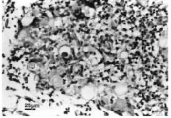
The infection was limited to the lungs. Epithelioid granulomas rich in fungi (Figure 1, left) were observed in the lungs 15 to 150 days after the infection. Giant cells, macro-phages and some polymorphonuclear cells, eosinophils and lymphocytes (Figure 1, right) were also found in the granulomas. Occa-sional necrosis was observed in a small num-ber of granulomas during the period of fol-low-up.
Tables 1, 2 and 3 show the mean and corresponding standard deviation (SD) of functional variables for hysteresis, elastance and resistance, respectively, measured for each period of time, as well as the corre-sponding output of ANOVA analysis for each variable.
Figure 1 - Compact granulomas in the lungs of Wistar rats 60 days after inoculation with P. brasiliensis (hematoxylin and eosin, left = 200X, right = 400X).
Table 1 - Effect of paracoccidioidomycosis infec-tion on pulmonary hysteresis.
P = 0.024, significance of paracoccidioidomycosis infection (ANOVA). P = 0.491 and P = 0.174, time of infection and interaction infection vs time (not significant, ANOVA).
Hysteresis (cmH2O/ml)
30 days 60 days Total
Control
Mean 0.76 1.39 1.13
Median 0.75 1.16 0.88
Standard deviation 0.26 0.99 0.81
Minimum 0.50 0.51 0.50
Maximum 1.09 3.34 3.34
No. of cases 5 7 12
Infected
Mean 1.79 1.61 1.69
Median 1.50 1.66 1.54
Standard deviation 0.79 0.88 0.80
Minimum 0.87 0.68 0.68
Maximum 2.82 3.23 3.23
No. of cases 6 7 13
Total
Mean 1.32 1.50 1.42
Median 1.09 1.40 1.16
Standard deviation 0.78 0.90 0.84
Minimum 0.50 0.51 0.50
Maximum 2.82 3.34 3.34
No. of cases 11 14 25
Increased hysteresis (P = 0.024) was ob-served in infected animals at 30 and 60 days after inoculation compared to control ani-mals. No difference in elastance or resis-tance was observed between groups during either period of infection.
node involvement by confluent granulomas (11). These changes might represent lesions which could later evolve to fibrosis. An ex-perimental model more similar to human pulmonary fibrosis seems to be represented by mice intranasally inoculated with conidia, as described by Restrepo et al. (12). How-ever, an intriguing difference between ex-perimental and human pulmonary disease is the long period of time between the initial infection and the development of chronic disease in human paracoccidioidomycosis.
We suggest that benign mycosis in isogenic Wistar-Furth rats provides a useful model to study changes in lung function during the early phases of paracoccidioido-mycosis, mainly due to the presence of pre-dominantly cellular granulomas in response the alveolar or interstitial level, as a
granulo-matous or an exudative pattern, which usu-ally does not lead to abnormalities in spiro-graphic tests. As a consequence of lymphatic spreading of the lesions along the peribron-chial tissue, centrifugal histological changes are commonly observed. These findings, sometimes with simultaneous fibrosis and alveolar destruction, may be responsible for the functional disturbances observed in pa-tients with chronic pulmonary paracoccidi-oidomycosis (10). Obstruction and restric-tion have been the most frequent spirographic patterns described in these patients.
Similarly to the human disease, hamsters infected by the intratracheal route presented with hilar bronchial involvement with cen-trifugal dissemination after regional lymph
Table 2 - Paracoccidioidomycosis (PBM) infection has no effect on pulmonary elastance.
Neither PBM infection (P = 0.283), time of infec-tion (P = 0.496) nor interacinfec-tion infecinfec-tion vs time (P = 0.561) were statistically significant (ANOVA).
Elastance (cmH2O/ml)
30 days 60 days Total
Control
Mean 2.37 2.91 2.68
Median 2.19 3.35 2.72
Standard deviation 0.81 0.92 0.88
Minimum 1.67 1.13 1.13
Maximum 3.73 3.79 3.79
No. of cases 5 7 12
Infected
Mean 2.99 3.09 3.04
Median 2.83 3.18 3.05
Standard deviation 0.82 1.02 0.90
Minimum 2.10 1.80 1.80
Maximum 4.00 4.38 4.38
No. of cases 6 7 13
Total
Mean 2.71 3.00 2.87
Median 2.38 3.27 3.05
Standard deviation 0.84 0.94 0.89
Minimum 1.67 1.13 1.13
Maximum 4.00 4.38 4.38
No. of cases 11 14 25
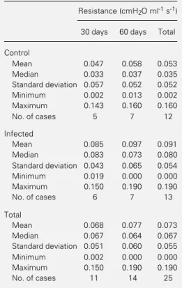
Table 3 - Paracoccidioidomycosis (PBM) infection has no effect on pulmonary resistance.
Neither PBM infection (P = 0.098), time of infec-tion (P = 0.609) nor interacinfec-tion infecinfec-tion vs time (P = 0.993) were statistically significant (ANOVA).
Resistance (cmH2O ml-1 s-1)
30 days 60 days Total
Control
Mean 0.047 0.058 0.053
Median 0.033 0.037 0.035
Standard deviation 0.057 0.052 0.052
Minimum 0.002 0.013 0.002
Maximum 0.143 0.160 0.160
No. of cases 5 7 12
Infected
Mean 0.085 0.097 0.091
Median 0.083 0.073 0.080
Standard deviation 0.043 0.065 0.054
Minimum 0.019 0.000 0.000
Maximum 0.150 0.190 0.190
No. of cases 6 7 13
Total
Mean 0.068 0.077 0.073
Median 0.067 0.064 0.067
Standard deviation 0.051 0.060 0.055
Minimum 0.002 0.000 0.000
Maximum 0.150 0.190 0.190
to the infectious agent and the absence of significant scarring. As a consequence of the granulomatous infection, a significant in-crease in hysteresis (P = 0.024) was ob-served at 30 days, which remained elevated up to the end of the experiment (60 days). The present study represents the first de-scription of increased tissue resistance inde-pendent of bronchoconstriction. Histopath-ological examination performed in three dif-ferent areas of the lung at day 30 and 60 after the infection showed fully developed
granu-lomas only in infected rats. These results indicate that changes in tissue mechanics due to granuloma formation along the axial compartment of lung connective tissue are an early event in experimental paracoccidi-oidomycosis. In addition, it is probable that the granuloma may act as a viscous or plastic element in the pulmonary interstitium caus-ing an increase of tissue pressure losses at the tissue level, which probably precedes the restrictive and/or obstructive functional pat-terns observed in chronic human disease.
References
1. Machado Filho J, Miranda JL & Teixeira GA (1965). Das sequelas da blastomicose sul americana. O Hospital,68:1346-1354. 2. Pereira JL & Silva AP (1982). The correc-tion of microstomia following South American blastomycosis: A case report. British Journal of Plastic Surgery, 35: 204-205.
3. Andrade DR, Hutzler RU, Carvalho SA, Rosenthal C, Carvalho MAB & Ferreira JM (1976). Hipoproteinemia em pacientes com paracoccidioidomicose do tubo digestivo e sistema linfático abdominal. Revista do Hospital das Clínicas da Faculdade de Medicina de São Paulo, 31: 174-179.
4. Shikanai-Yasuda MA, Segurado AAC, Pereira-Pinto W, Nicodemo AC, Sato M, Duarte AJS, Del Negro GB, Hutzler RU, Shiroma M & Amato-Neto V (1992). Im-munodeficiency secondary to juvenile paracoccidioidomycosis: associated infec-tions. Mycopathologia, 120:23-28.
5. Ratto OR & Afonso JE (1994). Pulmonary lesions: clinical and functional aspects. In: Franco MF, Lacaz CS, Restrepo-Moreno A & Del Negro G (Editors), Paracoccidioi-domycosis. CRC Press, Boca Raton, 271-278.
6. Fava Netto C, Vegas VS, Sciannaméa IM & Guarnieri DB (1969). Antígeno polissacarídeo do Paracoccidioides brasiliensis, necessário ao preparo do antígeno. Revista do Instituto de Medi-cina Tropical de São Paulo, 11: 177-181. 7. Berliner MD & Reca ME (1966). Vital
stain-ing of H. capsulatum with Janus green B. Sabouraudia, 5: 26-29.
8. Saldiva PHN, Zin WA, Santos RLB, Eidelman DH & Milic-Emili J (1992). Al-veolar pressure measurement in open-chest rats. Journal of Applied Physiology, 72:302-306.
9. Fredberg DF & Stamenovic D (1989). On the imperfect elasticity of lung tissue. Journal of Applied Physiology, 67: 2408-2419.
10. Tuder RM, El Ibrahim R, Godoy CE & De Britto T (1985). Pathology of human pul-monary paracoccidioidomycosis. Mycopa-thologia, 92: 179-188.
11. Tani EM, Franco MF, Peraçoli MTS & Montenegro MR (1987). Experimental pulmonary paracoccidioidomycosis in the Syrian hamster: morphology and correla-tion of lesion with immune response. Journal of Medical and Veterinary Mycol-ogy, 25:291-300.