Série Zoologia
Fundação Zoobotânica do Rio Grande do Sul Museu de Ciências Naturais
www.scielo.br/isz e-ISSN 1678-4766
Iheringia
Iheringia
Myxobolus saladensis sp. nov., a new species of gill parasite
of Mugil liza (Osteichthyes, Mugilidae) from Samborombón Bay,
Buenos Aires, Argentina
Paula Marcotegui & Sergio Martorelli
Centro de Estudios Parasitológicos y Vectores (CCT-La Plata), Boulevard 120 s/n entre av. 60 y calle 64 (1900) La Plata, Buenos Aires, Argentina.
Received 25 May 2016 Accepted 10 August 2016
DOI: 10.1590/1678-4766e2017026
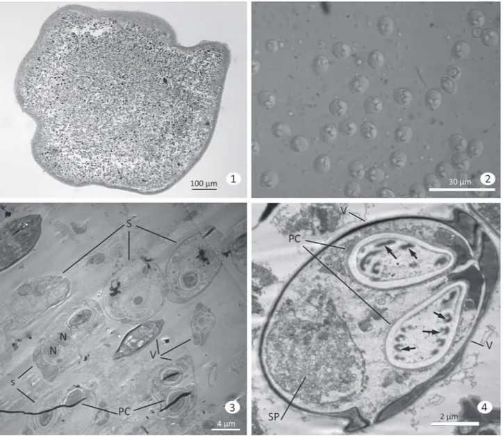
ABSTRACT. Myxosporean Myxobolus saladensis sp. nov. in the gills of Mugil liza Valenciennes, 1836 from Samborombón Bay was described by light and electron microscopy studies. Spores were pyriform and binucleated, measuring 10.63±0.36 µm (n=20) long, 9.24±0.50 µm (n=20) wide and 4.13±0.36 µm (n=20) thick, included in polysporic cyst-like plasmodia. Elongated pyriform polar capsules were of equal size (3.84±0.27 µm long and 2.30±0.12 µm wide). The sporoplasm contained some sporoplasmosomes. Each PC contained a polar fi lament with 4-5 coils obliquely arranged in relation to the polar capsules axis. The PC wall was composed of two layers of diff erent electron densities. Based on the morphological and ultrastructure diff erences of the spore to those of previously described species of Myxobolus, we describe a new species, Myxobolussaladensis sp. nov.
KEYWORDS. Myxosporean, mullets, Myxobolidae, gills parasites.
Members of Myxobolus Biitschli, 1882 are considered
cosmopolitan, with many species cited in mullets. According
to
Zatti
et al. (2015) approximately 90 myxosporean species
have been identifi ed in South America, of which 41 are
from the this genus, mostly from Brazil (
Eiras
et al., 2010;
Azevedo
et al., 2010; 2011; 2012). Mullets live in shallow
and brackish waters and are euryhaline, ranging from
hypersaline lagoons to freshwater. They utilize estuarine
nursery habitats where they feed largely on plant material
obtained by grubbing through bottom detritus (
Cervigón
et al., 1993).
The Mugilidae is represented by three species along
the coast of the Southwest Atlantic Ocean (
Cousseau
et al.,
2005). In Argentine waters, the only mullet permanently
present is the grey mullet Mugil liza Valenciennes, 1836
(see
Cousseau
et al., 2005) after synonymization of Mugil
platanus Günther, 1880 with M. liza (
Heras
et al., 2009). This
species is commercially exploited in Brazil and Argentina
by inshore artisanal fi sheries.
Helminths parasitizing M. liza have been reported
by many authors in the last years (
Knoff
&
Amato
, 1992;
Kohn
et al., 1994;
Knoff
et al., 1997;
Suriano
et al.,
2000;
Carnevia
&
Speranza
, 2003;
Abdallah
et al.,
2009;
Marcotegui
&
Marcotelli
, 2009a,b;
Siquier
&
Ostrowski
de
Nuñez
, 2009;
Alarcos
&
Etchegoin
, 2010;
Marcotelli
et al., 2012). However, gill Myxozoa have not
been reported from this host.
Eiras
et al. (2007) recorded
Myxobolus platanus Eiras et al., 2007 parasitizing pancreatic
tissue of Mugil platanus in Lagoa dos Patos, Brazil.
In a survey of fi sh parasites in estuarine areas of
Argentina a new myxosporidian species was found in Mugil
liza collected from Samborombón Bay. A light and electron
microscopic study on the new species is presented.
MATERIALS AND METHODS
A total of 206 specimens of Mugil liza (Valenciennes,
1836) (Mugilidae) were captured between April 2006 and
April 2009. These specimens ranging in total length from
2.8 to 32.0 cm and total weight from 0.22-331.41 g were
captured from the mouth of the Canal Aliviador of Salado
River (35°50’12.53”S, 57°25’22.28”W, Samborombón Bay,
Buenos Aires, Argentina) and examined for parasites. Live
fi sh were transported to the laboratory in containers fi lled with
estuarine water and were kept alive in oxygenated aquaria
until examination. Skin, fi ns, excised gills, and intestinal tract
from newly killed fi shes were analyzed under a dissecting
microscope to detect parasites.
Plasmodium (Pmd) containing numerous spores were
found in the gill fi laments and gill rakers (Fig. 1). The gills
infected were observed by DIC microscopy for fresh spore
measurements. For ultrastructural studies, fragments of gills
infected were excised and fi xed in 2.5% Glutaraldehyde in
0.2 M Sodium Cacodylate buff er (pH 7.2) for 12 h at 4°C,
washed in the same buff er 12 h at 4°C and post fi xed in 2%
The dimensions of the spores (in µm) were expressed as
the mean ±standard deviation. Length and width of spores were
obtained from 20 fresh specimens. Thickness were obtained from
20 additional spores. Measurements of polar capsules length and
width of the spores were obtained from ultrathin serial sections.
All measurements were taken from microphotographs using
Image J software (National Institutes of Health).
RESULTS
Myxobolus saladensis sp. nov.
(Figs 1-7)
urn:lsid:zoobank.org:act:51F1772D-C806-4B52-9A59-CB70F10D47A5
Diagnosis. Spherical Pmd containing numerous
spores located in the gill filaments and rakers. Pyriform
fresh spores tapering anteriorly 10.63±0.36 (range
10.05-11.13) µm long, 9.24±0.50 (range 8.42-9.79) µm wide,
and 4.13±0.36 (range 2,65-4,9) µm thick. Two equal-sized
pyriform PC measuring 3.84±0.27 (range 3.33-4.03) µm
long and 2.30±0.12 (2.14-2.43) µm wide. Polar filament
coiled in four or five turns.
Site of infection: spores located in epithelium of
gill rakers.
Etymology.
The specific epithet refers to the type
locality, Salado River.
Type material. One glass slide with semithin sections
of the cyst containing spores (Hapantotype) was deposited
in the Museo de La Plata collection, number MLP-Pr-095.
Figs 1-4. Light and transmission electron micrographs of the myxosporean Myxobolus saladensis sp. nov. infecting gills and gills rakers of Mugil liza
Type Locality. Salado River, Samborombón Bay,
Buenos Aires, Argentina.
Type Host: Mugil liza.
Prevalence: 12.7%.
Description.
Spores typical of Myxobolus Bütschli,
1882, rounded in valvular view and biconvex in sutural
view, shell valves smooth and without projections. Fresh
mature spores pyriform in shape (Fig. 2). Mean spore
measurements ± standard deviations as follows: 10.63±0.36
(range 10.05-11.13) long, 9.24±0.50 (range 8.42-9.79) wide,
and 4.13±0.36 (range 2.65-4.91) thick. Spore wall thin and
smooth comprising two symmetrical and equal shell valves
adhering together along the prominent longitudinal sutural
line (Figs 3-6). Internally, two equal and elongated pyriform
polar capsules (PCs), located side by side at the same level,
measured 3.84±0.27 (range 3.33-4.03) long, 2.30±0.12
(range 2.14-2.43) wide (Figs 3-6). Intercapsular appendix
not observed. Inside the PCs, polar filament coil displayed
four or five slightly oblique to the longitudinal axis (Fig. 6).
Apical end of the PCs contained a circular stopper formed by
electron-lucent material (Fig. 6). At the posterior pole of the
spore, a binucleated sporoplasm contained numerous light
vesicles, numerous sporoplasmosomes (Figs 4, 5).
Nuclei located at the same level, contained uniform
chromatin without evident nucleoli (Fig. 3). A schematic
drawing of spore morphology (Fig. 7) shows the arrangements
of the different structures and organelles.
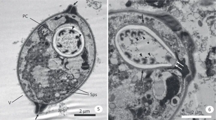
Figs 5, 6. Myxobolussaladensis sp. nov. parasite of Mugil liza Valenciennes, 1836: 5, sectioned spore showing valves (V) and their suture lines (arrows), polar capsules (PC), different sections of the polar filaments (arrowheads) and some sporoplasmosomes (Sps); 6, detail of apical region of the PCs showing the PC wall (arrows) composed of two layers and the apical stopper (double arrows) and the different sections of the polar filament (arrowheads).
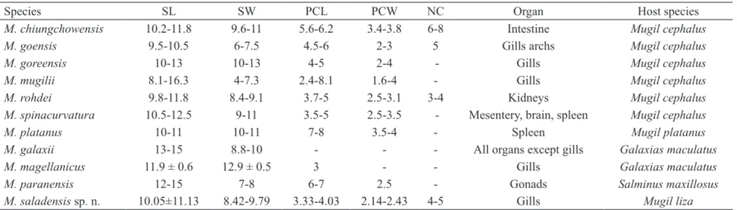
Tab. I.Comparative measurement (in µm) of the spores from Myxobolus spp. parasiting mugilid fishes which resembles the new species (SL, spore
length; SW, spore width; PCL, polar capsules length; PCW, polar capsules width; NC, number of coils on the polar filament).
Species SL SW PCL PCW NC Organ Host species
M. chiungchowensis 10.2-11.8 9.6-11 5.6-6.2 3.4-3.8 6-8 Intestine Mugil cephalus
M. goensis 9.5-10.5 6-7.5 4.5-6 2-3 5 Gills archs Mugil cephalus
M. goreensis 10-13 10-13 4-5 2-4 - Gills Mugil cephalus
M. mugilii 8.1-16.3 4-7.3 2.4-8.1 1.6-4 - Gills Mugil cephalus
M. rohdei 9.8-11.8 8.4-9.1 3.7-5 2.5-3.1 3-4 Kidneys Mugil cephalus
M. spinacurvatura 10.5-12.5 9-11 3.5-5 2.5-3.5 - Mesentery, brain, spleen Mugil cephalus
M. platanus 10-11 10-11 7-8 3.5-4 - Spleen Mugil platanus
M. galaxii 13-15 8.8-10 - - - All organs except gills Galaxias maculatus
M. magellanicus 11.9 ± 0.6 12.9 ± 0.5 3 - - Gills Galaxias maculatus
M. paranensis 12-15 7-8 6-7 2.5 - Gonads Salminus maxillosus
M. saladensis sp. n. 10.05±11.13 8.42-9.79 3.33-4.03 2.14-2.43 4-5 Gills Mugil liza
DISCUSSION
Of the Myxobolus species described from mullet,
those analyzed during the present study resemble M.
chiungchowensis Chen, 1998, M. goensis Eiras & D’Souza,
2004, M. goreensis
Fall
et al., 1977, M. mugilii Haldar
et al., 1996, M. rohdei Lom & Dykova, 1994, and M.
spinacurvatura Maeno et al., 1990 from the size of
spores. Nevertheless the new species proposed can be
distinguished from M. chiungchowensis, M. goensis, M.
goreensis, and M. mugilii, by having smaller size of the
polar capsules, and M. spinacurvatura and M. rohdei by
the number of turns of the filament polar capsules (see
Tab. I).
Eiras
et al. (2007) reported Myxobolus platanus
parasitizing pancreatic tissue of Mugil platanus (actually M.
liza
) in Lagoa dos Patos, Brazil. The new species differs from
M. platanus by having smaller polar capsules not exceeding
half the length of the spore (see Tab. I). Furthermore, the
infection site (pancreatic tissue) of the parasite described
by
Eiras
et al
. (2007) differs from that of
M. saladensis sp.
nov., which infects gill rakers in M. liza.
In Argentina M. galaxii Szidat, 1953 and M.
magellanicus Szidat, 1953 (
Flores
&
Viozzi
, 2001) have
been reported from Galaxias maculatus Jenyns, 1842,
and M. paranensis from Salminus maxillosus Bonetto &
Pignalberi, 1965. The specimens studied can be distinguished
from those species by having smaller spores (Tab. I).
Additionally,
Viozzi
(1996) has reported Myxobolus sp. from
Percichthys trucha Valenciennes, 1833, Galaxias maculatus
and Hatcheria macraei Girard, 1855 in Patagonian lakes,
and
Sardella
et al. (1998) reported Myxobolus sp. from
Genypterus brasiliensis Regan, 1903 in Argentine Sea. In
both reports, a formal description has not been undertaken,
preventing a thorough comparison with the specimens found
during this study.
According to
Molnar
(2002) knowledge of the
actual site of establishment of the parasite in the gill may
also facilitate the identification of parasite species; therefore,
indicating the precise location of plasmodium development
is indispensable for species descriptions. Host and organ
specificity and tissue tropism should be considered in
species identification (Liu
et al., 2013). The new species
plasmodium was found, in the epithelium of the gills rakers
of all infected fish. The site of the plasmodium coincides
with infections of the gill arch epithelium according to the
typical localization of the plasmodium given by
Molnar
(2002).
Some gills Myxobolus species have been reported as
pathogenic to fish (Camus
&
Griffin
, 2010;
Milanin
et al.,
2010;
Liu
et al., 2013). Heavy infections of myxozoan in
gill lamellae, gills filaments or blood vessels of the gill arch,
could produce severe gill changes affecting gas exchange.
Although the new species is located in the epithelium of
rakers this could affect the respiratory flow.
This is the second report of the genus Myxobolus from
Mugil liza
, and the fifth record of this genus in Argentina.
Acknowledgments. This work was funded by a fellowship grant from Consejo Nacional de Investigaciones Científicas y Tecnológicas (CONICET) to P. S. Marcotegui, and a research grant from Universidad Nacional de La Plata (UNLP: N504) to S. R. Martorelli.
REFERENCES
Abdallah, V. D.; Azevedo, R. K. & Luque, J. L. 2009. Four new species of Ligophorus (Monogenea: Dactylogyridae) parasitic on Mugilliza
(Actinopterygii: Mugilidae) from Guandu River, Southeastern Brazil. Journal of Parasitology 95:855-864.
Alarcos, A. J. & Etchegoin J. A. 2010. Parasite assemblages of estuarine dependent marine fishes from Mar Chiquita coastal lagoon (Buenos Aires Province, Argentina. Parasitology Research 107:1083-91. Azevedo, C.; Casal, G.; Mendonça, I.; Carvalho, E.; Matos, P. &
Matos, E. 2010. Light and electron microscopy of Myxobolus sciades
n. sp. (Myxozoa), a parasite of the gills of the Brazilian fish Sciades herzbergii (Block, 1794) (Teleostei: Ariidae). Memórias do Instituto Oswaldo Cruz 105(2):203-207.
Azevedo, C.; Casal, G.; Marques, D.; Silva, E. & Matos, E. 2011. Ultrastructure of Myxobolus brycon n. sp. (Phylum Myxozoa), parasite of the piraputanga fish Brycon hillari (Teleostei) from Pantanal (Brazil). Journal of Eukaryotic Microbiology 58:88-93.
Azevedo, C.; São Clemente, S. C.; Casal, G.; Matos, P.; Alves, A.; Al-Quraishy, S. & Matos, E. 2012. Myxobolus myleus n. sp. infecting the bile of the Amazonian freshwater fish Myleus rubripinnis (Teleostei: Serrasalmidae): morphology and pathology. Systematic Parasitology 82(3):241-247. Camus, A. C. & Griffin, M. 2010. Molecular Characterization and
Carnevia, D. & Speranz, G. 2003. Seasonal variations in parasites found in mullet (Mugil platanus Günther, 1880) juveniles captured on the Uruguayan coast of the River Plate. Bulletin European Association of Fish Pathologists 23:245-249.
Cervigón, F. 1993. Los peces marinos de Venezuela. Caracas, Fundación Científica los Roques, vol. 2. 425p.
Chen, Q. L. & Ma, C. L. 1998. Myxozoa, Myxosporea. Fauna Sinica. Beijing, Science Press, p. 292-528.
Cousseau, M.; González Castro, M.; Figueroa, D. & Gosztonyi, A. 2005. Does Mugil liza Valenciennes 1836 (Teleostei: Mugiliformes) occur in Argentinean waters? Revista de Biología Marina y Oceanografía 40:133-140.
Eiras, J. C.; Abreu, P. C.; Robaldo, R. & Pereira Jr., J. 2007. Myxobolus platanus n. sp. (Myxosporea, Myxobolidae), a parasite of Mugil platanus
Günther, 1880 (Osteichthyes, Mugilidae) from Lagoa do Patos, RS, Brazil. Arquivo Brasileiro de Medicina Veterinária e Zootecnia 59:895-898.
Eiras, J. C. & D’Souza, J. 2004. Myxobolus goensis n. sp. (Myxozoa: Myxosporea: Myxobolidae), a parasite of the gills of Mugil cephalus
(Osteichthyes, Mugilidae) from Goa, India. Parasite 11:243-248. Eiras, J.; Monteiro, C. M. & Brasil-Sato, M. C. 2010. Myxobolus
franciscoi sp. nov. (Myxozoa: Myxosporea: Myxobolidae), a parasite of Prochilodus argenteus (Actinopterygii: Prochilodontidae) from the Upper São Francisco River, Brazil, with a revision of Myxobolus spp. from South America. Zoologia 27(1):131-137.
Fall, M.; Kpatcha, T. K. & Diebakate, C. 1977. Observations sur des myxosporidies (Myxozoa) du genre Myxobolus parasites de Mugil cephalus (Poisson, Téléostéen) du Sénégal. Parasite 2:173-180. Flores, V. & Viozzi, G. 2001. Myxobolus magellanicus in Galaxias
maculatus. Seasonality, host-parasite relationship and distribution in Patagonian Andean lakes (Argentina). Acta Parasitologica 46:159-163. Haldar, D. P.; Samal, K. K. & Mukhopadhyaya, D. 1996. Studies on the protozoan parasites of fishes in Orissa: eight species of Myxobolus
Bütschli (Myxozoa: Bivalvulida). Journal of the Bengal Natural History Society 16:3-24.
Heras, S.; Roldán, M. & González Castro, M. 2009. Molecular phylogeny of Mugilidae fishes revised. Reviews in Fish Biology and Fisheries 19:217-231.
Knoff, M. & Amato, J. F. R. 1992. Nova especie do genero Phyllodistomum
Braun, 1899 (Gorgoderidae, Gorgoderinae) parasita de tainha, Mugil platanus Günther, 1880 da costa do estado do Rio de Janeiro, Brasil. Revista Brasileira de Biologia 52:51-56.
Knoff, M.; Luque, J. L. & Amato, J. F. R. 1997. Community ecology of the metazoan parasites of grey mullets, Mugil platanus (Ostheichthyes: Mugilidae) from the litoral of the state of Rio de Janeiro, Brazil. Revista Brasileira de Biologia 57:441-454.
Kohn, A.; Cohen, S. C. & Baptista-Farias, M. de F. D. 1994. A redescription of the morphology of Metamicrocotyla macracantha
(Alexander, 1954) Koratha, 1955 (Monogenea, Microcotylidae) from
Mugil liza in Brazil. Systematic Parasitology 27:127-132. Liu, Y.; Whipps, C. M.; Gu, Z. M.; Huang, M. J.; He, C.; Yang, H. L. &
Molnár, K. 2013. Myxobolus musseliusae (Myxozoa: Myxobolidae) from the gills of common carp Cyprinus carpio and revision of Myxobolus dispar recorded in China. Parasitology Research 112:289-296. Lom, J. & Dyková, I. 1994. Studies on protozoan parasites of Australian
fishes. 3. Species of the genus Myxobolus Bütschli, 1882. European Journal of Protistology 30:431-439.
Maeno, Y.; Sorimachi, M.; Ogawa, K. & Egusa, S. 1990. Myxobolus spinacurvatura sp. n. (Myxosporea: Bivalvulida) parasitic in deformed mullet, Mugil cephalus. Fish Pathology 25:37-41.
Marcotegui, P. S. & Martorelli, S. R. 2009a. Ligophorus saladensis
n. sp. (Monogenea: Acyrocephalidae) from Mugil platanus Günther in Samborombon Bay, Argentina. Systematic Parasitology 74:41-47. Marcotegui, P. S. & Martorelli, S. R. 2009b. Trichodinis (Ciliophora:
Peritrichida) of Mugil platanus (Mugiliformes: Mugilidae) and
Micropogonias furnieri (Perciformes: Sciaenidae) from Samborombon Bay, Argentina with the description of a new species. Folia Parasitologica 56:167-172.
Martorelli, S. R.; Lino, A.; Marcotegui, P.; Montes, M. M.; Alda, P. & Panei, C. J. 2012. Morphological and molecular identification of the fish-borne metacercaria of Ascocotyle (Phagicola) longa Ransom, 1920 in Mugilliza from Argentina. Veterinary Parasitology 190:599-603. Milanin, T.; Eiras, J.; Arana, S.; Maia, A.; Alves, A.; Silva, M.; Carriero, M.; Paulo, S.; Ceccarelli, P. & Adriano, E. 2010. Phylogeny, ultrastructure, histopathology and prevalence of Myxobolusoliveirai
sp. nov., a parasite of Bryconhilarii (Characidae) in the Pantanal wetland, Brazil. Memórias do Instituto Oswaldo Cruz 105:762-769. Molnar, K. 2002. Site preference of fish myxosporeans in the gill. Disease
Aquatic Organisms 48:197-207.
Sardella, N. H.; Avendaño, M. F. & Timi, J. T. 1998. Parasite communities of Genypterusblacodes and G. brasiliensis (Pisces: Ophidiidae) from Argentina. Helminthologia 35:209.
Siquier, G. F. & Ostrowski de Nuñez, M. 2009. Ligophorus uruguayense
sp. nov. (Monogenea, Ancyrocephalidae), a gill parasite from Mugil platanus (Mugiliformes, Mugilidae) in Uruguay. Acta Parasitologica 54:95-102.
Suriano, D. M.; Çuburu, M. L. & Labriola, J. B. 2000. Floridosentis mugilis (Machado Filho, 1951) (Acanthocephala: Neoechinorhynchidae)
from Mugil platanus Günther, 1880 (Mugiliformes: Mugilidae) in San Clemente del Tuyu, Buenos Aires Province, Atlantic Coast, Argentina. Research and Reviews in Parasitology 60:107-112.
Viozzi, G. P. 1996. Presencia de protozoos parásitos en peces autóctonos de Patagonia Argentina. Boletín Chileno de Parasitología 51:32-34. Zatti, S. A.; Naldonia, J.; Silvab, M.; Maiab, A. & Adriano, E. A. 2015. Morphology, ultrastructure and phylogeny of Myxobolus curimatae
n. sp. (Myxozoa: Myxosporea) a parasite of Prochilodus costatus