277 277 277 277 277 Mem Inst Oswaldo Cruz, Rio de Janeiro, Vol. 93(2): 277-282, Mar./Apr. 1998
The Ultrastructure of the Gastrodermis and the Nutrition of
the Gill Parasitic
Atriaster heterodus
Lebedev and Paruchin,
1969 (Platyhelminthes: Monogenea)
Cláudia P Santos/
+, Thais Souto-Padrón*,
Reinalda M Lanfredi**
Universidade Santa Úrsula, Instituto de Ciências Biológicas e Ambientais, Rua Fernando Ferrari 75, 22231-040 Rio de Janeiro, RJ, Brasil *Laboratório de Protozoologia I **Laboratório de Helmintologia, Instituto de Biofísica Carlos Chagas Filho, CCS, Bl. G, Universidade Federal do Rio de Janeiro, Ilha do Fundão, 21949-900
Rio de Janeiro, RJ, Brasil
The gastrodermis of Atriaster heterodus Lebedev & Paruchin, 1969 (Polyopisthocotylea), a gill parasite from Diplodus argenteus (Valenciennes, 1830), is composed of “U”-shape hematin cells and a connecting syncytium, both having cytoplasmic lamellae. These cells show outgrowths and bent folds which were seen to enclose lumen material. The trapped material was then subjected to endocytosis. The nature of ingested food material was comparatively analyzed by cytochemical and histochemical tests. Blood residues were detected in the gut but tests for mucins were negative. No intact erythrocytes were observed in the gut lumen.
Key words: Atriaster heterodus - Diplodus argenteus - Monogenea - fish - Brazil - nutrition
The functional morphology of the digestive system has been studied for different Monogenea (Smyth & Halton 1983, Bogitsh 1993) and the feed-ing in polyopisthocotyleans and monopistho-cotyleans is generally distinct with the former in-gesting blood (Llewelyn 1954, Halton 1982) and the latter, mucus and epithelial cells. However, blood pigments were observed in a few monopisthocotyleans (Uspenskaya 1962, Kearn 1963, Fournier 1978, Buchmann et al. 1987) while Allen and Tinsley (1989) demonstrated blood and epithelial cells at the same time in the gut lumen of a polyopisthocotylean species.
Atriaster heterodus Lebedev & Paruchin, 1969 (Polyopisthocotylea) was recently reported from Rio de Janeiro coast (Santos et al. 1996). Surface topography and ultrastructural aspects of the sper-matogenesis of this species have been described by Santos et al. (1997). The ultrastructure of the gastrodermis and the nutrition of this species are described herein. This paper represents the first ultrastructural study of gut caeca and nutrition of a marine monogenean from Brazil.
Financial support by CNPq (Conselho Nacional de Pesquisa) and PRONEX (Programa de Núcleos de Excelência).
+Corresponding author. Fax: +55-21-551.6446. E-mail: cpsantos@ax.apc.org
Received 19 November 1997 Accepted 8 January 1998
MATERIALS AND METHODS
Parasites - Sixty five specimens of A. heterodus were obtained from the gills of 25 Diplodus argenteus (Valenciennes, 1830) Sparidae, collected in Copacabana beach, Rio de Janeiro, Brazil.
Histochemistry - Monogeneans and gill fila-ments were fixed in 5% buffered formalin or 70% ethanol. Samples were embedded in paraffin and cut in 5 mM sections which were deparaffinized with xylene and prior to staining, hydrated in de-creasing concentrations of ethanol to water. His-tochemical reactions are indicated with the refer-ences and color of positive reactions as follows. Neutral carbohydrate complexes: PAS (MacManus & Mowry 1960) (+=various shades of purplish-red); acidic carbohydrates: Alcian Blue (AB) and AB-PAS techniques (Mowry 1963) (+=blue); he-moglobin and derived pigments: Turnbull and Perls modified by Gomori (McManus & Mowry 1960) (+=blue) and after Lilly and Fullmer (1976) were tested Perls (+=blue-green), Okajima (+=or-ange-red) and Dunn-Thompson (+= emerald green). The picric alcohol solubility test (Llewelyn 1954) was tested for hematin (+=solve).
278 278 278 278
278 Gastrodermis and Nutrition of A. heterodus Cláudia P Santos et al.
0.1M cacodylate buffer, dehydrated in acetone, and embedded in Epon. Ultrathin sections were picked up on uncoated 200-mesh copper grids, stained with uranyl acetate and lead citrate, and observed in a Zeiss 900 or Jeol JEM 100CX Electron Mi-croscope. Some ultrathin sections were subjected to the picric acid/alcohol solubility test for hema-tin (Halton et al. 1968). Control sections were in-cubated in acid-free alcohol.
Cytochemistry - For basic protein detection, parasites were fixed as for ultrastructural studies, dehydrated in ethanol and incubated in a solution containing 2% phosphotungstic acid in absolute ethanol (E-PTA) for 2 hr at room temperature (Bloom & Aghajanian 1968). Specimens were rinsed in absolute ethanol, incubated for 10 min in propylene oxide and embedded in Epon to be ob-served with no counter stain. To test the presence of mucopolysacharides (Benhnke & Zelander 1970), parasites were fixed for 1 to 18 hr at room temperature in a solution containing 4% glutaral-dehyde and 1% Alcian blue in sea water. Samples were washed twice for 10 min in sea water, and were post-fixed for 2 hr at room temperature in dark conditions in a solution containing 1% OsO4, 0.8% potassium ferricyanide and 5mM CaCl2 in 0.1M cacodylate buffer + 3% sucrose. Parasites were, then, dehydrated in acetone and embedded in Epon. Ultrathin sections were picked up on un-coated 200 mesh copper grids, stained with uranyl acetate and lead citrate.
RESULTS
The digestive caeca of A. heterodus are highly branched and interspersed within vitellaria. Their folded gastrodermis consists of isolated hematin cells in a connecting syncytium.
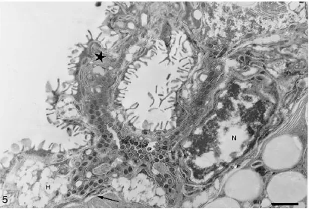
Hematin cells appear elongated, “U”-shaped in longitudinal sections or round with closed borders in cross section (Figs 1-2). The basal nuclei have conspicuous heterochromatin. Mitochondria and numerous vesicles of irregular shape and varying electrondensity are located in the cytoplasm. The membrane in contact with the gut lumen forms numerous long lamellae enlarging the external cell surface (Figs 2-3). The hematin cells are located at irregular intervals along the gut. They are merged in the connecting syncytium composed of cells with scattered elongate nuclei, thin cytoplasm and few organelles. The syncytium also surrounds the he-matin cells laterally and, in these areas, free lamel-lae, smaller than those of the hematin cells, can be observed on its surface (Fig. 3). Muscular fibers and parenchyma lie underneath the gastrodermis (Fig. 4).
Hematin cells and syncytium show outgrowths and bent folds which were seen to enclose lumen material with the help of the surface lamellae. The trapped material was then subjected to endocyto-sis (Fig. 4). Residual pigments remain inside of the vesicles in the external region of the cell. The way they are released from the cell was not ob-served. No free hematin cell was observed in the gut lumen.
The nature of hematin residues was confirmed by the histochemical test of picric acid in ethyl al-cohol. The Turnbull method comparatively applied in the parasite and fish gill filaments confirmed the distribution of ferrous iron in hematin cells. The methods of Perls, Okajima and Dunn-Thomp-son were not conclusive. There was no reaction for mucin using the PAS and Alcian blue methods. Hematin cells stained with E-PTA displayed a strongly positive reaction in nuclear heterochro-matin and in the small vesicles located at the ex-ternal edge of the cell. Vesicles containing hema-tin residues were not labeled. A collection of mem-brane tubules and flattened sacs extends through-out the cytoplasm of the hematin cells.
DISCUSSION
The gastrodermis of A. heterodus is quite simi-lar to the pattern described for other Polyo-pisthocotylea, with two cell types: hematin cell and connecting syncytium (Llewelyn 1954, Halton & Jennings 1965, Halton et al. 1968, Tinsley 1973, Halton 1975).
Longitudinal sections showed that the irregu-lar “U”-shaped hematin cells, are different from those previously reported for polyopisthocotyleans (Halton et al. 1968, Fournier 1978, Allen & Tinsley 1989). The feeding process is continuous, with lamellae looping back onto the surface to capture and endocyte food particles, in a process similar to that described by Halton (1974), actually called micropinocytosis. Fournier (1978) also mentioned the endocytosis of gut content but did not mention the importance of the lamellae in this process.
ex-279 279279 279279 Mem Inst Oswaldo Cruz, Rio de Janeiro, Vol. 93(2), Mar./Apr. 1998
Gut lumen of Atriaster heterodus. Fig. 1: E-PTA cytochemistry in a “U”-shape hematin cell with basal nucleus (N), lamellae (L),
280 280 280 280
280 Gastrodermis and Nutrition of A. heterodus Cláudia P Santos et al.
pected, the pigments were solubilized by the pi-cric acid and also reacted for the Turnbull method, it was stated that the pigment was in fact hematin. The histochemistry and cytochemistry tests, nega-tive for mucins and posinega-tive for iron residues, con-firmed the chemical nature as hematin pigments, indicating hematophagous food habit for this para-site. Host hemoglobin in vacuoles strongly sug-gest that at least intracellular disug-gestion occurs in these cells.
No intact erythrocytes were observed in the gut. This can be explained by fish handling. The mono-geneans were collected some hours after fish cap-ture and according to Llewellyn (1954) erythro-cytes can only be observed in their gut when fish are examined in less than one hour after capture, because a rapid haemolysis followed by phagocy-tosis normally occur. However, it cannot be ex-cluded that erythrocytes are lysed during the feed-ing process.
The structure of the syncytium cells with or without lamellae was previously reported (Halton et al. 1968, Tinsley 1973). Nevertheless, in both cases these monogeneans that feed on blood present a syncytium with few spread organelles,
function-ing mainly as a support for the hematin cells, on the contrary to those that present a mixed food habit like Polystomoides sp., where the syncytial cyto-plasm has numerous organelles (Allen & Tinsley 1989). The reduced syncytial cytoplasm of A. heterodus with few organelles presupposes a sup-port function but their role in the feeding process is still doubtful.
This is the first study on the ultrastructure of the gastrodermis of A. heterodus with detection of its haematophagous food habit.
ACKNOWLEDGEMENTS
To Dr Kurt Buchmann (Royal Veterinary and Agri-culture University, Denmark) and Dr Walter A Boeger (Universidade Federal do Paraná, Brazil) who kindly commented on the manuscript.
REFERENCES
Allen KM, Tinsley RC 1989. The diet and gastrodermal ultrastructure of polystomatid monogeneans infect-ing chelonians. Parasitology 98: 265-273.
Benhnke O, Zelander T 1970. Preservation of intercel-lular substances by the cationic dye Alcian blue in preparative procedures for electron microscopy. J Ultrastruct Res 31: 424.
281 281281 281281 Mem Inst Oswaldo Cruz, Rio de Janeiro, Vol. 93(2), Mar./Apr. 1998
Bloom FE, Aghajanian GK 1968. Fine structure and cytochemical analysis of the staining of synaptic junctions with phosphotungstic acid. J Ultrastruct Res 22: 361-375.
Bogitsh BJ 1993. A comparative review of the flatworm gut with emphasis on the Rhabdcoela and Neodermata. Trans Amer Microsc Soc 112: 1-9.
Buchmann K, Koie M, Prento P 1987. The nutrition of the gill parasitic monogenean Pseudactylogyrus anguillae. Parasitol Res 73: 532-537.
Fournier A 1978. Euzetrema knoepffleri: evidence for a
synchronous cycle of gastrodermal activity and an “apocrine-like” release of the residues of digestion.
Parasitology 77: 19-26.
Halton DW 1974. Hemoglobin absorption in the gut of a monogenetic trematode, Diclidophora merlangi. J Parasitology 60: 59-66.
Halton DW 1975. Intracellular digestion and cellular defecation in a monogenean Diclidophora merlangi. Parasitol 70: 331-340.
Halton DW 1982. X-ray microanalysis of pigment gran-ules in the gut of D. merlangi (Monogenoidea). Zeitsch Parasit 68: 113-115.
Halton DW, Jennings, JB 1965. Observations on the nutrition of monogenetic trematodes. Biol Bull 129:
257-272.
Halton DW, Dermott E, Morris GP 1968. Electron mi-croscope studies of Diclidophora merlangi
(Monogenea: Polyopisthocotylea). I. Ultrastructure of the cecal epithelium. J Parasitol 54: 909-916.
Kearn GC 1963. Feeding in some monogenean skin parasites: Entobdella solea on Solea solea and Acanthocotyle sp. on Raia clavata. J Mar Biol Ass United Kingdom 43: 749-766.
Lillie RD, Fullmer HM 1976. Histopathologic Tech-nique and Practical Histochemistry, 4th ed.,
MacGraw-Hill Book Company, New York, 850 pp. Llewellyn J 1954. Observations on the food and gut pigment of the Polyopisthocotylea (Trematoda: Monogenea). Parasitology 44: 428-437.
McManus JFA, Mowry RW 1960. Staining Methods. Histologic and Histochemical, Paul B, Hoeber, Inc.
Medical Division of Harper & Brothers, 423 pp. Mowry RD 1963. The special value of methods that color
both acidic and vicinal hydroxyl groups in the histhochemical study of mucins with revised direc-tions for the coloidal iron stain, the use of Alcian blue G8X and their combinations with the periodic acid-Schiff reaction. Ann NY Acad Sci 106: 402-423.
Santos CP, Lanfredi RM, Souto-Padrón T 1997. Ultra-structure of spermatogenesis of Atriaster heterodus
(Platyhelminthes, Monogenea, Polyopisthocotylea).
J Parasitol 83: 1007-1014.
Santos CP, Souto-Padrón T, Lanfredi RM 1996. Atriaster heterodus Lebedev & Paruchin, 1969 and Polylabris tubicirrus (Paperna & Kohn, 1964) (Monogenea)
from Diplodus argenteus (Val., 1830) (Teleostei:
Sparidae) from Brazil. J Helm Soc Washington 63:
181-187.
Smyth J, Halton DW 1983. The Physiology of Trema-todes, Cambridge University Press, Cambridge, 446
pp.
Tinsley RC 1973. Ultrastructural studies on form and function of the gastrodermis of Protopolystoma xenopi (Monogenoidea: Polyopisthocotylea). Biol Bull 144: 541-555.
282 282 282 282
