online | memorias.ioc.fiocruz.br
Comparison of HPV genotyping by type-specific PCr and sequencing
Nara de Oliveira Carvalho1/+, Dora Méndez del Castillo1, Carlos Perone1, José Nélio Januário1, Victor Hugo de Melo2,Geraldo Brasileiro Filho3
1Núcleo de Ações e Pesquisas em Apoio Diagnóstico 2Centro de Treinamento e Referência em Doenças Infecciosas e Parasitárias
Orestes Diniz 3Departamento de Anatomia Patológica e Medicina Legal, Faculdade de Medicina, Universidade Federal de Minas Gerais,
Belo Horizonte, MG, Brasil
Human papillomavirus (HPV) infection is the most common sexually transmitted disease worldwide and there is a strong link between certain high-risk viral types and cervical carcinogenesis. Although there are several typing methods, it is still unclear which test is the best. This study compared the effectiveness of type-specific PCR (TS-PCR) and sequencing, with a focus on their clinical application. A total of 260 cervical samples from HPV-positive patients were tested for types 6, 11, 16, 18, 31, 33 and 35 using TS-PCR and sequencing. The genotype was identified in 36% of cases by TS-PCR and in 75% by sequencing. Sequencing was four times more likely to identify the viral type in positive samples than TS-PCR (p = 0.00). Despite being more effective for virus genotyping, sequencing was unable to identify viral types in multiple infections. Combining both techniques resulted in highly sensitive detec-tion (87% of cases), showing that they are complementary methods. HPV genotyping is an important step in HPV management, helping to identify patients with a higher risk of developing cervical cancer and contributing to the development of type-specific vaccines.
Key words: HPV - PCR - sequencing - typing
Human papillomavirus (HPV) is the most common sexually transmitted virus in young and sexually active people of both sexes (Sanjose et al. 2007). Anogeni-tal HPVs, which are primarily mucosotropic, are clas- sified as high and low risk, according to their relation-ship with benign or malignant proliferative lesions (de Villiers et al. 2004).
Based on the viral DNA sequence, more than 230 HPV types are known (Haws et al. 2004); 118 genotypes are well-characterised according to biological niche, oncogenic potential and phylogenetic position (de Vil-liers et al. 2004). Approximately 40 HPV types infect the anogenital region and 15 of them - 16, 18, 31, 33, 35, 39, 45, 51, 52, 56, 58, 59, 68, 73 and 82 - are considered oncogenic or as high risk because they are associated with high grade squamous intraepithelial lesions or can-cer. Types 26, 53 and 66 are likely to be carcinogenic, whereas types 6, 11, 40, 42, 43, 44, 54, 61, 70, 72, 81 and candHPV89/Cp6108 are considered to be low risk (Munoz et al. 2003).
HPVs have been associated with many proliferative le-sions, with condyloma acuminatum being the most com-mon, as well as with different types of cancer, including cervical, vaginal, vulvar, penile, anal, oropharyngeal, buccal cavity and larynx (Bosch et al. 2002, Montaldo et al. 2007). Among them, uterine cervix carcinoma is par-ticularly important due to its high incidence and its high
Financial support: NUPAD, Faculdade de Medicina - UFMG + Corresponding author: [email protected]
Received 19 July 2009 Accepted 21 December 2009
mortality rate. In most cases, tumours evolve slowly and can be prevented by identifying precursor lesions in the cervical epithelium as early as possible, allowing for ef-fective treatment before local invasion and spread of the disease (Bezerra et al. 2005).
A strong association between HPV and cervical cancer stimulated the development of several diagnos-tic tests, pardiagnos-ticularly those based on molecular biology. There are currently two main approaches for molecular-ly detecting HPV: PCR with generic primers to amplify part of the L1 gene of the viral capsid, which is highly conserved among anogenital HPVs and the hybrid cap-ture test (HC2), which detects the main types of HPV by forming DNA-RNA hybrids (Iftner & Villa 2003, Gio-vannelli et al. 2004, Carestiato et al. 2006). As it is more sensitive, PCR has been largely used worldwide (Gravitti et al. 2000, Hubbard 2003, Kosel et al. 2003).
can characterise virus types in cases of multiple infection (Hubbard 2003, Sotlar et al. 2004, Carestianto et al. 2006, Fontaine et al. 2007, Lin et al. 2008). Selecting virus types to be tested should be based on epidemiological and prev-alence studies, as there is a wide variation in the genotype distribution in different regions around the world.
Over the last few years, virus genotyping has become an important way to approach cervical cancer. Several groups have searched for an effective genotyping test for HPV, due to its great contribution in the diagnosis of in-fections and to a better understanding of the relationship of HPV with carcinogenesis, in addition to contributing to the development of type-specific vaccines. This study compared two methods of HPV genotyping (TS-PCR and sequencing) to find an effective strategy for virus genotyping in clinical samples.
suBJeCts, Materials aND MetHODs
Study population - The present study was part of a multicentric research project entitled Multicentric Pro-gramme for Controlling and Preventing High Degree Cervical Lesions and Cervical Uterine Cancer in HIV-positive Women. Samples came from HIV-infected women attended at the Centro de Treinamento e Referên-cia em Doenças Infecciosas e Parasitária Orestes Diniz, in Belo Horizonte or at public gynaecology outpatient clinics in other cities (Betim, Barbacena, Divinópolis and Conselheiro Lafaiete), all of which were in state of Minas Gerais. From February 2006-February 2008, 463 samples were analysed; 260 were included in this study as they were HPV-positive and 203 were excluded (187 samples were HPV negative and 16 samples were globin negative). The research study was approved by the Re-search Ethical Committee of the Universidade Federal de Minas Gerais; all participants gave written consent.
Sample collection and processing - Cervical cells were obtained with an Ayre’s spatula and placed in a sterile tube containing 2 mL of physiological saline solu-tion (NaCl, 0.09%); samples were sent to the laboratory
within 24 h. Cytological results of the Papanicolaou test were not included.
DNA was extracted using Chelex 100 chelating res-in (BioRad), accordres-ing to the manufacturer’s protocol (Walsh et al. 1991). To control for DNA quality, the glob-in gene was amplified (Duggan et al. 1994) glob-in all sam-ples. PCR was performed in a final reaction volume of 50 µL, containing 10 µL of DNA, 5 µL buffer 10 x [100 mM Tris-HCl (pH 8,8) 500 mM KCl], 3 µL MgCl2, 1 µL dNTPs [200 µM], 2,5 µL of each primer at 10 pmol/µL and 2.5 U of Taq DNA polymerase. The PCR conditions were as follows: preheating for 1 min at 94ºC was fol-lowed by 30 cycles of 30 sec at 90ºC, 2 min at 54ºC and 1 min at 72ºC and a final extension of 10 min at 72ºC. HPV detection by PCR was carried out in a nested-PCR system, using the primers MY09/11 (Manos et al. 1989) and GP5+/6+ (de Roda Husman et al. 1995). For the first reaction, the same conditions were used as those for globin gene. Nested-PCR was performed in a final vol-ume of 50 µL, containing 1 µL of the first reaction, 5 µL buffer 10 x [100 mM Tris-HCl (pH 8,8) 500 mM KCl], 3 µL MgCl2, 1 µL dNTPs (200 µM), 2,5 µl of each primer at 10 pmol/µL and 2,5 U of Taq DNA polymerase. The PCR conditions were as follows: preheating for 4 min at 94ºC was followed by 40 cycles of 30 sec at 94ºC, 1 min at 45ºC and 1 min and 30 sec at 72ºC and the final exten-sion of 10 min at 72ºC.
TS PCR - DNA was amplified with specific primers for the following HPV types: 6, 11, 16, 18, 31, 33 and 35 (Arndt et al. 1994, Duggan et al. 1994) in independent reactions. PCR was performed in a final reaction volume of 50 µL, containing 5 µL of DNA, 5 µL buffer 10 x [100 mM Tris-HCl (pH 8.8), 500 mM KCl], 3 µL MgCl2, 1 µL dNTPs (200 µM), 2.5 µL of each primer at 10 pmol/ µL and 2.5 U of Taq DNA polymerase. Amplification conditions were the same as those for globin gene, ex-cept for annealing temperatures, which were as follows: for HPV types 16, 31 and 35: 2 min at 54ºC; for HPV type 6: 2 min at 56ºC; for HPV type 11: 2 min at 61ºC;
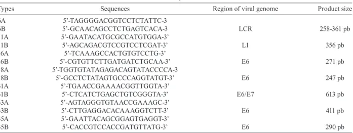
TABLE I
Primer sequences
Types Sequences Region of viral genome Product size
6A 5’-TAGGGGACGGTCCTCTATTC-3
6B 5’-GCAACAGCCTCTGAGTCACA-3 LCR 258-361 pb
11A 5’-GAATACATGCGCCATGTGGA-3’
11B 5’-AGCAGACGTCCGTCCTCGAT-3’ L1 356 pb
16A 5’-TCAAAGCCACTGTGTCCTG-3’
16B 5’-CGTGTTCTTGATGATCTGCAA-3’ E6 271 pb
18A 5’-TGGTGTATAGAGACAGTATACCCCA-3
18B 5’-GCCTCTATAGTGCCCAGGTATGT-3’ E6 247 pb
31A 5’-TGAACCGAAAACGGTTGGTA-3’
31B 5’-CTCATCTGAGCTGTCGGGTA-3’ E6/E7 613 pb
33A 5’-AGTAGGGTGTAACCGAAAGC-3’
33B 5’-CTTGAGGACACAAAGGTCTT-3’ E6 411 pb
35A 5’-GAATTACAGCGGAGTGAGGT-3’
for HPV type 18: 2 min at 58ºC; for HPV type 33: 2 min at 50ºC. All PCR products were submitted to agarose gel electrophoresis in a 2% gel, treated with ethidium bromide and analysed under UV light. Primer sequences and fragment sizes are shown in Table I.
Direct sequencing - Approximately 30 µL of the nested-PCR product of each sample was purified and sequenced using the BigDye Terminator kit version 3.1 (Applied Biosystems) and Gp6+ primer (4 pmol/µL), ac-cording to the manufacturer’s instructions. Sequences were read on a 3100-Avant Genetic Analyser ABI Prism sequencer (Applied Biosystems). Each sequence obtained was edited by selecting a segment of 30 nucleotides. The size and location of the L1-amplified region segment were chosen based on the degree of polymorphisms and according to published HPV sequencing methods (Feoli-Fonseca et al. 1998, Lee et al. 2007). Sequences of 30 nucleotides were aligned using the Bioedit programme (version 7.0) (Hall 1999) with HPV reference sequences obtained from the ICTVdB database (http://www.ictvdb. rothamsted.ac.uk/). A complementary analysis of sequences obtained from Blast was performed (http://www.ncbi.nlm. nih.gov/blast).
Data analysis - Data were tabulated using Microsoft Office Excel 2007. The Mcnemar statistical test was used to compare the effectiveness of both methods at identifying the virus type. Differences were considered significant at p < 0.05.
results
Using the two strategies (TS-PCR and sequencing), the HPV genotype was identified in 227 (87%) of the 260 samples. In 33 cases (13%), it was not possible to identify the virus genotype due to the presence of non-screened
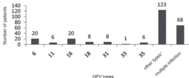
Fig. 1: frequency of the human papillomavirus virus (HPV) types identified.
Fig. 2: genotyping by type-specific PCR. HPV: human papillomavirus.
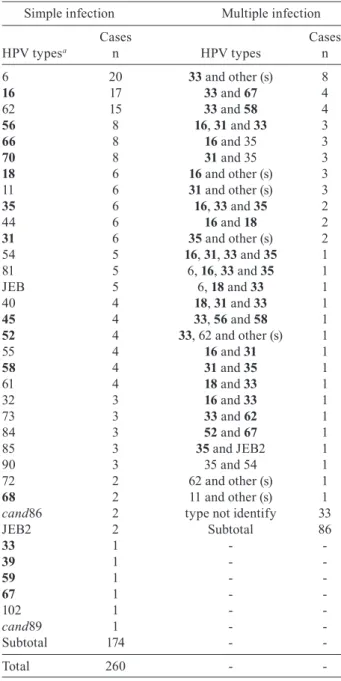
TABLE II
Human papillomavirus (HPV) genotypes in 260 cases
Simple infection Multiple infection
Cases Cases
HPV typesa n HPV types n
6 20 33 and other (s) 8
16 17 33 and 67 4
62 15 33 and 58 4
56 8 16, 31 and 33 3
66 8 16 and 35 3
70 8 31 and 35 3
18 6 16 and other (s) 3
11 6 31 and other (s) 3
35 6 16, 33 and 35 2
44 6 16 and 18 2
31 6 35 and other (s) 2
54 5 16, 31, 33 and 35 1
81 5 6, 16, 33 and 35 1
JEB 5 6, 18 and 33 1
40 4 18, 31 and 33 1
45 4 33, 56 and 58 1
52 4 33, 62 and other (s) 1
55 4 16 and 31 1
58 4 31 and 35 1
61 4 18 and 33 1
32 3 16 and 33 1
73 3 33 and 62 1
84 3 52 and 67 1
85 3 35 and JEB2 1
90 3 35 and 54 1
72 2 62 and other (s) 1
68 2 11 and other (s) 1
cand86 2 type not identify 33
JEB2 2 Subtotal 86
33 1 -
-39 1 -
-59 1 -
-67 1 -
-102 1 -
-cand89 1 -
-Subtotal 174 -
-Total 260 -
-a: boldface indicates high-risk type. Classification according de Villiers et al. (2004).
types in the panel investigated by the TS-PCR and/or to the occurrence of multiple-type infection, which cannot be typed by sequencing. In Fig. 1, the frequency of the 35 types identified in the study are shown, with HPV 16 (12.4%), HPV 33 (11.3%) and HPV 6 (8%) being the most frequent types identified. Table II shows the types identified for each case.
Direct sequencing of the amplified product identified the virus type in 196/260 (75%) cases. The types were distinct from those included in the panel surveyed by TS-PCR in 123 of the cases. The presence of more than one virus type, characterised by overlapping sequenc-es, was seen in 68/260 (26%) cassequenc-es, making it impos-sible to identify the types present in the great majority of cases; the types were identified in only four of these cases. Therefore, sequencing failed to identify the virus genotype in 64/260 (25%) cases. In addition to the seven types investigated by TS-PCR (types 6, 11, 16, 18, 31, 33 and 35), 28 other HPV types were identified by se-quencing (32, 39, 40, 44, 45, 52, 54, 55, 56, 58, 59, 62, 61, 66, 67, 68, 70, 72, 73, 81, 84, 85, 90, 102, cand86, cand89 and two types that still have not been classified taxonomically, the isolated JEB and type JEB2). These results are shown in Fig. 3.
Regarding multiple infections (n = 86), TS-PCR iden-tified more than one virus type in 21/86 (24%) cases, but it failed to identify multiple types in seven of the cases, recognising only one of the types present in the sample. In contrast, sequencing identified multiple infections in 68/86 (79%) samples and it was possible to identify mul-tiple types or at least one virus type in only 4/86 (4.6%). Considering the results of both techniques together, mul-tiple types or at least one type were identified in 53/86 (62%) cases of multiple infection. The typing result was different between the two methods employed in 12 cases. In the 19 cases of discordance between TS-PCR and sequencing (7 in which sequencing failed to identify multiple types and 12 in which typing was different in both methods), we considered that more than one virus type was present in the sample because sequencing may fail to identify multiple types.
When the effectiveness of both methods to identify HPV types present in samples was compared, sequenc-ing identified the genotype in more cases than TS-PCR for the seven types studied at the significance level. The estimated odds ratio showed that sequencing had a 4.2-fold greater chance of identifying the virus type present in a positive sample than TS-PCR for the seven types investigated (Table III).
DisCussiON
Molecular tests may accurately identify different types of HPV (of low and high cancer risk) in cells from cytological screening of cervical lesions and, due to their high sensitivities, have been the focus of attention of many studies (Gravitti et al. 2000, Hubbard 2003, Ko-sel et al. 2003). It is not always possible to identify the infecting virus type using PCR as a diagnostic method. The use of MY09/11 and GP5+/6+ primers in the nested-PCR system was proposed as a way to reduce this limita-tion, in addition to enhancing detection sensitivity, with a positivity rate 38.8% higher than that when MY09/11 was used alone; in this system, HPV can be detected in samples containing a low number of viral DNA copies (Pannier-Stockman et al. 2008). In the present study, the MY09/11 and GP5+/6+ strategy was employed and HPV90 and HPVcand86 were amplified. According to Terai and Burk (2002), these HPV types do not amplify
Fig. 3: genotyping by sequencing. a: human papillomavirus (HPV) types identified only by sequencing: 32, 39, 40, 44, 45, 52, 54, 55, 56, 58, 59, 62, 61, 66, 67, 68, 70, 72, 73, 81, 84, 85, 90, 102, cand86,
cand89, JEB and JEB2.
TABLE III
Analysis type-specific PCR (TS-PCR) vs. sequencing
TS-PCR
Sequencing Identified type Not identified type Total
Identified type 63 133 196
Not identified type 31 33 64
Total 94 166 260
confidence interval de 95% for odds ratio (OR): 2.90 ≤ ORestimated ≥ 6.34. OR: 133/31 = 4.29.
using MY09/11 alone. Speich et al. (2004) compared the genotyping results obtained when the MY09/11 and GP5+/6+ primers were used alone. They verified that MY failed to amplify types 30, 42, 43, 51, 59, 67, 74, 92 and 91 and that GP was unable to amplify types 61 and 62. In this study, types 59, 67, 90, 61 and 62 were ampli-fied and type 62 was the sixth most prevalent.
A total of 35 virus types were identified in this study, two of which have not yet been taxonomically classified: isolated JEB and type JEB2. Furthermore, 16 high risk genotypes were identified, two of which (HPV67 and HPV70) were not included in the set in-vestigated using the HC2 test. This data indicates that a large variety of virus is present in clinical samples, demonstrating the importance of PCR and sequencing as helpful tools for providing relevant information on the HPV infection. It was not possible to correlate the SiL/CIN grade of the lesion with the HPV DNA found in this study because its main goal was to compare the effectiveness of TS PCR (TS-PCR) and sequencing, with focuses on their clinical application.
types present in these patients. Sequencing identified vi-rus types in a larger number of cases (196/260, 75%) and recognised 28 types absent from the panel investigated by TS-PCR. Nevertheless, it was disadvantaged at iden-tifying genotypes in samples with multiple infections, in which viral sequences overlap and it is not possible to distinguish the various types, a finding also reported by others (Vernon et al. 2000, Serrano et al. 2003, Choi et al. 2005). In this study, sequencing was able to iden-tify types in only four cases of multiple infection, which were those having only two virus types in the sample. By sequencing, 25% of cases were not genotyped.
The greatest advantage of TS-PCR was its ability to identify multiple virus types or at least one virus type in cases of multiple infections (53/86, 62% of the samples in this series). However, this procedure requires several reactions for each sample and is more laborious, an opin-ion also shared by Lin et al. (2008). According to these authors, several reactions are required to investigate the great number of virus types, which makes this strategy non-viable for large-scale studies.
The results of the two methodologies were discrepant in 19 cases. In 10 of these cases, TS-PCR identified type 33 and sequencing identified types 58 (4 cases), 67 (4 cases), 62 (1 case) and 56/58 (1 case); in two cases, TS-PCR identified type 35 and sequencing identified types 54 (1 case) and JEB2 (1 case); in seven other cases, se-quencing identified only one virus type in the sample, whereas TS-PCR identified more than one type. Such discordances may be attributed to favouring the ampli-fication of these types in the PCR reaction, a phenom-enon that can occur when there is more than one virus type; in these cases, the type or types amplified are those existing in larger amounts in the sample. Kado et al. (2001) compared typing using sequencing (n = 107) with five different primers (GP17/18, MY09/11, L1C1/L1C2+L1C2M, pU1M-L/pU-2R and pU1M-L/ pU-2R-N) and they identified a different genotype in five cases, indicating a multiple infection. According to the authors, different types of HPV were preferably amplified depending on the primer used. Although this possibility is plausible, more studies are necessary to explain such differences.
As already reported in other studies (Serrano et al. 2003, Fontaine et al. 2007) and in spite of its limitations, sequencing has been considered the gold standard for HPV genotyping, due to the possibility of identifying virtually all virus types without mistaken classifications through cross-reactions among similar types, which can occur using tests based on hybridisation. This study demonstrated that sequencing was more effective in rec-ognising types of HPV, having identified 4.2 more cases than TS-PCR for the seven types studied and that both methods have advantages and disadvantages. Therefore, the best approach is the combination of both methods.
Based on our results, we suggest that both methods be employed as a genotyping strategy for HPV in clini-cal practice because they have been shown to be comple-mentary methods. Due to its great genotyping effective-ness, sequencing should be used in research studies or in those cases of recurrent/persistent/untreatable infections
not typed by TS-PCR. When choosing types to be in-vestigated by TS-PCR, a panel should include the most prevalent high risk types and those with greater clini-cal relevance. Some studies (Chow et al. 2000, Fontaine et al. 2007, Capra et al. 2008, Pannier-Stockman et al. 2008) have proposed to use TS-PCR after sequencing to identify HPV DNA, with consideration of the great variety of virus types, which are different in biological properties and carcinogenic risk.
aCkNOwleDGMeNts
To Prof. Arminda Siqueira Campos and Jacqueline Tibúr-cio, for their help with statistics.
reFereNCes
Ardnt A, Nottelmann K, Brock J, Neumann OG 1994. Das invert pa-pillom und seine assoziation mit dem humanen papa-pillomavirus (HPV). HNO 42: 670-676.
Astori G, Arzese A, Pipan C, de Villiers E-M, Botta GA 1997. Char-acterization of a putative new HPV genomic sequence from a cer-vical lesion using L1 consensus primers and restriction fragment length polymorphism. Virus Res 50: 57-63.
Bezerra SJS, Gonçalves PC, Franco ES, Pinheiro AKB 2005. Perfil de mulheres portadoras de lesões cervicais por HPV quanto aos fatores de risco para câncer de colo de útero. J Bras Doenças Sex Transm 17: 143-148.
Bosch FX, Lorincz A, Munoz N, Meijer CJLM, Shah KV 2002. The causal relation between human papillomavirus and cervical can-cer. J Clin Pathol 55: 244-265.
Capra G, Giovannelli L, Bellavia C, Migliore MC, Caleca MP, Perino A, Ammatuna P 2008. HPV genotype prevalence in cytologically abnormal cervical samples from women living in South Italy. Vi-rus Res 133: 195-200.
Carestiato FN, Silva KC, Balthazar DS, Silva L, Marinho M, Oliveira LHS, Cavalvanti SMB 2006. Analysis of molecular biology tech-niques for the diagnosis of human papillomavirus infection and cervical cancer prevention. Rev Soc Bras Med Trop 39: 428-432.
Choi Y-D, Jung W-W, Nam J-H, Choi H-S, Park C-S 2005. Detection of HPV genotypes in cervical lesions by the HPV DNA chip and sequencing. Ginecol Oncol 98: 369-375.
Chow VTK, Loh E, Yeo WM, Tan SY, Chan R 2000. Identification of multiple genital HPV types and sequence variants by consen-sus and nested type-specific PCR coupled with cycle sequencing.
Pathology 32: 204-208.
Clifford G, Franceschi S, Diaz M, Munoz N, Villa LL 2006. Chapter 3: HPV type-distribution in women with and without cervical neoplastic diseases. Vaccine 24S3: 26-34.
de Roda Husman A-M, Walboomers JMM, van den Brule AJC, Mei-jer CJLM, Snijders PJF 1995. The use of general primers GP5 and GP6 elongated at their 3’ ends with adjacent highly conserved sequences improves human papillomavirus detection by PCR.
J Gen Virol 76: 1057-1062.
de Villiers E-M, Fauquet C, Broker TR, Bernard H-U, zur Hausen H 2004. Classification of papillomaviruses. Virology 324: 17-27.
Duggan MA, Inoue M, Mcgregor SE, Stuart GCE, Morris S, Poon VG, Schepansky A, Honore L 1994. A paired comparison of dot blot hybridization and PCR amplification for HPV testing of cervical scrapes interpreted as CIN 1. Eur J Gynaec Oncol 15: 178-187.
Fontaine V, Mascaux C, Weyn C, Bernis A, Celio N, Lefevre P, Kauf-man L, Garbar C 2007. Evaluation of combined general primer-mediated PCR sequencing and type-specific PCR strategies for determination of human papillomavirus genotypes in cervical cell specimens. J Clin Microbiol45: 928-934.
Giovannelli L, Lama A, Capra G, Giordano V, Arico P, Ammatu-na P 2004. Detection of human papillomavirus DNA in cervical samples: analysis of the new PGMY-PCR compared to Hybrid Capture II and MY-PCR assay and two-step nested PCR assay. J Clin Microbiol 42: 3861-3864.
Gravitti PE, Peyton CL, Alessi TQ, Wheeler CM, Coutlee F, Hildesheim A, Schiffiman MH, Scott DR, Apple RJ 2000. Im-proved amplification of genital humam papillomaviruses. J Clin Microbiol 38: 357-361.
Hall TA 1999. Bioedit: a user-friendly biological sequence alignment editor and analysis program for Windows 95/98NT. Nucleic Acids Symp Ser 41: 95-98.
Haws ALF, He Q, Rady PL, Zhang L, Grady J, Hughes TK, Stisser K, Konig R, Tyring SK 2004. Nested PCR with the PGMY09/11 and GP5+/6+ primer sets improves detection of HPV DNA in cervical samples. J Virol Methods 122: 87-93.
Hubbard RA 2003. Human papillomavirus testing methods. Arch Pathol Lab Med 127: 940-945.
Iftner T, Villa LL 2003. Chapter 12: human papillomavirus technolo-gies. J Natl Cancer Inst Monographs 31: 80-88.
Kado S, Kawamata Y, Shino Y, Kasai T, Kubota K, Iwasaki H, Fu-kazawa I, Takano H, Nuoyama T, Mitsuhashi A, Sekiya S, Shi-rasawa H 2001. Detection of human papillomavirus in cervical neoplasias using multiple sets of generic polymerase chain reac-tion primers. Ginecol Oncol 81: 47-52.
Kleter B, van Doorn L-J, Schrauwen L, Molijn A, Sastrowijoto S, ter Schegguet J, Lindeman J, ter Harmsel B, Burger M, Quint W 1999. Development and clinical evaluation of a highly sensi-tive PCR-reverse hybridization line probe assay for detection and identification of anogenital human papillomavirus. J Clin Micro-biol 37: 2508-2517.
Kosel S, Burggraf S, Mommsen J, Engelhardt W, Olgemoller B 2003. Type-specific detection of human papillomaviruses in a routine laboratory setting - improved sensitivity and specificity of PCR and sequence analysis compared to direct hybridization. Clin Chem Lab Med 41: 787-791.
Lee SH, Vigliotti VS, Vigliotti JS, Pappu S 2007. Routine human papillomavirus genotyping by DNA sequencing in community hospital laboratories. Infect Agents Cancer 2: 1-11.
Lin C-Y, Chao A, Yang Y-C, Chou H-H, Ho C-M, Lin R-W, Chang T-C, Chiou J-Y, Chao F-Y, Wang K-L, Chien T-Y, Hsueh S, Huang C-C, Chen C-J, Lai C-H 2008. Human papillomavirus typing with a polymerase chain reaction-based genotyping array com-pared with type-specific PCR. J Clin Virol 42: 361-367.
Manos MM, Ting Y, Wright DK, Lewis AJ, Broker TR, Wolinsky SM 1989. Use of polymerase chain reaction amplification for the detec-tion of genital human papillomaviruses. Cancer Cells 7: 209-214.
Meijer CJ, Snijders PJ, Castle PE 2003. Clinical utility of HPV geno-typing. Gynecol Oncol 103: 12-17.
Mendez F, Munoz N, Posso H, Molano M, Moreno V, van den Brule AJC, Ronderos M, Meijer C, Munoz A 2005. Cervical coinfec-tion with human papillomavirus (HPV) types and possible im-plications for the prevention of cervical cancer by HPV vaccines.
J Infect Dis 192: 1158-1165.
Montaldo C, Mastinu A, Quartuccio M, Piras V, Denotti G, Pisano E, Orrú G 2007. Detection and genotyping of human papillomavirus DNA in samples from healthy Sardinian patients: a preliminary study. J Oral Pathol Med 36: 482-487.
Munoz N, Bosch FX, Sanjosé S, Herrero R, Castellsagué X, Shah KV, Snijders PJF, Meijer CJLM 2003. Epidemiologic classification of human papillomavirus types associated with cervical cancer.
N Eng J Med 348: 518-527.
Pannier-Stockman C, Segard C, Bennamar S, Gondry J, Boulanger J-C, Sevestre H, Castelain S, Duverlier G 2008. Prevalence of HPV genotypes determined by PCR and DNA sequencing in cer-vical specimens from French women with or without abnormali-ties. J Clin Virol 42: 353-360.
Sanjose S, Diaz M, Castellsague X, Clifford G, Bruni L, Munoz N, Bosch FX 2007. Worldwide prevalence and genotypes distribu-tion of cervical human papillomavirus DNA in women with nor-mal cytology: a meta-analysis. Lancet Infect Dis 7: 453-459.
Serrano ML, Correa M, Medina O, Melgarejo D, Bravo MM 2003. Tipi-ficación de vírus del papiloma humano mediante secuencia directa em mujeres com citologia normal. Rev Colomb Cancer 7: 18-24.
Sotlar K, Diemer D, Dethleffs A, Hack Y, Stubner A, Vollmer N, Menton S, Menton M, Dietz K, Wallwiener D, Kandolf R, Bult-mann B 2004. Detection and typing of human papillomavirus by E6 nested multiplex PCR. J Clin Microbiol 42: 3176-3184.
Speich N, Schimitt C, Bollmann R, Bollmann M 2004. Human papil-lomavirus (HPV) study of 2,916 cytological samples by PCR and DNA sequencing: genotype spectrum of patients from the West German area. J Med Microbiol 53: 125-128.
Terai M, Burk RD 2002. Identification and characterization of 3 nov-el genital human papillomaviruses by overlapping polymerase chain reaction: candHPV89, candHPV 90 and candHPV91.
J Infect Dis 185: 1794-1797.
Vernon SD, Unger ER, Willians D 2000. Comparison of human papil-lomavirus detection and typing by cycle sequencing, line blotting and hybrid capture. J Clin Microbiol 38: 651-655.
Verteramo R, Pierangeli A, Calzolari E, Patella A, Recine N, Mancini E, Marcone V, Masciangelo R, Bucci M, Antonelli G, Degener AM 2006. Direct sequencing of HPV DNA detected in gynaeco-logic outpatients in Rome, Italy. Microbes Infect 8: 2517-2521.