The Zymovars of
Vibrio cholerae:
Multilocus Enzyme
Electrophoresis of
Vibrio cholerae
Fernanda S Freitas, Hooman Momen, Carlos Andre Salles
Laboratório de Sistemática Bioquímica, Departamento de Bioquímica e Biologia Molecular, Instituto Oswaldo Cruz-Fiocruz, Av. Brasil 4365, 21045-900 Rio de Janeiro, RJ, Brasil
Zymovars analysis also known as multilocus enzyme electrophoresis is applied here to investigate the genetic
variation of
Vibrio cholerae
strains and characterise strains or group of strains of medical and epidemiological
interest.
Fourteen loci were analyzed in 171 strains of non-O1 non-O139, 32 classical and 61 El Tor from America, Africa,
Europe and Asia. The mean genetic diversity was 0.339.
It is shown that the same O antigen (both O1 and non-O1) may be present in several geneticaly diverse (different
zymovars) strains. Conversely the same zymovar may contain more than one serogroup.
It is confirmed that the South American epidemic strain differs from the 7th pandemic El Tor strain in locus LAP
(leucyl leucyl aminopeptidase). Here it is shown that this rare allele is present in 1
V. mimicus
and 4 non-O1
V.
cholerae
.
Non toxigenic O1 strains from South India epidemic share zymovar 14A with the epidemic El Tor from the 7th
pandemic, while another group have diverse zymovars.
The sucrose negative epidemic strains isolated in French Guiana and Brazil have the same zymovar of the
current American epidemic
V. cholerae.
Key words: Vibrio cholerae - enzyme electrophoresis - genetic diversity
The electrophoretic mobility of enzymes has been
used to detect allelic variation of the respective genes in
several microorganisms including
Vibrio cholerae
(Salles
& Momen 1991, Chen et al. 1991, Wachsmuth et al. 1993,
Beltran et al. 1999).
In our previous papers we focused on the problem of
finding a reliable genetic distinction between the
bio-types classical and El Tor and to evaluate the degree of
genetic variation prevalent among this species.
Then we defined a zymovar as the set of strains
having the same electromorphs among the loci
investi-gated and zymovar analysis the procedures required to
determine it. This approach is also known as
Multi-Lo-cus Enzyme Electrophoresis and zymovar as
electro-phoretic type, or ET.
The study of
V. cholerae
using molecular tools
intro-duced considerable change in our views affecting the
bacteriology and the epidemiology of cholera. Here we
present results obtained with 14 loci from a large sample
of strains.
MATERIALS AND METHODS
Geographical origin of strains
- Algeria, Australia,
Burma, Bangladesh, Brazil, Bolivia, Colombia, Chile, Czech
Republic, China, Sri Lanka, France, Ghana, Guatemala,
Guiana, Hong Kong, India, Indonesia, Israel, Italy, Japan,
Kuwait, Malaysia, Marroc, Mexico, Nepal, Nigeria,
Paki-stan, Peru, Philippines, Russia, Rwanda, Singapore,
Tan-zania, Thailand, Turkey, UK, USA, Venezuela, Zimbabwe.
Sources of strains
- B Davies, Peggy Pereira, P
Desmarchelier, R Colwell, Glenn Morris Jr, GB Nair, AC
Ghose, B Said, JV Lee, NICED (India), JM Fournier, L
Amenuvor, and our own collection.
Most of the general bacteriological, electrophoretical
and computational procedures have been described
pre-viously (Salles & Momen 1991).
The enzyme loci mentioned in the tables and text have
been described previously (Salles & Momen 1991). They
are: ACO (aconitase hydratase); ADH (alanine
dehydro-genase); IDH (isocitrate dehydrodehydro-genase); ME (malic
en-zyme); NSE (carboxilesterase); PGD (6-phosphogluconate
dehydrogenase); MDH (malate dehydrogenase); PGM
(phosphoglucomutase); GPI (glucose phosphate
isomerase); G6P (glucose-6-phosphatedehydrogenase) ;
PD (proline dipeptidase); P1 (peptidase
leucyl-leucyl-leu-cine); P2 (peptidase leucyl glycil glycine) with the
addi-tion of the locus LAP: leucyl leucyl aminopeptidase
(Pas-teur et al. 1990).
Genetic diversity was estimated according to Selander
et al. (1986). The genetic diversity per locus GDL is
calcu-lated from allele frequencies as
GDL= 1- [
Σ
Ai
2(n/n-1)]
were Ai is the frequency of the ith allele in locus A and n
the number of zymovars. The expression n/n-1 is a
cor-rection for small samples. Mean genetic diversity MGD is
the arithmetic average of GDL over all loci.
RESULTS
Table I shows the electromorphs of 134 zymovars
ob-tained from 155 strains of
V. cholerae
non O1 non O139
plus the zymovars of El Tor 7th pandemic (14A), ElTor S.
American pandemic (14B) and classical (13). For the
This project received financial support from Faperj.
+Corresponding author. Fax: +55-21-2598.3495. E-mails:
csalles@gene.dbbm.fiocruz.br ; caqps@ig.com.br Received 18 July 2001
TABLE I
Alleles present in each zymovar of Vibrio cholerae
ZYMO ACO ADH IDH ME NSE PGD MDH PGM GPI G6P PD P1 P2 LAP
N 001 1 1 1 2 2 3 4 2 3 3 4 3 3 2
N 002 1 1 1 2 5 3 4 2 3 3 4 3 1 2
N 003 1 1 1 2 5 3 4 2 3 2 5 3 1 2
N 004 1 1 1 2 5 3 3 2 3 5 4 2 1 2
N 005 1 1 1 2 5 3 3 2 3 1 3 2 1 1
N 006 1 1 1 2 5 3 3 2 3 4 4 2 1 2
2 007 1 1 1 2 5 3 3 1 3 4 2 2 1 2
N 008 1 1 2 2 5 3 3 2 3 4 3 2 1 2
N 009 1 1 1 2 5 6 3 0 3 4 5 2 1 1
2 010 1 1 1 2 5 5 3 1 3 4 5 2 1 2
2 011 1 1 1 2 5 4 3 2 3 2 5 2 1 2
2 012 1 1 1 2 5 4 3 2 2 2 5 2 1 2
1 013 1 1 1 2 4 2 3 2 3 5 4 2 1 2
N 015 1 1 1 2 5 3 3 2 3 3 1 1 1 2
2 016 1 1 1 2 5 1 3 2 3 6 2 2 1 2
2 017 1 1 1 2 5 5 4 1 3 5 3 2 1 2
2 018 1 1 1 2 6 3 1 2 1 4 4 2 1 1
N 022 1 1 1 2 3 3 3 2 3 4 5 2 1 1
N 023 1 1 1 2 5 4 3 2 3 4 4 2 1 2
N 024 1 1 1 2 4 1 3 2 3 5 4 2 1 2
2 025 1 1 1 2 5 3 3 2 3 2 5 2 1 2
2 026 1 1 1 2 5 3 3 2 2 2 6 2 1 2
2 027 1 1 1 2 4 3 4 2 3 4 4 3 1 2
2 028 1 1 1 2 5 3 4 2 3 2 5 2 1 2
N 029 1 1 1 2 4 3 3 3 2 4 3 2 1 2
N 030 1 1 1 2 5 3 4 2 3 4 4 2 1 2
N 031 1 2 1 2 5 3 3 2 3 4 5 2 1 2
N 032 1 1 1 2 5 4 3 0 3 5 4 3 1 2
N 033 1 1 1 2 4 3 3 1 3 4 4 3 1 1
N 035 1 1 1 2 5 3 3 2 4 4 4 3 3 1
N 036 1 1 1 2 5 3 3 2 2 5 4 2 2 2
N 037 1 1 1 2 5 3 3 2 2 5 2 3 1 1
N 038 1 1 1 2 4 3 3 2 3 5 4 2 1 2
N 039 1 1 1 2 5 3 3 2 3 5 4 2 1 1
N 041 1 1 1 2 5 4 4 2 3 5 4 2 1 2
N 042 1 1 1 3 5 3 4 2 3 5 3 3 1 1
N 043 1 1 0.5 2 5 4 3 2 3 4 1 3 1 2
N 045 2 1 1 2 5 3 3 2 3 5 3 2 1 1
N 046 1 1 1 2 0 3 3 2 3 4 4 3 1 1
N 047 1 1 1 2 5 3 4 2 3 5 3 3 1 2
N 048 1 1 1 2 5 3 4 2 3 5 4 2 1 2
N 049 1 1 1 2 5 3 4 2 3 5 5 2 1 2
N 050 1 1 1 2 5 3 4 2 3 5 6 3 1 2
N 051 1 1 1 2 5 3 4 2 3 5 5 3 1 2
N 052 1 1 1 2 5 3 4 1 3 5 4 2 1 2
N 053 1 1 1 2 5 3 3 2 3 5 4 3 0 1
2 054 1 1 1.5 2 5 4 3 1 3 5 5 2 1 2
N 055 1 1 1 2 5 3 3 2 3 5 5 2 1 2
N 056 1 1 1 2 5 3 3 2 3 4 4 3 1 2
N 057 1 1 1 2 5 3 3 2 3 4 3 3 1 1
N 058 1 1 1 2 5 3 3 2 3 4 5 3 1 2
N 059 1 1 1 2 5 3 3 2 3 4 2 3 1 0
N 060 1 1 1 2 5 4 3 2 3 4 6 2 1 3
N 061 1 1 1 2 5 5 3 2 3 4 5 0 0 2
N 062 1 1 1 2 5 3 4 2 3 4 3 2 1 2
N 063 1 1 1 2 5 3 4 2 3 4 2 2 1 1
N 064 1 1 1 2 5 3 4 2 3 5 4 4 1 2
N 065 1 1 1 2 5 3 4 2 3 4 4 3 1 2
N 066 1 1 1 2 5 5 3 2 3 4 4 2 1 2
N 067 1 1 1 2 5 4 4 2 2 4 4 2 2 0
N 068 1 1 1 2 5 5 3 2 3 4 4 3 1 0
ZYMO ACO ADH IDH ME NSE PGD MDH PGM GPI G6P PD P1 P2 LAP
N 070 1 1 1 2 5 3 3 2 2 4 6 3 2 3
2 071 1 1 1 2 5 3 3 2 2 5 4 2 1 2
N 072 0.5 1 1 2 5 1 3 2 3 5 4 2 1 2
N 073 1 1 1 2 5 3 4 2 3 4 4 3 2 2
N 074 1 1 1 2 5 5 3 2 3 4 5 3 1 2
N 075 1 1 1 2 5 5 3 2 3 4 5 3 1 1
N 076 1 2 1 2 5 3 4 2 3 4 4 3 1 2
N 077 1 1 1 2 6 4 3 2 3 3 4 2 1 2
N 078 1 3 3 2 6 2 3 2 3 3 3 1 1 3
N 079 1 3 3 2 6 2 4 2 3 4 5 2 1 0
N 080 1 1 1 2 5 5 3 2 3 4 5 3 1 2
N 080 1 1 1 2 5 5 3 2 3 4 5 3 1 2
N 081 1 1 1 2 5 3 3 0 4 2 3 3 1 2
N 082 1 1 1 2 5 3 3 2 3 4 3 2 1 2
N 083 1 1 1 2 5 3 3 2 2 4 3 2 1 1
N 084 1 1 1 2 5 3 4 2 3 3 4 1 1 1
N 085 1 1 1 2 5 3 4 1 3 4 4 3 1 1
N 086 1 1 1 2 5 2 3 2 3 4 2 3 1 2
N 087 1 1 1 2 5 3 4 2 2 4 4 3 1 2
N 088 1 1 1 2 5 3 3 2 2 4 3 4 1 2
N 089 1 1 1 2 5 4 4 2 3 4 5 2 1 2
N 090 1 1 1 2 5 3 3 1 3 5 5 2 1 0
N 091 1 1 1 2 5 3 4 2 3 4 3 3 2 2
N 092 1 1 1 2 5 3 4 2 3 4 3 2 1 2
N 093 1 1 1 2 5 3 4 2 3 4 3 3 1 2
N 094 1 1 1.5 2 5 3 4 1 2 5 5 1 1 2
N 095 1 1 1 2 5 3 4 3 3 4 3 2 1 1
N 096 1 1 1 2 5 3 4 2 3 4 5 3 1 2
N 098 1 1 1 2 5 3 3 2 4 4 4 3 1 2
N 099 1 1 1 2 5 3 3 2 3 5 3 2 1 2
N 100 1 1 1 2 5 2 3 1 3 5 4 2 1 0
N 101 1 1 1 2 5 3 4 1 3 5 6 2 1 2
N 102 1 1 1 2 5 3 4 1 3 5 4 3 1 2
N 103 1 1 1 2 5 3 3 2 2 4 4 3 1 2
N 104 1 1 1 2 5 3 3 2 3 5 4 1 1 2
N 105 1 1 1 2 5 3 3 2 3 5 2 2 1 2
N 106 1 1 1 2 5 2 3 1 3 5 4 2 1 2
N 107 1 1 1 2 5 3 3 2 3 5 4 3 1 2
N 108 1 2 1 2 5 4 3 2 3 5 5 2 1 2
N 109 1 1 1 2 5 4 3 1 3 3 5 2 1 2
N 110 1 1 1 2 5 3 3 3 3 5 3 2 1 2
N 111 1 1 1 2 5 3 3 1 2 4 4 2 1 1
N 112 1 1 1 2 5 4 3 2 3 4 4 2 1 1
N 113 1 1 1 2 4 3 3 2 3 4 4 2 1 2
N 114 1 1 1 2 5 3 3 1 3 4 4 3 1 1
N 116 1 1 1 2 5 3 4 1 2 4 4 4 1 2
N 117 0.5 1 1 2 5 4 3 1 2 4 5 1 1 2
N 118 1 1 2 1 5 3 3 2 3 5 3 2 2 1
N 119 1 1 1 2 5 3 4 2 3 4 4 2 1 1
N 120 1 1 1 2 5 3 4 2 2 5 4 3 1 2
N 121 1 1 1 2 5 3 4 2 3 5 4 3 1 0
N 122 1 1 1 2 5 3 4 2 2 4 4 2 1 2
N 123 1 1 1 2 5 3 3 1 3 5 4 2 1 1
N 124 1 1 1 2 5 4 4 1 2 5 5 2 1 2
N 141 1 1 1 2 5 2 3 1 3 4 5 1 2 2
N 142 1 1 1 2 5 4 4 2 3 4 5 2 2 2
N 143 1 1 1 2 5 2 4 2 3 4 2 1 2 2
N 144 0.5 1 1 2 5 3 4 2 3 4 4 3 2 2
N 145 2 1 1 2 5 4 3 2 3 4 5 2 2 2
N 146 1 1 1 2 5 4 3 2 3 4 4 2 2 2
N 147 1 1 1 2 5 2 3 3 3 4 4 2 2 2
1 14A 1 1 1 2 4 3 3 2 2 5 4 2 1 1
1 14B 1 1 1 2 4 3 3 2 2 5 4 2 1 3
2 152 1 1 1 2 5 4 3 2 4 2 5 2 1 5
ZYMO ACO ADH IDH ME NSE PGD MDH PGM GPI G6P PD P1 P2 LAP
N 154 2 1 1 2 5 3 3 2 3 5 3 2 1 2
N 155 1 1 1 2 5 4 3 1 3 4 3 3 1 3
N 156 1 1 1.5 2 5 3 3 1 3 4 5 3 1 1
N 157 1 1 1.5 2 5 4 3 1 3 4 3 2 1 2
N 158 1 1 1 2 5 3 3 1 3 4 4 2 1 2
N 159 1 1 1 2 5 4 4 1 3 4 5 3 1 2
N 160 1 1 1 2 5 4 3 2 2 4 4 2 1 2
N 161 1 1 1 2 5 3 4 1 2 4 4 2 1 2
N 162 1 1 1.5 2 5 3 4 2 2 5 4 1 2 2
N 163 1 1 1 2 5 3 3 2 2 4 4 2 1 2
N 164 1 1 1 2 5 3 3 2 3 4 5 3 2 2
N 165 1 1 1 2 5 3 3 2 3 4 3 3 1 2
N: non O1 non O139 zymovars; 1: O1 and O139 epidemic strains: zymovar 14A El Tor 7th pandemic Old World, zymovar14B El Tor American pandemic and zymovar 13 classical strains; 2: O1 strains non zymovar 13 or 14; ZYMO: zymovar; ACO (aconitase hydratase); ADH (alanine dehydrogenase); IDH (isocitrate dehydrogenase); ME (malic enzyme); NSE (carboxilesterase); PGD (6-phosphogluconate dehydrogenase); MDH (malate dehydrogenase); PGM (phosphoglucomutase); GPI (glucose phosphate isomerase); G6P (glucose-6-phosphatedehydrogenase) ; PD (proline dipeptidase); P1 (peptidase leucyl-leucyl-leucine); P2 (peptidase leucyl glycil glycine) with the addition of the locus LAP: leucyl leucyl aminopeptidase (Pasteur et al. 1990).
present purpose the O139 strains (Bengal) are equated
with El Tor 7th Pandemic with the same zymovar 14A.
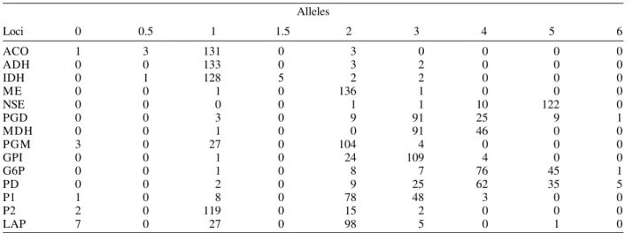
The genetic diversity per locus was as following:
ACO-0.092 ADH-0.064 IDH-0.133 ME-0.003 NSE-0.222
PGD-0.525 MDH-0.453 PGM-0.392 GPI-0.334 G6P-0.588
PD-0.690 P1-0.553 P2-0.314 LAP-0.461.
The mean genetic diversity (MGD) was 0.339, sd = 0.2.
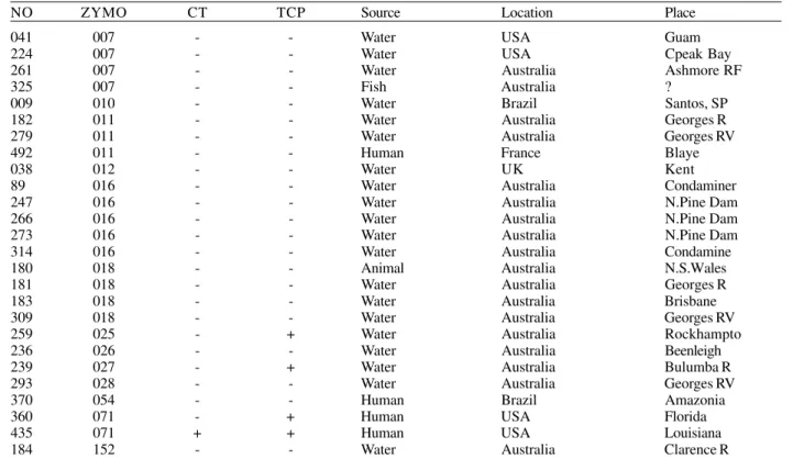
Table III list selected O1 serogroup strains isolated
from clinical and environmental sources with zymovars
other than 13 or 14 showing the correspondence or lack
of it between zymovar, serogroup, CT, TCP and sources.
DISCUSSION
The present estimate of MGD is 0.339. Our previous
estimated MGD was 0.326 using nearly half the strains of
the present study. Chen et al. (1991) found 0.311 and
TABLE II
Profiles for 14 loci in 138 zymovars
Alleles
Loci 0 0.5 1 1.5 2 3 4 5 6
ACO 1 3 131 0 3 0 0 0 0
ADH 0 0 133 0 3 2 0 0 0
IDH 0 1 128 5 2 2 0 0 0
ME 0 0 1 0 136 1 0 0 0
NSE 0 0 0 0 1 1 10 122 0
PGD 0 0 3 0 9 91 25 9 1
MDH 0 0 1 0 0 91 46 0 0
PGM 3 0 27 0 104 4 0 0 0
GPI 0 0 1 0 24 109 4 0 0
G6P 0 0 1 0 8 7 76 45 1
PD 0 0 2 0 9 25 62 35 5
P1 1 0 8 0 78 48 3 0 0
P2 2 0 119 0 15 2 0 0 0
LAP 7 0 27 0 98 5 0 1 0
ACO (aconitase hydratase); ADH (alanine dehydrogenase); IDH (isocitrate dehydrogenase); ME (malic enzyme); NSE (carboxilesterase); PGD (6-phosphogluconate dehydrogenase); MDH (malate dehydrogenase); PGM (phosphoglucomutase); GPI (glucose phosphate isomerase); G6P (glucose-6-phosphatedehydrogenase) ; PD (proline dipeptidase); P1 (peptidase leucyl-leucyl-leucine); P2 (peptidase leucyl glycil glycine) with the addition of the locus LAP: leucyl leucyl aminopeptidase (Pasteur et al. 1990).
Beltran et al. (1999) from a large sample of American
non-O1 isolates found a higher diversity of 0.436.
A possible source of variation in MGD seems to be the
choice of loci. Our choice was aimed at finding loci
diag-nostic of the species
V. cholerae
with low genetic diversity
GD and this may have biased in part the MGD. This
diver-sity is considerable and is of the same level of
Escherichia
coli
but still less than some Gram negative bacteria.
iso-TABLE III
Vibrio cholerae serogroup O1 with diverse zymovars
NO ZYMO CT TCP Source Location Place
041 007 - - Water USA Guam
224 007 - - Water USA Cpeak Bay
261 007 - - Water Australia Ashmore RF
325 007 - - Fish Australia ?
009 010 - - Water Brazil Santos, SP
182 011 - - Water Australia Georges R
279 011 - - Water Australia Georges RV
492 011 - - Human France Blaye
038 012 - - Water UK Kent
89 016 - - Water Australia Condaminer
247 016 - - Water Australia N.Pine Dam
266 016 - - Water Australia N.Pine Dam
273 016 - - Water Australia N.Pine Dam
314 016 - - Water Australia Condamine
180 018 - - Animal Australia N.S.Wales
181 018 - - Water Australia Georges R
183 018 - - Water Australia Brisbane
309 018 - - Water Australia Georges RV
259 025 - + Water Australia Rockhampto
236 026 - - Water Australia Beenleigh
239 027 - + Water Australia Bulumba R
293 028 - - Water Australia Georges RV
370 054 - - Human Brazil Amazonia
360 071 - + Human USA Florida
435 071 + + Human USA Louisiana
184 152 - - Water Australia Clarence R
NO: our strain no.; ZYMO: zymovar; CT: CTx cholera toxin gene; TCP: toxin co-regulated pili
lated from human sources, O37 and O5 (Donovan 1984).
This locus seems to be useful for a preliminary screening
of new strains.
Most genes investigated are housekeeping genes
necessarily conserved for the survival of the organism.
The know pathogenicity islands are phages or phage-like
mobile elements responsible for the lateral transfer of non
essential genes coding cholera toxin (CT) and toxin
co-regulated pilli (TCP) (Karaolis et al. 1999, Faruque et al.
1999) and genes coding somatic O antigens as in the
Bengal O139. This latter strain is genetically an El Tor 7th
pandemic zymovar 14 A which received from a non-O1
vibrio, genes coding a modified LPS (Johnson et al. 1994,
Bik et al. 1995). It is therefore unlikely that strong
correla-tion may be found between zymovars and antigenic or
virulence factors carried by these mobile elements.
The strain responsible for the American pandemic is
not the same strain causing the 7th pandemic. As show
by Wachsmuth et al. (1993) and our results, it differ in
locus LAP, with allele 3 while the Old World pandemic
strains have allele 1. The 7th pandemic in the Old World
is caused by zymovar 14A and the American pandemic
by zymovar 14B. Both pandemics are usually described
as the same 7th pandemic but the genetic distinction
be-tween the two agents may suggest an independent
ori-gin of the later. This poses an interesting epidemiological
problem, the origin of the American pandemic, the agent
and source of the first cases of cholera in Peru. LAP 3 is
not common in our sample and may be equally rare among
V. cholerae
in nature. Locus LAP has epidemiological
interest since cholera on the Atlantic coast may be due to
the prevalent clone but may be the result of import from
the Old World.
A novelty brought by the American pandemic was
the detection of epidemic
V. cholerae
O1 zymovar 14B
sucrose negative. It was first isolated in French Guiana
(JM Fournier, pers. commun.) and later spread to Brazilian
Amazon region (De Paula et al. 1997). The production of
acid from sucrose in cholera diagnostic medium TCBS is a
characteristic of most
V. cholerae
used for the initial
iso-lation and identification procedures. Although many
su-crose negative strains have been described by
Desmarchelier and Reichelt (1984), none were O1 and
epidemic. This may be the first occurrence of epidemic
O1 with such phenotypic variation.
Table III list few strains of O1 serogroup with
di-verse genetic profiles. Strains from South India and
Amazon Brazil have been studied in detail by Saha et al.
(1996) and Coelho et al. (1995) respectively. The India
strains with the same zymovar of the epidemic strain
(14A) seems to be derived from the later with loss of the
CT cluster but conserved TCP. The Amazon strains
belong to a diverse genetic group (zymovar 54) and do
not have CT and TCP. Therefore CT, TCP and the O1
antigen may not be linked to any particular zymovar.
accounts of cholera outbreaks and epidemics (Pollitzer
1960, Wachsmuth et al. 1994) must be reviewed with
cau-tion when the isolacau-tion of O1 organisms from the
envi-ronment is connected to clinical cases.
Attempts to type strains of
V. cholerae
have been
made involving detection of differences in the Vibrio’s
genome (RAPD, ribotyping etc). The RAPD applied to
protozoa gave results in agreement with multilocus data
(Tibayrenc et al. 1993) and may be a promising tool while
the shortcomings with ribotyping (Lan & Reeves 1998)
and the possibility of intense lateral gene transfer
be-tween organisms (Doolittle 1999) including
V. cholerae
(Cruz & Davies 2000) renders the interpretation of
rybotyping and similar techniques rather problematic. A
combination of multilocus enzyme electrophoresis with
DNA sequencing of the loci studied proposed by Maiden
et al. (1998) may show promising results.
ACKNOWLEDGEMENTS
To the scientists mentioned in Materials and Methods sec-tion for providing strains, their serogroup and clinical-epide-miological information. To Ana Carolina Vicente and Veronica Vieira (Genetic Department, IOC) for data on Cholera Toxin gene and Toxin Co-regulated Pili gene of several strains and helpful advice.
REFERENCES
Beltran P, Delgado G, Navarro A, Trujillo F, Selander RK, Cravioto A 1999. Genetic diversity and population struc-ture of Vibrio cholerae. J Clin Microbiol 37: 581-590. Bik EM, Bunschoten AE, Gouw RD, Mooi FR 1995.Genesis
of the novel epidemic Vibrio cholerae O139 strain: evidence for horizontal transfer of genes involved in polysaccharide synthesis. EMBO J 14: 209-216.
Coelho A, Andrade JRC, Vicente ACP, Salles CA 1995. New variant of Vibrio cholerae O1 from clinical isolates in Amazonia. J Clin Microbiol33: 114-118.
Chen F, Evins GM, Cook WL, Almeida R, Hargreett –Bean N, Wachsmuth K 1991. Genetic diversity among toxigenic and non-toxigenic Vibrio cholerae O1 isolated from the western hemisphere. Epidemiol Infect1: 225-233.
Cruz F, Davies J 2000. Horizontal gene transfer and the origin of species: lessons from bacteria. Trends Microb 8: 128-133.
De Paula RFL, Lins-Laison ZC, Da Silva EL, Proietti Jr AA, Mareco ML, Lamarao ML 1997. Cholera in north Brazil: on the occurrence of strains of Vibrio cholerae O1 which fail to ferment sucrose during routine plating on thiosulphate-citrate-bile salt-sucrose agar (TCBS). A new problem in diagnosis and control ? Ver Latinoam Microbiol 39: 141-144.
Donovan T 1984. Serology and serotyping of Vibrio cholerae. In RRColwell, The Vibrios, John Willey & Sons, New York,
p. 83-101.
Doolittle WF 1999. Phylogenetic classification and the univer-sal tree. Science284: 2124-2128.
Desmarchelier P, Reichelt JL 1984. A phenotypic and genetic study of sucrose nonfermenting strains of Vibrio mimicus
and Vibrio cholerae.Curr Microbiol10: 41-48.
Faruque MS, Rahman MM, Asaldugh, Islam NMK, Mekalanos JJ 1999. Lysogenic conversion of environmental Vibrio mimicus strains by CTX J Infec Immun 67: 5723-5729. Johnson JA, Salles CA, Panigrahi P, Albert JM, Wright AC,
Johnson RJ, Morris GJ Jr 1994. Vibrio cholerae O139 syn-onym Bengal is closely related to Vibrio cholerae El Tor but has important differences. Infect Immun62: 2108-2110. Karaolis DKR, Somara S, Maneval DR Jr, Johnson JA, Kaper
JB 1999. A bacteriophage encoding a pathogenicity island, a type IV pilus and a phage receptor in cholera bacteria.
Nature 399: 1-4.
Lan R, Reeves PR 1998. Recombination between rRNA oper-ons created most of the ribotype variation observed in the seventh pandemic clone of Vibrio cholerae. Microbiol 144: 1213-1221.
Maiden CJM, Bygraves JA, Feil E, Morelli G, Russell JE, Urwin R, Zhang Q, Zhou J, Zurth K, Caugant DA, Feavers IM, Achtman M, Spratt BG 1998. Multilocus sequence typing: a portable approach to the identification of clones within populations of pathogenic microorganisms. Proc Natl Acad Sci USA95: 3140-3145.
Pasteur N, Pasteur G, Bonhomme F, Catalan J, Britton-Davidian J 1990. Practical Isozyme Genetics. Chichester: Ellis Horwood, 215 pp.
Pollitzer R 1960. Le Cholera, Organisation Mondiale de la Sante, serie de monographies No.43, Geneve, 1065 pp.
Selander RK, Caugant DA, Ochman H, Mosser JM, Gilmour MN, Whittan TS 1986. Methods of multilocus enzyme electrophoresis for bacterial population genetics and sys-tematics. App Env Microbiol 51: 873-884.
Saha PK, Koley H, Mukhopadhyay AK, Bhattacharya SK, Nair GB, Ramakrishman BS, Krishnan S, Takeda T, Takeda Y 1996. Non toxigenic Vibrio cholerae O1 serotype Inaba Biotype El Tor associated with a cluster of cases of cholera in Southern India. J Clin Microbiol 34: 1114-1117. Salles CA, Momen H 1991. Identification of Vibrio cholerae by
enzyme electrophoresis. Trans R Soc Trop Med Hyg85: 544-547.
Tibayrenc M, Neubauer K, Barnabe C, Guerrini F, Skarecky D, Ayala FJ 1993. Genetic characterization of six parasitic protozoa: parity between random-primer DNA typing and multilocus enzyme electrophoresis. Proc Natl Acad Sci USA 90: 1335-1339.
Wachsmuth IK, Evins MG, Fields I, Olsvik O, Popovic T, Bopp CA, Wells JG, Carrillo C, Blake Paul A 1993. The molecular epidemiology of cholera in Latin America. J In-fect Dis 167: 621-626.
Wachsmuth IK, Blake PA, Olsvik O 1994. Vibrio cholerae and