Typing of
Streptococcus mutans
strains isolated from caries free and susceptible
subjects by multilocus enzyme electrophoresis
Arezoo Tahmourespour
1, Abdolreza Nabinejad
2, Hannaneh Shirian
3,
Edvaldo Antonio Ribeiro Rosa
4, Sanaz Tahmourespour
5 1Department of Basic Medical Sciences, School of Dentistry, Khorasgan-Isfahan Branch, Islamic Azad University, Isfahan, Iran.
2
Razi Vaccine & Serum Research Institute, Isfahan Branch, Vet Dept of Agriculture, Amirhamzeh, Isfahan, Iran.
3
Biotechnology Lab, Khorasgan, Isfahan branch, Islamic Azad University, Isfahan, Iran. 4
Pontifícia Universidade Católica do Paraná, Curitiba, PR, Brazil. 5
Department of Pediatric Dentistry, School of Dentistry, Khorasgan-Isfahan branch, Islamic Azad University, Isfahan, Iran.
Submitted: May 24, 2012; Approved: November 13, 2012.
Abstract
This study was evaluated the clonal diversity ofStreptococcus mutansin caries-free and caries-active subjects using MLEE. Strains from caries-free subjects were grouped in a single taxon. Unrooted dendrogram showed that different strains clustered in four different clades, also showed that more than one clonal type can be found in a same individual.
Key words:Streptococcus mutans, multilocus enzyme electrophoresis, caries free, caries suscepti-ble, typing.
Streptococcus mutans, which is the primary cario-genic species, plays an important role in the generation of caries. This bacterium is widely investigated in order to de-termine its role in the cariogenic microbiota (Arakawaet al., 2004; Bacaet al., 2008; Beckeret al., 2002; Kamiyaet al., 2005; Rosaet al., 2006).S. mutansis widely dissemi-nated not only in populations with moderate or high caries prevalence (Alaluusuaet al., 1987; Beightonet al., 1987; Liet al., 2011; Napimogaet al., 2004) but also in popula-tions having no or low caries experience (Carlsonet al., 1985; Liet al., 2011; Mateeet al., 1993; Napimogaet al., 2004).
For epidemiological purposes, establishment of some criteria that may separate two or more different genetic types, so-called clones, as distinct entities with their partic-ularities is necessary. Intra-species genetic heterogenecity can be confirmed by DNA-based methods, such as restric-tion enzyme analysis (Caulfield et al., 1989), ribotyping (Alaluusuaet al., 1996), arbitrary primed polymerase chain reaction (Gronroos and Alaluusua, 2000) and pulsed field
gel electrophoresis (Jordan and LeBlane, 2002). Multilocus enzyme electrophoresis (MLEE) has been applied to study the epidemiology of several genera of pathogenic microor-ganism such as gram-positive, gram-negative bacteria (Baptistet al., 1971; Caugantet al., 1986; Rosaet al., 2006; Schableet al., 1991), mycoplasms (O’Brineet al., 1981; Redmoet al., 2003; Rosaet al., 2005), filamentous fungi (Araujoet al., 1997), yeasts (Naumovet al., 1997; Rosaet al., 2006), and protozoa (Meloniet al., 1988). Gilmouret al.(1987) proposed the use of MLEE to differentiate oral streptococci in some groups as mutans streptococci and sanguinis streptococci. However, the potential of MLEE forS. mutansspecimen differentiation was evaluated in a few cases (Napimogaet al., 2004; Redmoet al., 2003; Rosa
et al., 2005). In this work, such potential was explored, as well as many electrophoresis/revelation systems were screened for the optimization of enzymatic bands detec-tion.The aims of the present study were to evaluate the clonal diversity ofS. mutansin caries-free and caries-active
Send correspondence to A. Tahmourespour. Assistant Professor of Microbiology, Department of Basic Medical Sciences, School of Dentistry, Khorasgan-Isfahan branch, Islamic Azad University, Isfahan, Iran. E-mail: [email protected].
subjects and to compare some virulence traits between iso-lates of these two groups.
The study groups consisted of seven young adults with the mean age of 23 years without regard to sex. There was no history of antibiotic treatment at least 1 month prior to study. Caries-free individuals [DMF (Decayed, Missing, Filled) = 0] contained three subjects and the group of car-ies-susceptible individuals [DMF = 8±2] contained four subjects.
Volunteers were informed not to brush their teeth dur-ing the preceddur-ing 12 h and not to drink or eat anythdur-ing for 2 h before sampling. Pooled samples of dental plaque were taken with sterile dental curettes from three surfaces of the anterior and posterior teeth. The samples were immediately transferred to 3 mL of cold brain heart infusion broth (Difco Laboratories, Detroit, Mich.) and were vigorously shaken with a Vortex mixer to disperse the bacteria, then were transferred to a laboratory on ice and cultured immediately.
The serial dilutions of the samples were cultured on Mitis Salivarius Agar (Merck, Germany) supplemented with 2% sucrose (Synth) and 2.8mg/mL of bacitracin (Sig-ma, USA). Plates were incubated at 37 °C for 48 h in an at-mosphere of 10% CO2. After purification of S. mutans isolates, their identification was done according to Gram staining, Catalase and sugar fermentation tests also Rap ID STR kit (Remel, USA). Standard strain of S. mutans
ATCC®35688 was also studied for comparison.
In order to obtain sufficient concentrations of pro-teins, 1011cells of each isolate were grown in 100 mL Brain Heart Infusion (BHI) for 18 h at 37 °C with shaking (160 rpm) in a shaker incubator (Heidolph, Germany). Cells were collected by centrifugation (10,000 x g for 15 min) and washed three times in 40 mM potassium phos-phate (pH 7.5) with 3 mM dithiothreitol, 10 mM L-cysteine HCl, 0.06 mM MnSO4 (Selanderet al., 1986). Cells were normally lysed by sonication (Hielscher, Germany) with microtip for 30 to 60 s, with ice-bath cooling. After lysis and centrifugation at 30,000 x g for 20 min, 0.5 mL of the several milliliters of lysate (supernatant) were transferred to three or four culture tubes and stored at -20 °C until used for electrophoresis.Protein concentrations were also deter-mined by the Lowry method (Lowryet al., 1951).
Electrophoresis was carried out in 10% (w/v) poly-acrylamide resolving gels (1.5 M Tris-HCl pH 8.9, 10% acrylamide + bisacrylamide) and 2.5% stacking gels (0.5 M Tris-HCl pH 6.9, 3% acrylamide + bisacrylamide) by using 0.75 mm thick slab gels and Tris/HCl buffer (pH 6.8). The amount of 5mL of Samples were diluted (total concentra-tion of 50 g protein in diluted samples) in an equal volume of sample buffer (1 g sucrose, 500mLb-mercaptoethanol, 0.002 g bromophenol blue, 1 mL of 1 M Tris/HCl). A marker of known molecular mass (Fermentas SM 0661 protein ladder) was also loaded (20mL) along with the sam-ples. The apparatus was connected with constant electric current (30 mA) till the bromophenol blue (BPB) reached
the bottom of the plate. The gel was run in a cold room at 5 °C. After running (120 min, 30 mA), gels were revealed for enzyme active band detection of alcohol dehydrogenase (ADH), malic enzyme (ME), glucose dehydrogenase (GDH), glucose-6-phosphate dehydrogenase (G6PD), glu-cosyltransferase (GTF), lactate dehydrogenase (LDH), ma-late dehydrogenase (MDH), superoxide dismutase (SOD), and glutamic-oxaloacetic transaminase (GOT).
For enzymatic band revelation, stain solution [100 mL Tris Glycine buffer 1X pH8, the specific substrate of each enzyme, coenzyme (100 mg NAD), Dye (50 mg NBT) and catalyst (20 mg PMS) and 40 mg MgCl2] was applied to the gel in an agar overlay. Gels were incubated at 37 °C in the dark until bands appear (45-60 min). The stain solution was then poured off and fixed in a 5% acetic acid solution.
After appearing enzymic bands, they were scored ac-cording to their respective relative mobility’s generating binary data (Schableet al., 1991).
Genetic diversity for a locus is calculated as
h= [1-åxi2][n/(n-1)], wherexiis the frequency ofith allele at the locus, n is the number of isolatesn/(n-1)is the correc-tion for bias in small samples (Nei, 1978; Rosaet al., 2005; Selanderet al., 1986).
A dendrogram was generated after the overall gel analysis using Euclidean Distance coefficient) calculated by the NTSYS 1.70 packages (Applied Biostatistics, Inc.). A tree of genetic distance was generated by the unweighted pair-group arithmetic average (UPGMA) clustering method (Rosaet al., 2005; Sneath and Sokal, 1973).
In This Study, twelveS. mutansisolates were ana-lyzed (Table 1). Six isolates were isolated from caries-susceptible subjects and, five other were isolated from car-ies-free persons.
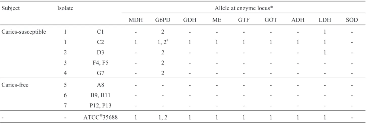
All analyzed enzyme systems (ADH, GDH, G6PD, LDH, MDH, ME, GOT, GTF) showed no activity for anyS. mutansisolates of caries-free subject group (Table 1). Glu-cose-6-phosphate dehydrogenase (G6PD) showed thin band zones that did not vary among all of theS. mutans iso-lates of caries susceptible group and ATCC®35688. Two alleles (allele 1 and allele2) for G6PD were visualized in C2 and ATCC®35688 strains while in other isolates only the second allele was observed and no locus of G6PD was seen in caries free isolates (Figure 1). For lactate dehydrogenase (LDH), the unexpected electrophoretic bands appeared only in C1, C2 and D3 (Table 1).
Isolates #C1, #C2, #D3, #F4, #F5, and #G7 were iso-lated from caries susceptible subjects; Strain #S was S. mutansATCC®35668; Slot E is the pure enzyme that was added as an electrophoretic control.
As it is shown in Table 1, isolate C1 also differed from C2 and they were isolated from the same individual (subject #1) and their enzymatic patterns suggest that they belong to different clones. Indeed, average genetic diver-sity between both clones was determined as 0.468. In this study, genetic diversities for the eight evaluated loci were, 0.59, 0.88, for G6PD and LDH respectively and 0.097 for other loci. The mean diversity for the loci was 0.228.
To determine the genetic distance among isolates as well as their clustering behavior, a non-rooted dendrogram was built (Figure 2). Such tree shows that different isolates could be clustered in four different taxa with genetic dis-tances varying from 0.2398 to 0.9592.
All isolates from free caries subjects as listed in Ta-ble 1 belong to clade C as shown in Figure 2 (genetic dis-tance d³0.4796).
Although in different taxa, different isolates of same individuals (patients #3, #6 and #7) presented no diver-gences (d = 0.0000). Patient #1, however, presented two isolates separated one to each other with a d = 0.9592, showing that more than one clonal type can be found in a same individual.
The differentiation ability of MLEE for cariogenic and non cariogenicS. mutansisolates was confirmed in this study. Isolates of caries susceptible group differed from isolates of caries free group.
According to the results, it is evident that caries-sus-ceptible subjects bear more genotypes than caries-free. It is in agreement with earlier reports (Alaluusuaet al., 1996; Napimogaet al., 2004). Redmo-Emanuelssonet al.(2003), in a study enrolling young adults (mean age of 25.2 years) found a maximum of seven genotypes in subjects who had previous caries experience and Napimogaet al.(2004) found a maximum of eleven and eight genotypes in caries-active subjects using MLEE and AP-PCR respectively.
We obtained up to three genotypes in caries-sus-ceptible subjects (4 subjects; 7 isolates) using MLEE. In contrast, the results of Kreulenet al.(1997) showed a nega-tive relationship between caries activity and genotype di-versity. Redmo-Emanuelssonet al.(2003) suggested that the larger number of genotypes found could be because of the larger number of isolates analyzed, which increases the possibility of detecting different genotypes.
Table 1- Electromorphotypes ofS. mutansisolates of caries-free and caries susceptible individuals.
Subject Isolate Allele at enzyme locus*
MDH G6PD GDH ME GTF GOT ADH LDH SOD
Caries-susceptible 1 C1 - 2 - - - 1
-1 C2 1 1, 2a 1 1 1 1 1 1
-2 D3 - 2 - - - 1
-3 F4, F5 - 2 - - -
-4 G7 - 2 - - -
-Caries-free 5 A8 - - -
-6 B9, B11 - - -
-7 P12, P13 - - -
-- - ATCC®35688 1 1, 2 1 1 1 1 1 1
-*Ac*according to the anodal migration. a
: Two alleles (allele 1 and allele 2) for G6PD were visualized while for others only one locus was seen.
Figure 1- Multilocus enzyme electrophoresis analysis ofS. mutans iso-lates of caries free & caries susceptible subjects.
In this study, all analyzed enzyme systems revealed no activity for anyS. mutansisolates of caries free subject group (3 subject, 5 isolates) so, they belong to one clade. Perhaps, such enzymes are either produced in smaller quan-tities than the method can detect or they are not produced by those isolates. Alaluusuaet al.(1996) related that six car-ies-free children (3 to 7 strains per volunteer) only har-boured one ribotype ofS.mutans. According to these, it is probable that from plaque samples of caries free subjects the only primary strains were found and other strains, if they existed were below the detection level.
Getting activity for G6PD (inS. mutans isolates of caries susceptible group and ATCC®35688) and LDH (in C1, C2 and D3) in this study was in contrast with previous researches that showed no activity for the major part of dehydrogenases (ADH, GDH, G6PD, LDH, MDH, ME) in
S. mutansstrains (Napimogaet al., 2004; Rosaet al., 2006). These differences are probably due to the pH, type of buffer system used in preparation of enzyme extract or type and concentration of used gel. Occasionally, the relative mobil-ities of certain electromorphs are reversed in different buffer system (Selanderet al., 1986). According to Wunder and Bowen (2000), glucosyltransferases are enzymes that act at extracellular environment. This may, at least in part, explain why glucosyltransferase activity was not observed in all of the cases and it is only appeared in C2 and ATCC®35688.
A non-rooted dendrogram was shown that different isolates of this study could be clustered in four different taxa. All isolates from three caries-susceptible patients were grouped in a single taxon (taxon C). Patient #1, how-ever, presented two isolates separated one to each other (d = 0.9592), showing that more than one clonal type can be found in a same individual. Rosaet al.(2006) also could isolate two clusters ofS. mutansisolates from a same indi-vidual, and their enzymatic patterns suggest that they be-long to different clones. The isolates of caries free subjects, presented no divergences (d = 0.0000) although they were isolated from non-related individuals.
In parallel to the discrimination ability, MLEE has proven to be a useful tool for establishing genetic diversity, even in small or subdivided (Rosaet al., 2005) populations. This method detects allelic frequencies prompter than other methodologies, such as RAPD and RFLP. These facts sup-port the premise that MLEE may be used in surveys in which intra-species determination ofS. mutansis required. In conclusion, the results here presented support the propo-sition of MLEE employment for clonal differentiation of
Streptococcus mutans.Three clonal types and one clonal type were observed in caries-susceptible and caries-free subjects, respectively.
Acknowledgments
This work is the part of research project that was sup-ported by Islamic Azad University Khorasgan- Isfahan
branch. The authors thank the President, and the Educa-tional and the Research Vice- Presidents of Islamic Azad University, Khorasgan branch, Isfahan, Iran (Dr A.A. Foroughiabari, Dr M. Hoodaji and Dr P. Najafi) for their help and support.
References
Alaluusua S, Kleemola-Kujala E, Nystrom M (1987) Caries in the primary teeth and salivaryStreptococcus mutansand lacto-bacillus levels as indicators of caries in permanent teeth. Paediatr Dent 9:126-130.
Alaluusua S, Matto J, Gronroos L, Innila S, Torkko H, Asikainen S (1996) Oral colonization by more than one clonal type of mutans streptococci in children with nursing-bottle dental caries. Arch Oral Biol 41:167-173.
Arakawa H, Karasawa K, Igarashi T, Suzuki S, Goto N, Maeda M (2004) Detection of cariogenic bacteria genes by a combina-tion of allele-specific polymerase chain reaccombina-tions and a no-vel bioluminescent pyrophosphate assay. Anal Biochem 15:296-302.
Araújo JV, Junghans TG, Alfenas AC, Gomes AP (1997) Isoen-zyme analysis of Arthrobotrys a nematode-trapping fungus. Braz J Med Biol Res 30:1149-1152.
Baca P, Castillo AM, Baca AP, Liebana MJ, Junco P, Liebana J (2008) Genotypes ofStreptococcus mutansin salivavs. den-tal Plaque, Arch Oral Boil 53:751-754.
Baptist JN, Shaw CR, Mandel M (1971) Comparative zone elec-trophoresis of enzymes ofPseudomonas solanacearumand
Pseudomonas cepacia. J Bacteriol 108:799-803.
Becker MR, Paster BJ, Leys EJ, Moeschberger ML, Kenyon SG, Galvin JL, Boches SK, Dewhirst FE, Griffen AL (2002) Mo-lecular analysis of bacterial species associated with child-hood caries. J Clin Microbiol. 40:1001-1009.
Beighton D, Rippon HR, Thomas HEC (1987) The distribution of
Streptococcus mutansserotypes and dental caries in a group of 5- to 8-year-old Hampshire schoolchildren. Braz Dent J 162:103-106.
Carlsson P, Olsson B, Bratthall D (1985) The relationship be-tween the bacteriumStreptococcus mutansin the saliva and dental caries in children in Mozambique. Arch Oral Biol 30:265-268.
Caugant DA, BfVre K, Gaustad P, Bryn K, Holten E, HfIby EA (1986) Multilocus genotypes determined by enzyme electro-phoresis of Neisseria meningitidis isolated from patients with systemic disease and from healthy carriers. J Gen Microbiol 132:641-652.
Caulfield PW, Walker TM (1989) Genetic diversity within Strep-tococcus mutansevident from chromosomal DNA restric-tion fragment polymorphism. J Clin Microbiol 27:274-278. Gilmour MN, Whittam TS, Kilian M, Selander RK (1987) Ge-netic relationships among the oral streptococci. J Bacteriol 169:5247-5257.
Gronroos L, Alaluusua S (2000) Site specific oral colonization of mutans streptococci detected by arbitrarily primed PCR fin-gerprinting. Caries Res 34:474-80.
Jordan C, LeBlanc DJ (2002) Influences of orthodontic appliances on oral populations of mutans streptococci. Oral Microbiol Immunol 17:65-71.
geno-types isolated from caries-affected and caries-free individu-als. Oral Microbiol Immunol 20:20-24.
Kreulen CM, de Soet HJ, Hogeveen R, Veerkamp JS (1997)
Streptococcus mutans in children using nursing bottles. ASDC J Dent Child 64:107-111.
Li M, Lai G, Wang J (2011) The prevalence of virulent clonal strains of mutans streptococciin vivoand co-culture succes-sion of the strainsin vitro.Open J Stomatol 1:18-24. Lowry OH, Rosebrough NJ, Farr AL, Randall RJ (1951) Protein
measurement with the Folin phenol reagent. J Biol Chem 193:266-267.
Matee MIN, Mikx FHM, de Soet JS, Masell SY, de Graff J, van Palenstein Helderman WH (1993) Mutans streptococci in caries-active and caries-free infants in Tanzania. Oral Microbiol Immunol 8:322-324.
Meloni BP, Lymberi AJ, Thompson RCA (1988) Isoenzyme elec-trophoresis of 30 isolates of Giardia from humans and fe-lines. Am J Trop Med Hyg 38:65-73.
Napimoga MH, Kamiya RU, Rosa TR, Rosa EAR, Hofling JF (2004) Genotypic diversity and virulence traits of Strepto-coccus mutansin caries-free and caries active Individuals. J Med Microbiol 53:697-703.
Naumov GI, Naumova ES, Sniegowiski PD (1997) Differentia-tion of European and Far East Asian populaDifferentia-tions of
Saccharomyces paradoxusby allozyme analysis. Int J Syst Bacteriol 47:341-344.
Nei M (1978) Estimation of average heterozygocity and genetic distance from a small samole of individuals. Genetics 89:583-90.
O’Brien SJ, Simonson JM, Grabowski MW, Barile MF (1981) Analysis of multiple isoenzyme expression among twenty-two species of Mycoplasma and Acholeplasma. J Bacteriol 146:222-232.
Redmo Emanuelsson IM, Carlsson P, Hamberg K, Bratthall D (2003) Tracing genotypes of mutans streptococci on tooth sites by random amplified polymorphic DNA (RAPD) anal-ysis.Oral Microbiol Immunol 18:24-29.
Rosa RT, Napimoga MH, Hofling JF, Gonçalves RB, Rosa EAR (2006) Clonal characterization of Streptococcus mutans
strains by multilocus enzyme electrophoresis. Braz J Micro-biol 37:17-19.
Rosa RT, Napimoga MH, Hofling JF, Gonçalve RB, Rosa EAR (2005) Clonal Diversity ofStreptococcus Clarke(1924) in caries-free adults.Estud Biol 27:49-51.
Schable B, Villarino ME, Favero MS (1991) Application of multi-locus enzyme electrophoresis to epidemiologic investiga-tions of Xanthomonas maltophilia. Infect Control Hosp Epidemiol 12:163-167.
Selander RK, Caugant DA, Ochman H, Musser JM, Gilmour MN, Whittam TS (1986) Methods of multilocus enzyme electro-phoresis for bacterial population genetics and systematics. Appl Environ Microbiol 51:873-884.
Sneath PHA, Sokal RQ (1973) Numerical taxonomy. San Fran-cisco: Freeman, 482 pp.
Wunder D, Bowen WH (2000) Effects of antibodies to glucosyltransferase on soluble and insolubilized enzymes. Oral Dis 6:289-296.