Quim. Nova, Vol. 40, No. 2, 154-157, 2017
A
rti
g
o
http://dx.doi.org/10.21577/0100-4042.20160159
*e-mail: [email protected]
IONTOPHORESIS ON MINOXIDIL SULPHATE-LOADED CHITOSAN NANOPARTICLES ACCELERATES DRUG RELEASE, DECREASING THEIR TARGETING EFFECT TO HAIR FOLLICLES
Breno N. Matos, Larissa M. A. Melo, Maíra N. Pereira, Thaiene A. Reis, Marcílio Cunha-Filho, Taís Gratieri and Guilherme M. Gelfuso*
Departamento de Farmácia, Faculdade de Ciências da Saúde, Universidade de Brasília, 70910-900 Brasília – DF, Brasil
Recebido em 21/06/2016; aceito em 08/08/2016; publicado na web em 26/08/2016
The experiments described in this paper tested the hypothesis whether iontophoresis applied on a chitosan nanoparticle formulation could combine the enhanced drug accumulation into the follicular casts obtained using iontophoresis and the sustained drug release, reducing dermal exposure, provided by nanoparticles. Results showed that even though iontophoresis presented comparable minoxidil targeting potential to hair follicles than passive delivery of chitosan-nanoparticles (4.1 ± 0.9 and 5.3 ± 1.0 µg cm-2, respectively), it
was less effective on preventing dermal exposure, since chitosan-nanoparticles presented a drug permeation in the receptor solution of 15.3 ± 4.3 µg cm-2 after 6 h of iontophoresis, while drug amounts from passive nanoparticle delivery were not detected. Drug release
experiments showed particles were not able to sustain the drug release under the influence of a potential gradient. In conclusion, the application of MXS-loaded chitosan nanoparticles remains the best way to target MXS to the hair follicles while preventing dermal exposure.
Keywords:chitosan; iontophoresis; minoxidil sulphate; nanoparticle; nanosphere; skin.
INTRODUCTION
Minoxidil application is already established as the standard treat-ment for male and female androgenic alopecia. It is the only topical therapy with FDA’s approval and several products based on this drug are available in the market. Even though the exact mechanism of action remains uncertain, research has demonstrated that minoxidil is able to stimulate hair follicles, reversing their progressive minia-turization associated with androgenic alopecia.1
Clinical trials have demonstrated that the success rate of the treatment with 5% minoxidil barely exceeds 40%.2 To elevate these
indices, last researches have focused on developing more efficient formulations, i.e., formulations capable of targeting the active compound to hair follicles and sustaining its release.3 For this, solid
lipid nanoparticles,4 penetration enhancer-containing vesicles5 and
squarticles nanoparticles, formed from sebum-derived lipids,6 have
been proposed. Although many studies have indeed demonstrated improved skin permeation, it is important to consider that minoxi-dil, a pyridine-derivative, is a potent antihypertensive agent; hence, dermal exposure and a consequent systemic effect should be avoided to minimize adverse side effects.7
Our research group has recently reported a two-fold minoxi-dil sulphate (MXS, Figure 1) increase into hair follicles with the application of MXS loaded in chitosan nanoparticles (MXS-NP) instead of the drug solution, which provided a sustained drug release and prevented dermal exposure.8 In this study, we have prepared
and characterized MXS-NP and demonstrated that after passive treatment of porcine skin with this formulation, an enhancement in MXS penetration in hair follicles was observed compared to a simple solution of the drug. Moreover, it was not observed in any experiment significant concentrations of the drug in deeper skin layers.8 Other study of our group has observed that application of
iontophoresis in a gel formulation containing the free drug, i.e., non-encapsulated MXS, increased by 5-fold the amount of drug reaching the follicular infundibula when compared with passive
delivery, resulting therefore in a more pronounced target effect to the hair follicles.9 The drawback of this last approach, however,
was that the increased targeting effect was also followed by a very significant drug transdermal transport, once a significant concen-tration of MXS was recovered from epidermis and dermis in both tested skin models (porcine and rat skin).
Iontophoresis is a technique classically studied to increase and control the transport of hydrophilic and charged molecules into and through skin, by application of a mild direct current (of no more than 0.5 mA/cm2).10,11 It has been currently used for topical and transdermal
delivery of a great variety of drugs.12-16 The two main mechanisms
involved in iontophoretic delivery of drugs are electromigration and electroosmosis. Both positively or negatively charged molecules are typically delivered by electromigration, as the charged drug stands in the same charged compartment, while neutral molecules can be transported via electroosmosis.10,11 As hair follicles and other skin
ap-pendices present lower electrical resistance for drug transport through the skin, it is well established in literature that molecules transported by iontophoresis follow the appendageal path.17
Some authors have recently evaluated the application of ion-tophoresis to enhance skin permeation of drugs encapsulated in nanostructured particles,18-21 but the feasibility of this approach still
remains inconclusive.
Figure 1. Chemical structure of MXS (2,4-diamine-6-piperidine-
Iontophoresis on minoxidil sulphate-loaded chitosan nanoparticles accelerates drug release 155
Vol. 40, No. 2
In this way, the aim of the experiments presented in this paper was to evaluate whether the application of iontophoresis to the MXS-NP formulation could result in a maximum MXS delivery into the hair follicles, at the same time that could minimize transdermal drug absorption of the encapsulated drug.
EXPERIMENTAL
Material
MXS (99%) was kindly provided by Galena Química e Farmacêutica Ltda. (Campinas, Brazil). Low molecular weight chitosan (75–85% of deacetylation), acetic acid and sodium tripoly-phosphate (TPP), used to prepare nanoparticles, were purchased from Sigma–Aldrich (Steinheim, Germany). Silver (Ag) wire (99.99%, ø = 1.5 mm), silver chloride (AgCl, 99.99%) and platinum wire (ø = 1.0 mm) used to prepare electrodes were purchased from Sigma–Aldrich (Steinheim, Germany). Scotch book tape (3M, St Paul, MN, USA) was used for tape stripping and cyanoacrylate superglue (Henkel Loctite, Dublin, Ireland) was used for follicles biopsies. Cellulose acetate membranes (cut off = 0.22 µm) were purchased from Fisher Scientific (Leicestershire, UK). HEPES salt and sodium chloride were from Acros (Berkeley, NJ, USA). The solvents used for extraction and chromatographic analysis were all of HPLC grade. The water used in all preparations was of Milli-Q grade (Millipore, Illkirch-Graffenstaden, France).
Skin
Porcine ears were gently donated by a local abattoir (Bonasa Alimentos, São Sebastião, Brazil) and were obtained less than 1 h post-sacrifice of the animal. The whole skin was removed from the outer region of the ear, separated from its underlying layer, and used “full-thickness” to guarantee the intactness of the hair follicles. The skin was stored frozen at −20 °C for a maximum of 1 month before use.
Preparation of nanoparticles
MXS-NP were prepared by inducing the gelation of chitosan solu-tions with a crosslink agent (TPP), as previously described.8 Briefly,
8.75 mg of MXS was added to 5 mL of an acidified chitosan solution (containing 8.75 mg chitosan, 50 mg acetic acid), pH 5. Then, 2 mL of a TPP solution (at 1 mg mL-1) was added under constant magnetic
stirring (700 rpm), resulting in the nanoparticles dispersion. The MXS-NP formed presented mean diameter of 200 ± 50 nm (PDI = 0.25 ± 0.03), positive zeta potential of +23 ± 4 mV and entrapment efficiency of 72%.
Permeation studies protocol
Modified Franz diffusion cells were assembled with the skin of the porcine’s ear separating the donor to the receptor compartment. The receptor compartment was filled with 15 mL of HEPES buffer (pH 7.4). In the donor compartment, it was added 1 mL of (i) MXS-NP formulation (1.25 mg mL-1 MXS, 0.53 mg mL-1 NaCl, pH 5.5),
or (ii) MXS aqueous solution (1.25 mg mL-1 MXS, 0.53 mg mL-1
NaCl, pH 5.5). Positive Ag electrode was placed in donor chamber (area = 1.3 cm2) in contact with formulation, while negative AgCl
electrode was placed in receptor solution. A direct constant current of 0.65 mA (0.5 mA cm-2) was applied with the aid of a power source
connected to the positive and negative electrodes (Sinewave generator, Model GCC20200, Piracicaba, Brazil). Passive experiments were also
conducted with the same formulations but without the insertion of the electrodes. All experiments were conducted for 6 h, when receptor solution was withdrawn from the diffusion cells and analysed for MXS content in HPLC.
Recovery of MXS from skin layers
At the end of permeation experiments, the skin was removed from the diffusion cell and placed onto a flat surface with the stra-tum corneum facing up. The skin was cleaned with a water-soaked gauze pad and tape-stripped 15 times, using Scotch book tapes. MXS content in the tapes was determined as described below after exhaustive extraction of the drug with methanol over a 24 h period. A drop of cyanoacrylate superglue was applied to the stripped skin area and covered with a further tape-strip using light pressure. After total polymerisation of the glue (∼5 min), this tape-strip was then removed and the skin surface biopsy obtained in this way contained follicular casts, from which MXS was extracted with methanol and quantified. Finally, reminiscent skin was cut into small pieces and placed in plastic tube along with methanol also over a period of 24 h for drug extraction. The resulting suspensions were filtered on 0.22 µm filters and quantified by HPLC.
The efficiency of recovery of MXS from the stratum corneum, follicular material and viable epidermis was previously determined, with MXS recovery values higher than 90%.
Influence of iontophoresis in MXS release
In this series of experiments, modified Franz-type diffusion cells were mounted with hydrophilic cellulose membranes separating the donor and receptor compartment, which was filled with 15 mL of HEPES buffer (pH 7.4). The cellulose membranes served here only as a support to the formulations. The release of MXS from 1 mL of MXS-NP formulation containing the equivalent to 1.25 mg/mL MXS (pH 5.5) was determined in vitro under the influence of anodal ion-tophoresis or not over a period of 6 h. The ionion-tophoresis experiment followed the protocol described in the previous section. Diffusion of MXS from an aqueous solution (1.25 mg mL-1, pH 5.5) through
the cellulose acetate membrane was also determined as a control. Release rates of MXS from MXS-NP with or without ionto-phoresis application, as well as, from a simple MXS solution, were calculated from the slope of linear curves obtained by plotting amount of MXS released (µg cm-2) versus time (h), considering the 6 time
points of each experiment. All release profiles followed zero order kinetics, with linear correlation coefficients (r) higher than 0.9 when amount of drug released is related with time.
HPLC analysis
The quantification of MXS was performed by a reversed-phase chromatographic method using a HPLC with UV detection, set at 285 nm (model LC-20AD, Shimadzu, Kyoto, Japan). The operating conditions of the method were the following: 50 µL of injection volume; reverse-phase C18 column (Shimadzu, 4.6 mm × 15 mm, 5µm); water/
acetronitrile (80:20, v/v) as an isocratic mobile phase; and flow rate of 1.0 mL/min. The method was previously validated in accordance to FDA guidelines, and proved to be selective and linear (r = 0.9997). The LOQ and LOD were 0.5 µg mL-1 and 0.05 µg mL-1, respectively.
Statistical studies
Matos et al.
156 Quim. Nova
deviations. The data were analysed by ANOVA followed by a non-parametric Tukey’s test. The statistical significance was fixed at P < 0.05.
RESULTS AND DISCUSSION
The hypothesis to be tested in this paper was whether iontopho-resis applied to a nanoparticle formulation could target the nanopar-ticles to the follicular casts and whether, once nanoparnanopar-ticles were within the casts, they could sustain drug release, preventing dermal exposure. The rationale for this approach has been partially proven by our former publications: iontophoresis clearly targeted MXS to the follicular route, as it is the pathway with lower resistance under the influence of a potential gradient,9-11,17 and MXS-nanoparticles
sustained drug release about twice more than previous prepared MXS-loaded microparticles.8,22
Figure 2 shows recovery of MXS from stratum corneum tape strips and follicular casts after 6 h of either passive or iontophoresis driven diffusion from the two tested formulations – free-drug solution and MXS-NP. The passive delivery study of MXS from the chitosan nanoparticles reflects previously published results of our group8 and
is depicted here simply to serve as a control.
MXS-NP have a positive zeta potential (+23 ± 4 mV), as a result of amino groups of the polymer chitosan, which at the acid pH of the formulation is positively charged. It was thereby supposed that an electrical current would improve accumulation of these positively charged nanoparticles into the hair follicles when the formulation was put in contact with the positive electrode in the anode compart-ment. Present results, however, showed that the combination of these techniques did not provide the expected cumulative targeting effect. MXS accumulation in follicle casts following iontophoresis of the nanoparticles (4.1 ± 0.9 µg cm-2) was not higher (p>0.05) than drug
amounts obtained after passive permeation of the MXS chitosan
nanoparticles (5.3 ± 1.0 µg cm-2) or the iontophoretic delivery from
the drug solution (4.7 ± 0.9 µg cm-2). This last result contrasts a study
previously conducted by our group,9 in which the iontophoresis
in-creased 5 times the penetration MXS follicles from a gel formulation. However, besides it is a different formulation (poloxamer gel formula-tion instead of a simple aqueous soluformula-tion), the drug concentraformula-tion in that gel formulation was 10-fold higher than this used here, which was prepared containing 1.25 mg mL-1 MXS to serve as a control for
the formulation of nanoencapsulated drug.
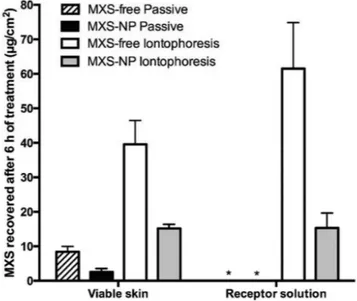
Concerning dermal exposure, which is represented by drug amounts retained in viable skin and receptor solution (Figure 3), although it was significantly lower when the iontophoresis was applied to the nanoparticle formulation (15.2 ± 1.2 µg cm-2 and
15.3 ± 4.3 µg cm-2, respectively) in comparison to the free drug
solution (39.6 ± 6.9 µg cm-2 and 61.5 ± 13.4 µg cm-2, respectively)
(p<0.05), the values were still higher than those obtained with the passive nanoparticle formulation delivery (2.5 ± 1.0 µg cm-2 in viable
skin and no drug detected in receptor solution).
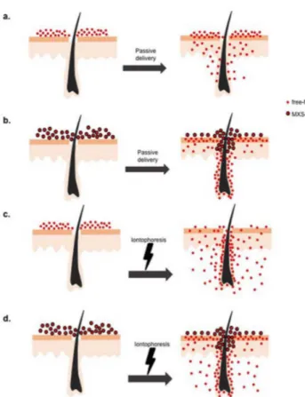
Results, therefore, show that there is no advantage on applying iontophoresis for alopecia treatment using these formulations. In fact, as dermal exposure is increased upon the application of the potential gradient, the risk of side effects is higher. A schematic representation of the results is presented in Figure 4.
Taveira et al.21 have explored the combination of iontophoresis
with solid lipid nanoparticles loading doxorubicin and also observed that twice more drug was recovered from deeper layers of skin with this association compared to the passive delivery of the drug from the nanoparticles. In contrast to the rigid chitosan nanoparticles, how-ever, solid lipid nanoparticles are deformable particles, which tend to accumulate in skin surface forming an occlusive film. This film increases stratum corneum hydration and, consequently, skin perme-ability for encapsulated drugs.23 Furthermore; there is no evidence
that lipid nanoparticles under influence of iontophoresis penetrate hair follicles, and the authors believe that under the influence of ionto-phoresis, doxorubicin was released from the solid nanoparticles and Figure 2. MXS recovered from stratum corneum, hair follicles, after 6 h of
either passive treatment with a simple MXS water solution (1.25 mg mL-1
MXS, pH 5.5), passive treatment with MXS-loaded chitosan nanoparticles (1.25 mg mL-1 MXS, pH 5.5, 0.53 mg mL-1 NaCl), anodal iontophoresis applied
in a MXS water solution (1.25 mg mL-1 MXS, pH 5.5, 0.53 mg mL-1 NaCl),
anodal iontophoresis of MXS-loaded chitosan nanoparticles (1.25 mg mL-1
MXS, pH 5.5, 0.53 mg mL-1 NaCl)
Figure 3. MXS recovered from viable dermis and receptor solution, after 6 h
of either passive treatment with a simple MXS water solution (1.25 mg mL-1
MXS, pH 5.5), passive treatment with MXS-loaded chitosan nanoparticles (1.25 mg mL-1 MXS, pH 5.5, 0.53 mg mL-1 NaCl), anodal iontophoresis applied
in a MXS water solution (1.25 mg mL-1 MXS, pH 5.5, 0.53 mg mL-1 NaCl),
ano-dal iontophoresis of MXS-loaded chitosan nanoparticles (1.25 mg mL-1 MXS,
Iontophoresis on minoxidil sulphate-loaded chitosan nanoparticles accelerates drug release 157
Vol. 40, No. 2
permeated the skin under the influence of electroosmotic flow,23 i.e.,
a convective water flow towards the skin formed under the influence of an electric potential application.10,11
Indeed, our results can be explained by release rate of MXS from MXS-NP dispersion under the influence of iontophoresis. It has been clearly demonstrated in Table 1 that, under the influence of anodal iontophoresis, the nanoparticles are no longer able to sustain drug release, i.e. MXS release flux from nanoparticles formulation under influence of electric potential was not statistically different from free-drug diffusion through the cellulose membrane (p>0.05), but is considerably different from MXS release from chitosan nanoparticles without current application (p<0.05).
In this way, the use of an additional technology, as iontophore-sis, which could increase therapy cost, is not justified for chitosan--nanoparticles loading MXS. Further studies employing different nanosystems are required to better understand nanoparticle behaviour under the influence of a potential gradient.
CONCLUSION
Even though iontophoresis presents comparable MXS targeting potential to hair follicles as nanoencapsulation, it is less effective on preventing dermal exposure, since the electric current accelerates drug release from the nanoparticles. The passive application of MXS-loaded chitosan nanoparticles remains the best way to target MXS to the hair follicles while avoiding possible side effects.
ACKNOWLEDGEMENTS
The authors would like to thank CAPES (Coordenação de Aperfeiçoamento de Pessoal de Nível Superior), CNPq (Conselho Nacional de Desenvolvimento Científico e Tecnológico) and the University of Brasília (Edital FUB/DPP 10/2011 and 02/2013) for funding this research. We would also like to thank “Bonasa Alimentos” for providing porcine skin.
REFERENCES
1. Messenger, A. G.; Rundegren, J.; Br. J. Dermatol.2004, 150, 186. 2. Olsen, E. A.; Whiting, D.; Bergfeld, W.; Miller, J.; Hordinsky, M.;
Wan-ser, R.; Zhang, P.; Kohut, B.; J. Am. Acad. Dermatol.2007, 57, 767. 3. Fang, C. L.; Aljuffali, I. A.; Li, Y. C.; Fang, J. Y.; Ther. Delivery2014,
5, 991.
4. Padois, K.; Cantiéni, C.; Bertholle. V.; Bardel, C.; Pirot, F.; Falson, F.;
Int. J. Pharm.2011, 416, 300.
5. Mura, S.; Manconi, M.; Fadda, A. M.; Sala, M. C.; Perricci, J.; Pini, E.; Sinico, C.; Pharm. Dev. Technol.2013, 18, 1339.
6. Aljuffali, I. A.; Sung, C. T.; Shen, F. M.; Huang, C. T.; Fang, J. Y.; AAPS J.2014, 16, 140.
7. Han, J. H.; Kwon, O. S.; Chung, J. H.; Cho, K. H.; Eun, H. C.; Kim, K. H.; J. Dermatol. Sci.2004, 34, 91.
8. Matos, B. N.; Reis, T. A.; Gratieri, T.; Gelfuso, G. M.; Int. J. Biol. Ma-cromol.2015, 75, 225.
9. Gelfuso, G. M.; Gratieri, T.; Delgado-Charro, M. B.; Guy, R. H.; Vianna Lopez, R. F.; J. Pharm. Sci. 2013,102, 1488.
10. Gratieri, T.; Gelfuso, G. M.; Lopez, R. F. V.; Quim. Nova2008, 31, 1490. 11. Gratieri, T.; Kalia, Y. N.; Adv. Drug Delivery Rev. 2013, 65, 315. 12. Sylvestre, J. P.; Díaz-Marín, C.; Delgado-Charro, M. B.; Guy, R. H.; J.
Control. Release2008, 131, 41.
13. Gelfuso, G. M.; Gratieri, T.; Souza, J. G.; Thomazine, J. A.; Lopez, R. F.; Eur. J. Pharm. Biopharm.2011, 77, 249.
14. Djabri, A.; Guy, R. H.; Delgado-Charro, M. B.; Int. J. Pharm.2012, 435, 27.
15. Bhatia, G.; Banga, A. K.; Biomed. Res. Int.2014, 2014, 537941. 16. Gratieri, T.; Pujol-Bello, E.; Gelfuso, G. M.; de Souza, J. G.; Lopez, R.
F.; Kalia, Y. N.; Eur. J.Pharm. Biopharm.2014, 86, 219. 17. Cullander, C.; Guy, R. H.; J. Invest. Dermatol.1991, 97, 55.
18. Tomoda, K.; Terashima, H.; Suzuki, K.; Inagi, T.; Terada, H.; Makino, K.; Colloids Surf., B2011, 88, 706.
19. Tomoda, K.; Watanabe, A.; Suzuki, K.; Inagi, T.; Terada, H.; Colloids Surf., B2012, 97, 84.
20. Tomoda, K.; Terashima, H.; Suzuki, K.; Inagi, T.; Terada, H.; Makino, K.: Colloids Surf., B2012, 92, 50.
21. Taveira, S. F.; De Santana, D. C.; Araújo, L. M.; Marquele-Oliveira, F.; Nomizo, A.; Lopez, R. F.; J. Biomed. Nanotechnol.2014, 10, 1382. 22. Gelfuso, G. M.; Gratieri, T.; Simão, P. S.; de Freitas, L. A.; Lopez, R. F.;
J. Microencapsulation2011, 28, 650.
23. Müller, R. H.; Petersen, R. D.; Hommoss, A.: Pardeike, J.; Adv. Drug Deliv. Rev.2007, 59, 522.
Table 1. MXS release rate calculated based on drug release profile from MXS--free solution, MXS-NP, and MXS-NP under the influence of iontophoresis (0.5 mA cm-2)
Formulation Release rate (µg cm-2 h-1) r
MXS-free solution 188.9 (± 6.0) 0.98
MXS-NP 35.4 (± 1.8) 0.91