O
RIGINALA
RTICLE Revista Brasileira de FisioterapiaBreathing pattern and thoracoabdominal
motion in mouth-breathing children
Padrão respiratório e movimento toracoabdominal de crianças respiradoras orais
Brant TCS1, Parreira VF1, Mancini MC2, Becker HMG3, Reis AFC4, Britto RR1
Abstract
Objective: To characterize the breathing pattern and thoracoabdominal motion of mouth-breathing children aged between eight and ten years and to compare these characteristics with those of nose-breathing children of the same ages. Methods: This observational study was carried out in a university laboratory. The sample size of 50 subjects was estimated based on the results of a pilot study with ten children in each group (total of 20 children) and considering a significance level of 0.05 and statistical power of 0.80. Twenty-six mouth-breathing and 25 nose-mouth-breathing children participated. Calibrated respiratory inductive plethysmography was used to analyze the following variables, among others: respiratory frequency (f), rib cage contribution towards tidal volume (%RC/Vt), phase angle (PhAng) and the ratio between time taken to reach peak inspiratory flow and total inspiratory time (PifT/Ti). Peripheral oxygen saturation of hemoglobin (SpO2)
was measured using pulse oximetry. Statistical analysis was performed using the Student’s t test for independent groups or the Mann-Whitney U test, according to the sample distribution of the variables. Results: A total of 4,816 respiratory cycles were analyzed: 2,455 from mouth-breathers and 2,361 from nose-breathers, with a mean of 94 cycles per child. No statistically significant differences were observed between the groups, for the variables studied (f=20.00±2.68 versus 20.73±2.58, p=0.169; %RC/Vt=39.30±11.86 versus38.36±10.93,
p=0.769; PhAng=14.53±7.97versus13.31±7.74, p=0.583; PifT/Ti=57.40±7.16 versus 58.35±5.99, p=0.610; SpO
2=96.42±1.52% versus
96.88±1.01%, p=0.208; respectively). Conclusions: These results suggest that mouth-breathing children show breathing patterns and thoracoabdominal motion that are similar to those of nose-breathing children in the same age group.
Key words: mouth breather; nose breather; breathing pattern; chest physical therapy; children.
Resumo
Objetivo: Caracterizar o padrão respiratório e o movimento toracoabdominal de crianças respiradoras orais, na faixa etária entre oito e dez anos, e compará-lo ao de seus pares respiradoras nasais. Métodos: Estudo observacional realizado em laboratório universitário. O número amostral calculado com base em um estudo piloto com dez crianças em cada grupo, perfazendo um total de 20 crianças, foi de 50 para um nível de significância de 0,05 e um poder estatístico de 0,80. Participaram do estudo 26 crianças respiradoras orais e 25 respiradoras nasais. A pletismografia respiratória por indutância calibrada foi o instrumento utilizado para a análise das seguintes variáveis, entre outras: freqüência respiratória (FR), contribuição da caixa torácica para o volume corrente (%CT/Vc), ângulo de fase (Angfase) e a razão entre o tempo para alcançar o pico de fluxo inspiratório e o tempo inspiratório (PifT/Ti). A saturação periférica da hemoglobia em oxigênio (SpO2) foi medida pela oximetria de pulso. A análise estatística foi realizada por meio do teste t de Student para grupos independentes e
do teste U de Mann-Whitney, em função da distribuição das variáveis. Resultados: No total, 4.816 ciclos respiratórios foram analisados, sendo 2.455 de respiradores orais e 2.361 de respiradores nasais, com média de 94 ciclos por criança. Não houve diferença significativa entre os grupos nas variáveis estudadas (FR=20,00±2,68 versus 20,73±2,58, p=0,169; %CT/Vc=39,30±11,86 versus 38,36±10,93,
p=0,769; Angfase=14,53±7,97versus13,31±7,74, p=0,583; PifT/Ti=57,40±7,16 versus 58,35±5,99, p=0,610; SpO
2=96,42±1,52% versus
96,88±1,01%, p=0,208; respectivamente). Conclusões: Estes resultados sugerem que as crianças respiradoras orais apresentam padrão respiratório e movimento toracoabdominal semelhantes às de respiradores nasais de mesma faixa etária.
Palavras-chave: respirador oral; respirador nasal; padrão respiratório; fisioterapia respiratória; criança.
Received: 14/05/2008 – Revised: 27/08/2008 – Accepted: 09/09/2008
1 Department of Physical Therapy, School of Physical Education, Physical Therapy and Occupational Therapy (EEFFTO), Universidade Federal de Minas Gerais (UFMG) – Belo Horizonte (MG), Brazil 2 Department of Occupational Therapy, EEFFTO, UFMG
3 Department of Otorhinolaryngology of the School of Medicine, UFMG
4 Graduate Program in Rehabilitation Sciences, Department of Physical Therapy and Occupational Therapy, EEFFTO, UFMG
Correspondence to: Verônica Franco Parreira, Departamento de Fisioterapia, Escola de Educação Física, Fisioterapia e Terapia Ocupacional, Universidade Federal de Minas Gerais, Avenida Presidente Antônio Carlos, 6.627, Pampulha, CEP 31270-901, Belo Horizonte (MG), Brazil, e-mail: [email protected] / [email protected]
Introduction
he human physiological breathing pattern is nasal, regard� less of age1,2, because the nose has three important functions:
heating, iltering, and moistening the air that is inhaled3,4. Any
factor that leads to upper respiratory tract (URT) obstruction causes nose breathing to be replaced by mouth breathing1,5. Ac�
cording to the literature, mouth breathing may cause changes in the respiratory pattern, such as an increase in respiratory frequency, associated with a reduced amplitude6,7 and the need
to use accessory inspiratory muscles to overcome the high na� sal resistance8.
Mayer et al.9 analyzed thoracoabdominal synchrony in
healthy children (aged three to ive) by means of plethysmo� graphy. hhe authors concluded that this procedure success� e authors concluded that this procedure success� fully evaluated the synchronism of thoracoabdominal motion and suggested that this variable should be integrated to the evaluation measurements of the respiratory function of chil� dren with all types of respiratory disorders.
The search for scientific information that may guide clinical practice has been considered relevant by health pro� fessionals10. In the literature, there are no studies that eva�
luate the respiratory pattern of mouth�breathing children6,7.
A systematic study focusing on this particular issue might contribute to its evaluation as well as to the therapeutic approach.
hus, the aim of this study was to characterize the respi� ratory pattern and the thoracoabdominal motion of school children aged eight to ten with a clinical diagnosis for mouth� breathing, and compare them to those of nose�breathing children.
Methods
Sample
he sample number was calculated based on a pilot study involving 20 children, ten of which were mouth�breathers (MB) and ten nose�breathers (NB). he sample size was es� timated at 50 for both groups together considering a α=0.05 level of signiicance (non�directional analysis) and a 0.80 statistical power. Children aged eight to ten were selected non�randomly and allocated into two groups: group 1 com� prised 26 children with a clinical diagnosis of mouth�brea� thing, selected from the Mouth�Breather Out�Patient Clinic of Universidade Federal de Minas Gerais (UFMG) and from the local community; group 2 comprised 25 NB children, se� lected from the local community. he criteria for inclusion
in the MB group were: mouth�breathing predominance conirmed by clinical tests, medical diagnosis of URT obs� truction, interview with the parents and direct observation of lip closure loss. For the NB group, the criteria for inclusion were: nose�breathing predominance conirmed by clinical tests, interview with the parents and direct observation of the presence of lip closure11. All children had a body mass
index (BMI=kg/m2) below the 95th percentile12, a report of
respiratory pathology in the lower respiratory tract, such as asthma or bronchiectasis, and no previous or current physi� cal therapy intervention.
he weight and height were measured by means of a scale equipped with a stadiometer (Filizola, São Paulo, Bra� zil); the heart rate (HR) and the peripheral oxygen saturation of hemoglobin (SpO2) were measured with a pulse oximeter (Datex�Ohmeda, Louisville, CO, USA). he children’s parents or guardians were informed and instructed about the procedures, which were only carried out after they had read and signed the consent form. he study was approved by the Research Ethics Committee of UFMG (ETIC 265/04).
Instruments
In order to carry out the research, an inductance ple� thysmography system was used (Respitrace, Nims, Miami, FL, USA). It is a highly reliable instrument used to monitor the volume and timing of breathing patterns and the tho� racoabdominal motion through changes in the transverse section of the compartments of the rib cage (RC) and abdo� men (AB)13�15. The calibration procedure has been described
in previous studies16,17. In this study, the following variables
of the respiratory cycle were analyzed by the plethysmogra� phy: tidal volume (Vt), respiratory frequency ( f ), minute ventilation (VE), ratio of time to peak inspiratory flow to inspiratory time (PifT/Ti), mean inspiratory flow (Vt/Ti), rib cage contribution towards Vt(%RC/Vt), and phase angle (PhAng).
Procedures
he respiratory pattern variables and the thoracoabdominal motion variables were recorded by means of plethysmography for 20 minutes. he irst ten minutes were set aside for calibra� tion and signal stabilization and the remainder of the time for the analysis itself.
Statistical analysis
The statistical analysis consisted of descriptive measu� rements, displayed as means and standard deviation, for the different variables relating to the sample characterization and to the respiratory pattern. In order to test the nor� mality of each variable, we used the Kolmogorov�Smirnov test. When the variable had a normal distribution both in the mouth� and nose�breathing groups, the t test for in� dependent samples was applied. When the variable had a distribution that was not normal in each group of children separately, or in one of them, we applied Mann�Whitney U test for the inferential comparative analysis between the groups18. A α=0.05 level of significance was adopted in the
analyses. The statistical analyzes were carried out with the software Statistical Package for Social Sciences 11.0(SPSS, Chicago, IL, USA).
Results
Sixty children were initially invited to take part in the study. Seven of them were excluded: four for having a BMI above the 95th percentile; one due to a URT infection; one because of a
medical diagnosis for bronchitis; and one for having a congeni� tal heart murmur. hus, the sample was reduced to 53 children. However, it was not possible to analyze the plethysmographic record of two of these children, due to its degree of irregularity. herefore, the data for these children were excluded, and the study was carried out with a total of 51 children, 26 of which were MB, and 25 NB. All of the children were able to perform all of the procedures.
he MB group consisted of seven girls (three with allergic rhinitis, three with adenoid hypertrophy, one with allergic rhinitis associated with adenoid hypertrophy) and 19 boys (seven with allergic rhinitis, four with adenoid hypertrophy, two with adenoid and tonsil hypertrophy, two with unde� termined cause) with mean age of 8.81±0.80 years, mass of 31.71±6.02kg, height of 1.35±0.07m, BMI of 17.47±2.24kg/m2,
SpO2 of 96.42±1.52% and HR of 87.11±9.57bpm.
he NB group was comprised of 12 girls and 13 boys, with mean age of 9.08±0.81 years, mass of 32.61±7.47kg, height of 1.36±0.09m, BMI of 17.37±2.09kg/m2, SpO
2 of 96.88±1.01%, and
HR of 87.40±9.42bpm. he age, mass, SpO2 and BMI variables did not have normal distribution in any of the groups, leading us to include the Mann�Whitney statistical U test in the analy� sis. he t test for independent groups was used to analyze the height and HR. here was no signiicant diference between both groups with regard to age, mass, height, BMI, SpO2, or HR (p=0.230, p=0.873 p=0.470, p=0.932, p=0.208 and p=0.915, respectively).
During data collection, there was a technical problem with the Vitatrace table spyrometer used for the calibration of the tidal volume, which made it impossible to use the data of the calibrated Vt and of the variables that used Vt for cal� culation, namely the minute volume and the mean inspira� tory low. his occurred as we collected the data from the last 18 children (eight MB and ten NB). herefore, the data from these variables were analyzed in a group of 33 children (18 MB and 15 NB). he MB group consisted of three girls and ifteen boys with a mean age of 8.72±0.75 years, mass of 30.84±4.03 kg, height of 1.34±0.05m, BMI of 17.23±1,88kg/m2 and SpO
2
of 96.27±1.56 %. he NB group comprised nine girls and six boys with mean age of 9.20±0.77 years, mass of 33.41±8,05kg, height of 1.36±0.09m, BMI of 17.65±2.17kg/m2 and SpO
2 of
96.80±0.86%.
Mass and height showed normal distribution in both groups, therefore the t test for independent groups was ap� plied. Age, BMI, and SpO2 did not have a normal distribution, therefore the Mann�Whitney U test was applied. here was no signiicant diference between the two groups regarding age, mass, height, BMI, or SpO2 (p=0.083, p=0.245, p=0.356, p=0.406 and p=0.356, respectively).
In the present study, a total of 4,816 respiratory cycles were analyzed, 2,455 of which corresponded to MB and 2,361 to NB, with a mean of 94 cycles per child. Table 1 displays the variables in the respiratory pattern and thoracoabdominal motion of these 33 children. In the comparison between the MB and NB groups, there was no signiicant diference in any of the analyzed variables. Table 2 shows the variables relative to the time component of respiratory pattern and thoracoab� dominal motion, evaluated in the 51 children. hese variables are independent from volume calibration in the plethysmo� graph record, i.e. f, %RC/Vt, PhAng, and PifT/Ti. here was no signiicant diference between the two groups in any of the variables.
Discussion
According to the authors’ knowledge, this is the irst study to evaluate the respiratory pattern and thoracoabdominal mo� tion of mouth�breathing children in a systematized manner. he main indings were that the mouth�breathing children behaved as the nose�breathing children with regard to volume and time of the respiratory pattern, as well as thoracoabdomi� nal motion.
he literature is very scarce with regard to the study of the respiratory pattern in children. No previous studies were found concerning the evaluation of the volume variables in children’s respiratory pattern; thus, the values for Vt, VE and Vt/Ti observed in this study will be discussed in relation to those found by Feltrim in 40 adults19. he Vt was the value
that showed the greatest discrepancy (42 and 46%), and may be related to the fact that the children’s lungs are still
in a developmental stage, unlike adults, whose lungs are fully developed20. Proportionally, the data relative to the VE ob�
served in this study (77 and 81%) showed a greater similarity to those relative to the Vt. One must consider that the VE is the product of Vt and f. Because the f at rest observed in the children was relatively higher than that observed in young adults, there may have been a partial compensation21,22. One
can suggest that the increased f would be a physiological compensatory mechanism for the diminished Vt, resulting in VE values close to those in adults. In regard to Vt/Ti, which relects the action of the respiratory center, i.e. the ventila� tory drive, the discrepancy between children and adults was the smallest observed (89 and 90%). In the absence of neu� rological lesions that might interfere with the action of the respiratory center, it can be assumed that the behavior of the ventilatory drive, i.e., the urge to breathe, is similar in adults and children23.
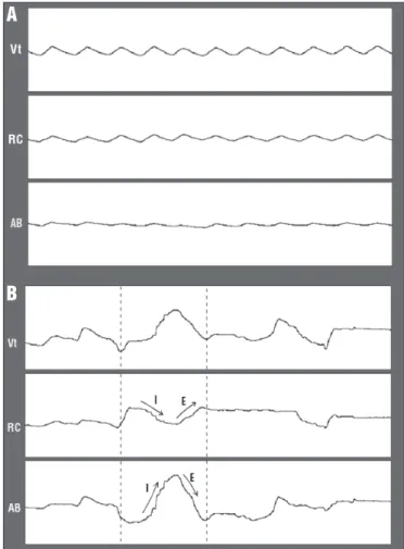
A
B
Figure 1. Waveform traces from Vt, from rib cage displacement (RC) and abdomen (AB). A. rest breathing, without asynchrony signs. B. active nasal aspiration, showing asynchrony. The arrows indicate the direction of motion: during inspiration (I), RC moves down while AB moves up; during expiration (E), RC moves up and AB moves down. The traces sections (A and B) refer to 30 seconds of the records of mouth-breathing children.
Data are presented as mean ± SD. MB refers to mouth-breathing children; NB to nose-breathing children; Vt to tidal volume; f to respiratory frequency; VE to minute ventilation; Vt/Ti to mean inspiratory flow; PifT/Ti to the ratio of time to reach peak inspiratory flow to inspiratory time; %RC/Vt to rib cage contribution towards Vt and PhAng to Phase Angle that measures asynchrony between thoracic and abdominal compartments. The Mann-Whitney U test was used to analyze the following variables: Vt, f and VE, and the t test for independent groups was used to analyze Vt/Ti, %RC/Vt, PhAng and PifT/Ti.
Table 1. Breathing pattern and thoracoabdominal motion data from 18 mouth-breathing and 15 nose-breathing children.
Variables MB NB p value
Vt (ml) 203.43±90.26 200.93±55.87 0.735
f (bpm) 19.76±2.32 20.92±3.02 0.294
VE (l/min) 3.93±1.62 4.12±1.04 0.381
Vt/Ti (ml/s) 175.31±70.04 182.00±42.07 0.381
PifT/Ti (%) 57.97±6.74 58.74±6.50 0.741
RC/Vt (%) 35.65±8.09 37.84±11.90 0.674
PhAng (°) 14.87±8.58 15.28±8.91 0.556
Variables MB NB p value
f (bpm) 20.00±2.68 20.73±2.58 0.169
PifT/Ti (%) 57.40±7.16 58.35±5.99 0.610
RC/Vt (%) 39.30±11.86 38.36±10.93 0.769
PhAng (°) 14.53±7.97 13.31±7.74 0.583 Table 2. Breathing pattern (time variables) and thoracoabdominal motion data from 26 mouth-breathing and 25 nose-breathing children.
One of the limitations of this study was the fact that there was no equivalence between the number of girls and boys in the MB and NB groups, despite the eforts to achieve that and the fact that there was no signiicant diference in age between the MB and NB groups. According to the literature, there is no diference between men and women concerning the respira� tory pattern in adults19,24. Another limitation was the impos�
sibility to analyze the components of the respiratory pattern volume of the 51 children. One must take into account the fact that variables such as time and thoracoabdominal motion of the respiratory pattern had very similar mean values in the groups of 33 and 51 children.
Mayer et al.9 studied thoracoabdominal time and syn�
chrony variables in 50 children, analyzing a range of 24 and 45 respiratory cycles per child. In the present study, there was a considerably higher number of respiratory cycles, which guarantees a greater consistency in the values observed. It is worth noting that both studies analyzed children belonging to diferent age groups. One may question whether the ab� sence of a signiicant diference between the groups, made evident in the present study, might be related to the fact that the variables for volume were studied in 33 children, there� fore not reaching the sample calculation. Nonetheless, for the analysis of the data relative to the time variables ( f, PhAng, and PifT/Ti) the data relative to the 51 children were used as an object of study, thus reaching the number predicted in the sample calculation. he mean values found in the groups of 33 children and of 51 children (Tables 1 and 2) were similar. his suggests that the absence of a signiicant diference in the variables of the respiratory pattern volume, analyzed in only 33 children, may have had a similar behavior had they been analyzed with the data of the 51 children. herefore this would not constitute a type�2 error, that is, the absence of sta� tistical signiicance may indeed indicate a probable absence of this efect in the sample18.
he f had mean values which corroborate the literature related to children of the same age group21,22. he PifT/Ti re�
lects the inspiratory time necessary to achieve peak inspira� tory low, i.e. a percentage of the inspiratory time. According to Sackner13,14, contacted via personal electronic mail, there are
currently no reference values for breathing at rest. However, in his investigations and clinical observations carried out in adults, he observed values ranging from 40 to 60%. he mean of the PifT/Ti values were close to the high�end limit in both groups evaluated in this study. here was also no diference between the groups and, therefore, one may speculate about whether this variable relects a partial URT obstruction. he variables of the thoracoabdominal motion also showed a beha� vior similar to that of the respiratory pattern, i.e., very similar mean values in the groups of 33 and 51 children. his reinforces
the consideration made previously in relation to the sample calculation.
We observed a smaller %RC/Vt, and this result is different if compared to those found by other authors. In the study by Verschakelen andDemedts24, the subgroup of individuals
aged ten to 20 years had a higher %RC/Vt in the seated po� sition. This discrepancy may be due to the low compliance of the abdominal wall in that position, which favors the motion of the rib cage because the abdominal wall is stiffer from a biomechanical point of view25. According to Krakauer
and Guilherme2, the abdominal compliance is greater until
age eight due to the immaturity of the abdominal muscles. At this age, the process of muscle maturation begins. The children of both groups evaluated in the present study sho� wed a greater abdominal predominance even though these muscles had already started the process of development2. It
is also worth mentioning that the children are undergoing a process of alveolar multiplication and bone mineralization of the RC20.
In the ten to 20 age group, the abdominal muscles have al� ready matured, but the number of alveoli is still on the rise as is the process of bone mineralization, which may allow a gre� ater displacement of the rib cage. In the study by Feltrim19, the
mean age of the adult individuals was close to 30, both genders showed a similar proportion of RC and AB contribution to the Vt. In this age group, both the process of alveolar formation and mineralization of the rib cage is inished, and the abdo� minal muscles are mature. his might favor the proportional displacement between the RC and AB during calm breathing, as the RC and AB have a similar rigidity from a biomechanical point of view25.
he thoracoabdominal motion was almost synchronic in both groups of children, as can be observed by the low PhAng values, showing indexes ranging from 0 (synchronic) to 180° (asynchronic)26. hese results are similar to those found by
Mayer et al.9, when the children analyzed were seated. he
literature mentions that the PhAng is increased when there is an obstruction of the airways, when there is an increase in respiratory overload and when diseases afect the thoracic wall27�29. Seeing that these values remained low in the present
study, one may argue whether URT obstruction could interfere with thoracoabdominal synchronism.
Considering the indings in the respiratory pattern and in the thoracoabdominal motion, one can infer that MB children develop compensatory strategies to deal with the consequen� ces of mouth breathing, without the appearance of noticeable adaptations in the analyzed variables.
1. Stokes N, Della Mattia D. A student research review of the mouthbreathing habit: discussing measurement methods, manifestations and treatment of the mouthbreathing habit. Probe. 1996;30(6):212-4.
2. Krakauer LH, Guilherme A. Relationship between mouth breathing and postural alterations of children: a descriptive analysis. Int J Orofacial Myology. 2000;26:13-23.
3. Emerson MFE, Cordeiro NGB. Respiração bucal em crianças com rinite alérgica: a ponta de um iceberg. Rev Bras Alerg Imunopatol. 1993;16(2): 51-64.
4. Saffer M, Rasia Filho AA, Lubianca Neto JF. Efeitos sistêmicos da obstrução nasal e da respiração oral persistente na criança. Revista AMRIGS. 1995;39(3):179-82.
5. Vig PS, Sarver DM, Hall DJ, Warren DW. Quantitative evaluation of nasal airflow in relation to facial morphology. Am J Orthod. 1981;79(3):263-72.
6. Aragão W. Respirador bucal. J Pediatr. 1988;64(8):349-52.
7. Aragao W. Aragao‘s functional regulator, the stomatognathic system and postural changes in children. J Clin Pediatr Dent. 1991;15(4):226-31.
8. Ribeiro EC, Marchiori SC, Silva AM. Electromyographic analysis of trapezius and sternocleidomastoideus muscles during nasal and oral inspiration in nasal- and mouth-breathing children. J Electromyogr Kinesiol. 2002;12(4):305-16.
9. Mayer OH, Clayton RG Sr, Jawad AF, McDonough JM, Allen JL. Respiratory inductance plethysmography in healthy 3- to 5-year-old children. Chest. 2003;124(5):1812-9.
10. Sampaio RF, Mancini MC, Fonseca ST. Produção científica e atuação profissional: aspectos que limitam essa integração na fisioterapia e na terapia ocupacional. Rev Bras Fisioter. 2002;6(3):113-8.
11. Bresolin D, Shapiro PA, Shapiro GG, Chapko MK, Dassel S. Mouth breathing in allergic children: its relationship to dentofacial development. Am J Orthod. 1983;8(4):334-40.
12. Oliveira RG. Black book: Manual de referências de pediatria medicamentos e rotinas médicas. 2ª ed. Belo Horizonte: Black book; 2002.
13. Sackner MA. Monitoring ventilation without a physical connection to the airway. In: Sackner MA, editor. Diagnostic Techniques in Pulmonar Disease. New York: Marcel Dekker, Inc.; 1980.p. 503-37.
14 Sackner MA, Watson H, Belsito AS, Feinerman D, Suarez M, Gonzalez G, et al. Calibration of respiratory inductive plethysmography during natural breathing. J Appl Physiol. 1989;66(1):410-20.
15. Tobin MJ, Chadha TS, Jenouri G, Birch SJ, Gazeroglu HB, Sackner MA. Breathing patterns. 1. Normal subjects. Chest. 1983;84(2):202-5.
16. Parreira VF, Tomich GM, Britto RR, Sampaio RF. Assessment of tidal volume and thoracoabdominal motion using volume and flow-oriented incentive spirometers in healthy subjects. Braz J Med Biol Res. 2005;38(7): 1105-12.
17. Parreira VF, Coelho EM, Tomich GM, Alvim AMA, Sampaio RF, Britto RR. Avaliação do volume corrente e da configuração toracoabdominal durante o uso de espirômetros de incentivo a volume e a fluxo, em sujeitos saudáveis: influência da posição corporal. Rev Bras Fisioter. 2004;8(1):45-51.
18. Portney L, Watkins M. Foundations of Clinical research: applications to practice. New Jersey: Prentice Hall; 2000.
19. Feltrim M. Estudo do padrão respiratório e da configuração toracoabdominal em indivíduos normais, nas posições sentada, dorsal e laterais, com o uso de pletismografia respiratória por indutância. [dissertação]. São Paulo: Unifesp; 1994.
20. Miyoshi MH, Guinsburg R. Desenvolvimento e Crescimento Pulmonar Perinatal. In: Kopelman BI, Miyoshi MH, editors. Distúrbios Respiratórios do Recém-Nascido. São Paulo: Atheneu; 1998. p. 3-14.
21. Leão E, Viana M, Corrêa E. Roteiro de anamnese e exame físico. In: Leão E, Viana M, Corrêa E, Mota J, editors. Pediatria Ambulatorial. 3ª ed. Belo Horizonte: COOPMED Editora Médica; 1998. p. 35-40.
records of two children, coinciding with the presence of active nasal aspiration, probably due to the presence of signiicant posterior dripping, i.e., the draining of secretion in the nasal� pharyngeal airway30. One may speculate that this ‘maneuver’
to displace the secretion, with posterior swallowing, could be related to the need to generate a sub�atmospheric pressure du� ring the inspiration. his would overcome the increased resis� tance of the nasal�pharyngeal airway, although the oral airway
itself contributes to reduce the resistance to air intake4. his
situation occurred in the absence of any exacerbation of the state of health, respecting the criteria for inclusion.
In short, the results of the present study that evaluated chil� dren at rest showed no signiicant diference between MB and nose�breathers. Future studies should consider the evaluation of MB children in maximal efort tests as well as the investiga� tion of speciic URT changes.
22. Fontes M. Sistema respiratório. In: López M, Medeiros-Laurentys J, editors. Semiologia médica. As bases do diagnóstico clínico. 4ª ed. Rio de Janeiro: Revinter; 2001. p. 1340-4.
23. Guyton AC. Fisiologia humana e mecanismos das doenças. 5ª ed. Rio de Janeiro: Guanabara Koogan; 1993.
24. Verschakelen JA, Demedts MG. Normal thoracoabdominal motions. Influence of sex, age, posture, and breath size. Am J Respir Crit Care Med. 1995;151(2 Pt 1):399-405.
25. Sahrmann S. Diagnosis and treatment of movement impairment syndromes. 1ª ed. St Louis: Mosby; 2002.
26. Wolfson MR, Greenspan JS, Deoras KS, Allen JL, Shaffer TH. Effect of position on the mechanical interaction between the rib cage and abdomen in preterm infants. J Appl Physiol. 1992;72(3):1032-8.
27. Allen JL, Greenspan JS, Deoras KS, Keklikian E, Wolfson MR, Shaffer TH. Interaction between chest wall motion and lung mechanics in normal infants and infants with bronchopulmonary dysplasia. Pediatr Pulmonol. 1991;11(1):37-43.
28. Deoras KS, Greenspan JS, Wolfson MR, Keklikian EN, Shaffer TH, Allen JL. Effects of inspiratory resistive loading on chest wall motion and ventilation: differences between preterm and full-term infants. Pediatr Res. 1992;32(5):589-94.
29. Perez A, Mulot R, Vardon G, Barois A, Gallego J. Thoracoabdominal pattern of breathing in neuromuscular disorders. Chest. 1996;10(2):454-61.