Reliability of different methodologies of infrared image
analysis of myofascial trigger points in
the upper trapezius muscle
Almir V. Dibai-Filho, Elaine C. O. Guirro, Vânia T. K. Ferreira, Hugo E. Brandino, Maíta M. O. L. L. Vaz, Rinaldo R. J. Guirro
ABSTRACT | Background: Infrared thermography is recognized as a viable method for evaluation of subjects with myofascial pain. Objective: The aim of the present study was to assess the intra- and inter-rater reliability of infrared image analysis of myofascial trigger points in the upper trapezius muscle. Method: A reliability study was conducted with 24 volunteers of both genders (23 females) between 18 and 30 years of age (22.12±2.54), all having cervical pain and presence of active myofascial trigger point in the upper trapezius muscle. Two trained examiners performed analysis of point, line, and area of the infrared images at two different periods with a 1-week interval. The intra-class correlation coeficient (ICC2,1) was used to assess the intra- and inter-rater reliability. Results: With regard to the intra-rater reliability, ICC values were between 0.591 and 0.993, with temperatures between 0.13 and 1.57 °C for values of standard error of measurement (SEM) and between 0.36 and 4.35 °C for the minimal detectable change (MDC). For the inter-rater reliability, ICC ranged from 0.615 to 0.918, with temperatures between 0.43 and 1.22 °C for the SEM and between 1.19 and 3.38 °C for the MDC. Conclusion: The methods of infrared image analyses of myofascial trigger points in the upper trapezius muscle employed in the present study are suitable for clinical and research practices.
Keywords: myofascial pain syndromes; thermography; skin temperature; physical therapy.
HOW TO CITE THIS ARTICLE
Dibai-Filho AV, Guirro ECO, Ferreira VTK, Brandino HE, Vaz MMOLL, Guirro RRJ. Reliability of different methodologies of infrared image analysis of myofascial trigger points in the upper trapezius muscle. Braz J Phys Ther. 2015 Mar-Apr; 19(2):122-128. http://dx.doi.org/10.1590/bjpt-rbf.2014.0076
Programa de Pós-graduação em Reabilitação e Desempenho Funcional, Departamento de Biomecânica, Medicina e Reabilitação do Aparelho
Locomotor, Faculdade de Medicina de Ribeirão Preto, Universidade de São Paulo (USP), Ribeirão Preto, SP, Brazil Received: May. 21, 2014 Revised: July. 18, 2014 Accepted: Oct. 02, 2014
Introduction
Myofascial trigger points are structures found in skeletal muscles that present with some type
of dysfunction. Conceptually, they are nodules
hypersensitive to palpation due to pain, change in muscular activity, limitation in joint mobility, and autonomic manifestations1. In addition, the literature
suggests that the physiopathological mechanism of myofascial trigger points is related to changes in muscular activity and the repercussions for blood circulation and local metabolism2,3.
Myofascial trigger points may present as active or latent. The active points are hypersensitive points that trigger pain recognized as familiar by the patient during the application of compressive force; in turn, latent points are clinically quiescent with respect to spontaneous pain, generating unfamiliar pain2.
The active myofascial trigger points also differ by the presence of different algesic substances, such as
bradykinin, substance P, and serotonin3.
Recent studies4-6 have used the criteria set
by Simons et al.7 for the correct diagnosis of myofascial trigger points based on muscle palpation.
However, other authors highlight that palpation
requires a combination of skill, training, and critical clinical practice8. In addition, other instruments
may be applied for evaluation of subjects with myofascial trigger points, such as ultrasonography9, sonoelastography10, and electromyography11.
However, despite advances in diagnostic technology,
physical examination remains the most accepted method of evaluation of myofascial trigger points due to the limited clinical applicability of the new instruments.
Within this context and considering both autonomic and metabolic repercussions resulting from the presence of myofascial trigger points2, infrared
thermography is recognized as another viable method for the evaluation of subjects with myofascial pain,
and Haddad et al.13. This is a non-invasive method for
evaluating the behavior of body skin temperature14,
which is dependent on microcirculatory, metabolic, and autonomic activities15,16.
In general, infrared images can be evaluated in two ways: qualitatively, in which an experienced examiner gives an opinion based on the visual analysis of the image17,18; and quantitatively, in which body skin regions of interest are measured by means
of speciic software. According to the literature, the
latter is the most used form13,19-21. However, despite the studies using infrared thermography in subjects with myofascial pain, there is a lack of standardization in the method of infrared image analysis, as reported
by Costa et al.21.
In light of this, the objective of the present study was to assess the intra- and inter-rater reliability of infrared image analyses of myofascial trigger points in the upper trapezius muscle. The hypothesis tested herein is that the methodologies for the analysis of infrared images show reliability that endorses its use in clinical practice and research.
Method
Sample
A sample size calculation was performed with
a confidence coefficient of 0.95 and a range of the confidence interval (CI) for the intraclass correlation coefficient (ICC) of 0.30. Fleiss’s22
coeficients were also calculated to detect substantial reliability (ICC=0.76)22. Therefore, a sample size
of 24 volunteers was estimated. The sample size calculation was based on the study conducted by
Bonett23.
The target population of this study was recruited
from the university community of Ribeirão Preto, SP, Brazil, by means of verbal invitation and posters.
The inclusion criteria were the following: age group between 18 and 30 years old; both genders; presence of neck pain24, anatomically deined as pain within
the region limited by the superior nuchal line, the lateral margins of the cervical vertebrae and an
imaginary transverse line immediately above the irst
thoracic spinous process25, which was identiied by
a Neck Disability Index (NDI) score ≥5 points26,27
and a score ≥3 points according to the Numeric Pain Rating Scale28; use of computer for at least 2 hours
daily29; and the presence of active myofascial trigger point, unilateral and of central location30 in the upper
trapezius muscle on the same side of the dominance of the upper limb.
The diagnosis of the myofascial trigger point was performed only once according to the criteria established by Simons et al.7 and Gerwin et al.31:
1) presence of a palpable taut band in a skeletal muscle, 2) presence of a hypersensitive tender spot within the taut band, 3) local twitch response elicited by the snapping palpation of the taut band, and 4) reproduction of referred pain in response to myofascial trigger point compression. These criteria were found to have good levels of inter-rater reliability31. Myofascial trigger point was considered
active if local and spontaneous pain evoked by digital compression was recognized as familiar pain by the volunteer32.
The exclusion criteria were the following: volunteers with history of cervical trauma; surgery of the head, face or neck; cervical disc disease; degenerative diseases of the spine; physical therapeutic treatment in the past 3 months; use of
analgesics, anti-inlammatories or muscle relaxants
in the past week; presence of systemic diseases;
diagnosis of ibromyalgia; body mass index (BMI)
greater than 25 kg/m2.
The procedures of the present study were approved
by the Research Ethics Committee of Hospital das Clínicas da Faculdade de Medicina de Ribeirão Preto da Universidade de São Paulo (USP), Ribeirão Preto, SP, Brazil, according to protocol number
030643/2013. Each volunteer signed a consent form.
Infrared thermography
Myofascial trigger points do not show a pattern
of identiication when analyzed by infrared imaging.
Therefore, to ensure that the skin temperature was measured precisely on the myofascial trigger points,
we initially performed palpation and identiication of
the myofascial trigger point centrally located30 in the
upper trapezius muscle according to the diagnostic criteria of Simons et al.7 and Gerwin et al.31; next,
four Styrofoam markers measuring 8 mm in diameter were used because of their isolating characteristic, positioned equidistantly at a distance of 25 mm from the center of the myofascial trigger point (Figure 1); after these procedures, the volunteers remained seated and at rest for 15 minutes in a room with
controlled environment at a temperature of 22°±2°C and humidity of 50%, as established by Roy et al.33;
and inally, three infrared images were sequentially
perpendicular to the myofascial trigger point12,21, thus
allowing the muscle to be framed.
The room used for the thermographic examination
was lit with luorescent lamps, without the presence
of electrical equipment generating heat and no
incidence of sunlight or airlow on the volunteer33.
The subjects were instructed to avoid taking a hot bath or shower, using topical agents such as creams or talc, practicing physical exercises, and ingesting stimulating substances such as caffeine, nicotine or chocolate during the two hours before data collection19,21.
During the collection procedures, the volunteers
remained seated on a bench, with their trunk erect, hands on the thighs, and staring ahead. They were asked to let the region of the muscle being evaluated free of clothes or personal items, such as earrings or necklaces, in addition to keeping their hair tied up.
A thermal camera (T300, FLIR Systems, Wilsonville, OR, USA) was used to capture infrared images, operating with precision of up to 0.05 °C, emissivity of 0.98. The device was stabilized for
10 min prior to the reading.
Analysis of infrared images
All analyses were conducted by using the
QuickReport software, version 1.2 (FLIR Systems).
Two examiners, who had previously received training with infrared thermography, performed the measurements of the images twice with a 1-week interval34, thus making it possible to assess the
intra- and inter-rater reliability of the infrared image analyses.
Based on the analysis features of the software used
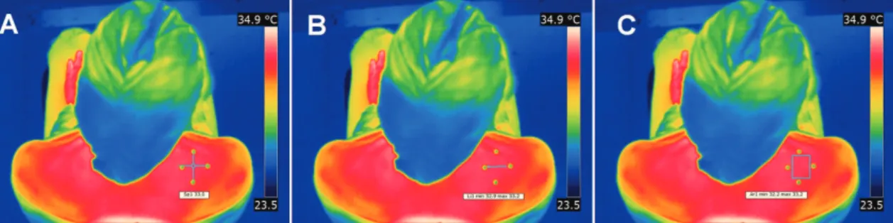
in the present study, three forms of measurement of the skin temperature were performed over the myofascial trigger point: point analysis, in which the temperature of the central point of the area delimited by the markers was measured
(Figure 2A); line analysis, in which a straight-line linking two markers was drawn across the region where the trigger point was located (Figure 2B); and area analysis, in which the area delimited by the four markers was established (Figure 2C). Initially, the mean values of skin temperature for the three analyses were calculated. Next, minimum and maximum values were considered for line and area analyses.
Statistical analysis
Intraclass correlation coefficient (ICC2,1) was
used to determine the intra- and inter-rater reliability,
with its respective 95% confidence interval (CI 95%) , standard error of measurement (SEM), and minimum detectable change (MDC)35. Interpretation
of ICC values was based on that suggested by Fleiss22. For values less than 0.40, the reliability was
considered low; between 0.40 to 0.75, moderate; between 0.75 to 0.90, substantial; and inally, values greater than 0.90, excellent. All statistical analyses were performed using the SPSS software, version 17.0 (Chicago, IL, USA).
Figure 1. Styrofoam markers used to delineate the myofascial trigger point in the upper trapezius muscle.
Results
According to the eligibility criteria, twenty-eight volunteers were recruited from the university
community. However, four volunteers were excluded from the study due to an NDI score of less than 5 points, thus resulting in a inal sample of 24 subjects
of both genders (23 females), 22 right-handed,
mean age of 22.12 (SD=2.54) years, mean BMI of 21.04 (SD=1.95) kg/cm2, mean NDI score of
10.41 (SD=3.18) points, mean pain intensity of 5.00 (SD=1.66) points, and mean duration of cervical pain of 39.33 (SD=33.26) weeks.
Table 1 lists the values of intra-rater reliability, showing excellent reliability for point analysis (mean value), line analysis (mean and maximum values), and area analysis (mean, minimum, and maximum
values), with ICC values ranging between 0.943 and 0.993. In addition, there was a moderate reliability for line analysis (minimum value), with ICC value equal to 0.591, and substantial reliability for area analysis (minimum value), with ICC value equal to
0.821. With respect to the SEM, there was variation
in values between 0.13 and 1.57 °C. In turn, the MDC values ranged from 0.36 to 4.35 °C.
Table 2 lists the values of inter-rater reliability, showing excellent reliability for point and line
analysis (mean value), with ICC values equal to 0.908 and 0.918, respectively. For the other measures, there was moderate to substantial reliability, with ICC values ranging from 0.615 to 0.894. With respect to
the SEM, there was variation in values between 0.43
and 1.22 °C. In turn, the MDC values ranged from 1.19 to 3.38 °C.
Discussion
In the present study, the intra- and inter-rater reliability of infrared image analyses by using point, line, and area approaches had substantial to excellent
ICC values, except the minimum value for line analysis, as moderate ICC values were observed for
intra- and inter-rate analyses.
The results of the present study are in partial
accordance with those reported by Costa et al.21,
who found excellent intra- and inter-rater reliability for point and line analyses regarding the masseter, temporalis anterior, suprahyoid, and upper trapezius muscles in individuals with or without
temporomandibular disorder. However, it should
Table 1. Intra-rater reliability of the minimum, maximum, and mean values of skin temperature for point, line, and area analyses in the myofascial trigger point.
Analysis Values ICC 95% CI SEM MDC
Point Mean 0.955* 0.928-0.972 0.34 0.94
Line Minimum
Maximum Mean
0.591*
0.963*
0.993*
0.346-0.744 0.942-0.977 0.989-0.996
1.57
0.28 0.13
4.35
0.78
0.36
Area Minimum
Maximum Mean
0.821*
0.943*
0.947*
0.714-0.888 0.909-0.964 0.915-0.967
0.66 0.32 0.16
1.83
0.89
0.44
ICC: Intra-class correlation coeficient; CI: Conidence interval; SEM: Standard error of measurement (in °C); MDC: Minimum detectable change (in °C). *p<0.001.
Table 2. Inter-rater reliability of the minimum, maximum, and mean values of skin temperature for point, line, and area analyses in the myofascial trigger point.
Analysis Values ICC 95% CI SEM MDC
Point Mean 0.908* 0.853-0.942 0.48 1.33
Line Minimum
Maximum Mean
0.615*
0.864*
0.918*
0.348-0.759 0.783-0.915 0.869-0.949
1.22 0.52 0.43
3.38 1.44
1.19
Area Minimum
Maximum Mean
0.809*
0.851*
0.894*
0.695-0.880 0.762-0.907 0.831-0.934
0.62 0.50 0.44
1.72 1.39
be pointed out that these authors were not assessing myofascial trigger points in the skeletal muscles in question as their aim was to investigate skin temperature on the muscle belly.
Point analysis was also employed by Rodrigues-Bigaton et al.36 for assessment of skin temperature
in the temporomandibular joint of individuals with
and without arthralgia, with ICC values ranging from 0.841 to 0.874. Rodrigues-Bigaton et al.37 also
used area analysis of the masseter and temporalis anterior muscle belly in both individuals with temporomandibular disorder and controls, reporting
ICC values ranging from 0.945 to 0.998.
Some studies assessed the reliability of the infrared thermography in other clinical conditions, reporting results similar to those found in the present study. In the analysis of skin temperature regarding the
paraspinal region, McCoy et al.38 found excellent
intra- and inter-rater reliability. Choi et al.39 observed
a high inter-rater reliability in the assessment of individuals with complex regional pain syndrome. In addition to these studies, Zaproudina et al.40 found
high ICC values for inter-rater reliability in healthy subjects, however these authors identiied reasonable ICC values when considering the temperature of the
extremities on different days.
The studies conducted by Costa et al.21,
Rodrigues-Bigaton et al.36, and Rodrigues-Bigaton et al.37 were
based on the mean value of analyses performed for measurement of the skin temperature. Within this
context, Klamann et al.41 assessed the intra-rater
reliability of the temperature analysis of ocular
surface, reporting ICC values of 0.947, 0.949, and 0.955 for minimum, maximum, and mean values,
respectively. In the present study, not only the mean value was used but also minimum and maximum values of line and area analyses.
Regarding the values of SEM and MDC,
published studies that evaluated the reliability of infrared thermography showed no such statistical measures21,37-40. In the present study, when considering
the intra-rater reliability, higher SEM and MDC were observed for the minimum value of line (1.57 and 4.35 °C) and area (0.66 and 1.83) analyses. For
inter-rater reliability, similar results were found, with
higher SEM and MDC for the minimum value of the line (1.22 and 3.38 °C) and area (0.62 and 1.72 °C)
analyses.
Thus, in general, mean (point, line, and area analyses) and maximum (line and area analyses)
measures are the most reliable (intra-rater, ICC
between 0.943 and 0.993; inter-rater, ICC between 0.851 and 0.918) and with less error (intra-rater, SEM between 0.13 and 0.34 °C, and MDC between 0.36 and 0.94 °C; inter-rater, SEM between 0.43 and 0.52 °C, and MDC between 1.19 and 1.44 °C). Moreover, in a more rigorous analysis of ICC values, considering the lower limit of the CI 95% and
excepting the minimum values of the area and line
analyzes, excellent intra-rater reliability (ICC values between 0.909 and 0.989) and substantial inter-rater reliability (ICC values between 0.762 and 0.869)
were observed. These results give more robustness to the applicability of the methods of analyses (mean and maximum values) of the infrared images.
Considering the relevance of SEM and MDC in
reliability studies, Tucci et al.35 evaluated a speciic
test for identification of shoulder impingement syndrome and also found that previously published studies in the same subject did not consider these statistical measures in reliability analysis. In addition, these authors emphasize the importance of knowing
the values for SEM and MDC as these numbers give
a good indication of the minimal score difference between evaluations that could be considered as real improvement.
Finally, infrared thermography has been employed
for the evaluation of different musculoskeletal conditions42-44. Therefore, the aim of the present study
was to standardize the infrared image analyses of myofascial trigger points, thus making it possible to support the use of infrared thermography in clinical practice and research for either mapping the skin temperature of a given site or even for assessing the effects of therapeutic resources in musculoskeletal dysfunctions4,6,12.
The present study had the limitation of not including volunteers with latent myofascial trigger points, since these differ from the active ones due to the presence of algesic substances, among other features3. Moreover, we suggest that future studies
assess the reliability of the entire procedure of collecting thermographic data: patient preparation, instrumentation, recording, and analysis of the infrared images.
Conclusion
which supports the use of these methodologies in clinical and research practices.
Acknowledgements
To Fundação de Amparo à Pesquisa do Estado de São Paulo (FAPESP, grants 2013/19368-8 and
2013/09753-1) and Coordenação de Aperfeiçoamento de Pessoal de Nível Superior (CAPES), Brazil, for their inancial support of this study.
References
1. GeHY, Arendt-Nielsen L. Latent myofascial trigger points.
Curr Pain Headache Rep. 2011;15(5):386-92.http://dx.doi.
org/10.1007/s11916-011-0210-6. PMid:21559783 2. BronC, DommerholtJD. Etiology of myofascial trigger
points. Curr Pain Headache Rep. 2012;16(5):439-44.http://
dx.doi.org/10.1007/s11916-012-0289-4. PMid:22836591 3. Shah JP, Gilliams EA. Uncovering the biochemical milieu
of myofascial trigger points using in vivo microdialysis: an application of muscle pain concepts to myofascial pain syndrome. J Bodyw Mov Ther. 2008;12(4):371-84.http:// dx.doi.org/10.1016/j.jbmt.2008.06.006. PMid:19083696 4. Montañez-Aguilera FJ, Valtueña-Gimeno N, Pecos-Martín
D, Arnau-Masanet R, Barrios-PitarqueC, Bosch-MorellF.
Changes in a patient with neck pain after application of
ischemic compression as a trigger point therapy. J Back
Musculoskelet Rehabil. 2010;23(2):101-4. PMid:20555123. 5. Alonso-BlancoC, Fernández-de-las-PeñasC, Fernández-Mayoralas DM, de-la-Llave-Rincón AI, ParejaJA, Svensson
P. Prevalence and anatomical localization of muscle referred pain from active trigger points in head and neck musculature in adults and children with chronic tension-type headache. Pain Med. 2011;12(10):1453-63.http://dx.doi.org/10.1111/j.1526-4637.2011.01204.x.
PMid:21812909
6. Tekin L, Akarsu S, DurmuşO, Cakar E, Dinçer U, Kıralp MZ. The effect of dry needling in the treatment of myofascial pain syndrome: a randomized double-blinded placebo-controlled trial. Clin Rheumatol. 2013;32(3):309-15.http://
dx.doi.org/10.1007/s10067-012-2112-3. PMid:23138883
7. Simons DG, Travell J, Simons LS. Myofascial pain and dysfunction: the trigger point manual. 2nd ed. Baltimore:
Lippincott Williams & Wilkins; 1999.
8. Thomas K, Shankar H. Targeting myofascial taut bands by ultrasound. Curr Pain Headache Rep. 2013;17(7):349.http://
dx.doi.org/10.1007/s11916-013-0349-4. PMid:23793988
9. Sikdar S, Ortiz R, Gebreab T, Gerber LH, Shah JP. Understanding the vascular environment of myofascial trigger points using ultrasonic imaging and computational modeling. Conf Proc IEEE Eng Med Biol Soc. 2010;2010:5302-5.
10. BallynsJJ, Shah JP, HammondJ, Gebreab T, Gerber
LH, Sikdar S. Objective sonographic measures for characterizing myofascial trigger points associated with cervical pain. J Ultrasound Med. 2011;30(10):1331-40.
PMid:21968483.
11. Ibarra JM, GeHY, Wang C, Martínez VizcaínoV, Graven-Nielsen T, Arendt-Graven-Nielsen L. Latent myofascial trigger points are associated with an increased antagonistic muscle activity during agonist muscle contraction. J
Pain. 2011;12(12):1282-8.http://dx.doi.org/10.1016/j.
jpain.2011.09.005. PMid:22078789
12. Hakgüder A, Birtane M, Gürcan S, Kokino S, Turan FN. Efficacy of low level laser therapy in myofascial pain syndrome: an algometric and thermographic evaluation.
Lasers Surg Med. 2003;33(5):339-43.http://dx.doi. org/10.1002/lsm.10241. PMid:14677161
13. Haddad DS, BrioschiML, Arita ES. Thermographic and clinical correlation of myofascial trigger points in the masticatory muscles. Dentomaxillofac Radiol. 2012;41(8):621-9.http://dx.doi.org/10.1259/dmfr/98504520.
PMid:23166359
14. Szentkuti A, KavanaghHS, Grazio S. Infrared thermography and image analysis for biomedical use. Period Biol. 2011;113:385-92.
15. BrioschiML, Macedo JF, Macedo RAC. Skin thermometry: new concepts. J Vasc Bras. 2003;2:151-60.
16. HoleyLA, DixonJ, Selfe J. An exploratory thermographic investigation of the effects of connective tissue massage on autonomic function. J Manipulative Physiol Ther. 2011;34(7):457-62.http://dx.doi.org/10.1016/j. jmpt.2011.05.012. PMid:21875520
17. GrattBM, Sickles EA, RossJB, Wexler CE, GornbeinJA. Thermographic assessment of craniomandibular disorders: diagnostic interpretation versus temperature measurement analysis. J Orofac Pain. 1994;8(3):278-88. PMid:7812225. 18. Kontos M, Wilson R, Fentiman I. Digital infrared
thermal imaging (DITI) of breast lesions: sensitivity
and specificity of detection of primary breast cancers.
Clin Radiol. 2011;66(6):536-9.http://dx.doi.org/10.1016/j.
crad.2011.01.009. PMid:21377664
19. Dibai FilhoAV, PackerAC, CostaAC, Berni-Schwarzenbeck
KC, Rodrigues-BigatonD. Assessment of the upper trapezius muscle temperature in women with and without neck pain.
J Manipulative Physiol Ther. 2012;35(5):413-7.http://dx.doi. org/10.1016/j.jmpt.2012.04.006. PMid:22608286 20. Dibai-FilhoAV, CostaAC, PackerAC, Rodrigues-Bigaton
D. Correlation between skin surface temperature over masticatory muscles and pain intensity in women with myogenous temporomandibular disorder. J Back
Musculoskelet Rehabil. 2013;26(3):323-8. PMid:23893148. 21. CostaAC, Dibai FilhoAV, PackerAC, Rodrigues-BigatonD.
Intra and inter-rater reliability of infrared image analysis of masticatory and upper trapezius muscles in women with and without temporomandibular disorder. Braz J
Phys Ther. 2013;17(1):24-31.http://dx.doi.org/10.1590/ S1413-35552012005000058. PMid:23117649
22. FleissJ. The design and analysis of clinical experiments.
New York: Wiley; 1986.
23. Bonett DG. Sample size requirements for estimating intraclass correlations with desired precision. Stat Med. 2002;21(9):1331-5.http://dx.doi.org/10.1002/sim.1108.
PMid:12111881
24. Muñoz-Muñoz S, Muñoz-García MT,
Myofascial trigger points, pain, disability, and sleep quality in individuals with mechanical neck pain. J Manipulative
Physiol Ther. 2012;35(8):608-13.http://dx.doi.org/10.1016/j.
jmpt.2012.09.003. PMid:23158466
25. Bogduk N. The anatomy and pathophysiology of neck pain. Phys Med Rehabil Clin N Am. 2003;14(3):455-72, v.http://dx.doi.org/10.1016/S1047-9651(03)00041-X.
PMid:12948338
26. CookC, Richardson JK, BragaL, Menezes A, Soler X,
KumeP, et al. Cross-cultural adaptation and validation of
the Brazilian Portuguese version of the Neck Disability Index and Neck Pain and Disability Scale.Spine (Phila Pa 1976). 2006;31(14):1621-7.http://dx.doi.org/10.1097/01.
brs.0000221989.53069.16. PMid:16778699
27. VernonH, Mior S. The Neck Disability Index: a study of reliability and validity. J Manipulative Physiol Ther.
1991;14(7):409-15. PMid:1834753.
28. Ferreira-Valente MA, Pais-Ribeiro JL, Jensen MP.
Validity of four pain intensity rating scales. Pain. 2011;152(10):2399-404.http://dx.doi.org/10.1016/j.
pain.2011.07.005. PMid:21856077
29. Oliveira-Campelo NM, de Melo CA, Alburquerque-Sendín
F, Machado JP. Short- and medium-term effects of manual therapy on cervical active range of motion and pressure pain sensitivity in latent myofascial pain of the upper trapezius muscle: a randomized controlled trial. J
Manipulative Physiol Ther. 2013;36(5):300-9.http://dx.doi. org/10.1016/j.jmpt.2013.04.008. PMid:23769263
30. Ziaeifar M, Arab AM, Karimi N, Nourbakhsh MR. The effect of dry needling on pain, pressure pain threshold and disability in patients with a myofascial trigger point in the upper trapezius muscle. J Bodyw Mov Ther. 2014;18(2):298-305.http://dx.doi.org/10.1016/j. jbmt.2013.11.004. PMid:24725800
31. Gerwin RD, Shannon S, HongCZ, HubbardD, Gevirtz
R. Interrater reliability in myofascial trigger point examination. Pain. 1997;69(1-2):65-73.http://dx.doi.
org/10.1016/S0304-3959(96)03248-4. PMid:9060014 32. Alburquerque-SendínF, CamargoPR, Vieira A, Salvini
TF. Bilateral myofascial trigger points and pressure pain thresholds in the shoulder muscles in patients with unilateral shoulder impingement syndrome: a blinded, controlled study. Clin J Pain. 2013;29(6):478 -86.http://dx.doi.org/10.1097/AJP.0b013e3182652d65.
PMid:23328323
33. Roy RA, BoucherJP, Comtois AS. Digitized infrared segmental thermometry: time requirements for stable recordings. J Manipulative Physiol Ther. 2006;29(6):468. e1-10.http://dx.doi.org/10.1016/j.jmpt.2006.06.007.
PMid:16904493
34. Van MaanenCJ, Zonnenberg AJ, Elvers JW, Oostendorp
RA. Intra/interrater reliability of measurements on body posture photographs. Cranio. 1996;14(4):326-31.
PMid:9110628.
35. Tucci H, Martins J, Sposito G, CamariniP, Oliveira A. Closed
Kinetic Chain Upper Extremity Stability test (CKCUES
test): a reliability study in persons with and without shoulder impingement syndrome. BMC Musculoskelet
Disord. 2014;15(1):1. http://dx.doi.org/10.1186/1471-2474-15-1. PMid:24387196
36. Rodrigues-BigatonD, Dibai FilhoAV, CostaAC, Packer
AC, de Castro EM. Accuracy and reliability of infrared thermography in the diagnosis of arthralgia in women with temporomandibular disorder. J Manipulative Physiol Ther. 2013;36(4):253-8.http://dx.doi.org/10.1016/j. jmpt.2013.04.006. PMid:23719519
37. Rodrigues-BigatonD, Dibai-FilhoAV, PackerAC, Costa
AC, de Castro EM. Accuracy of two forms of infrared image analysis of the masticatory muscles in the diagnosis of myogenous temporomandibular disorder. J Bodyw Mov Ther. 2014;18(1):49-55.http://dx.doi.org/10.1016/j. jbmt.2013.05.005. PMid:24411149
38. McCoy M, Campbell I, Stone P, Fedorchuk C, Wijayawardana S, Easley K. Intra-examiner and inter-examiner reproducibility of paraspinal thermography.
PLoS One. 2011;6(2):e16535.http://dx.doi.org/10.1371/ journal.pone.0016535. PMid:21347290
39. Choi E, LeePB, Nahm FS. Interexaminer reliability of infrared thermography for the diagnosis of complex regional pain syndrome. Skin Res Technol. 2013;19(2):189
-93.http://dx.doi.org/10.1111/srt.12032. PMid:23331254 40. Zaproudina N, VarmavuoV, Airaksinen O, Närhi M.
Reproducibility of infrared thermography measurements in
healthy individuals. Physiol Meas. 2008;29(4):515-24.http://
dx.doi.org/10.1088/0967-3334/29/4/007. PMid:18401069 41. KlamannMK, Maier AK, GonnermannJ, KleinJP, Pleyer
U. Measurement of dynamic ocular surface temperature in healthy subjects using a new thermography device. Curr
Eye Res. 2012;37(8):678-83.http://dx.doi.org/10.3109/0271
3683.2012.674610. PMid:22559822
42. Dibai FilhoAV, PackerAC, Costa AC,
Rodrigues-BigatonD. Accuracy of infrared thermography of the masticatory muscles for the diagnosis of myogenous temporomandibular disorder. J Manipulative Physiol Ther. 2013;36(4):245-52.http://dx.doi.org/10.1016/j.
jmpt.2013.04.007. PMid:23706912
43. Zaproudina N, Airaksinen O, Närhi M. Are the infrared thermography findings skin temperature-dependent? a study on neck pain patients. Skin Res Technol. 2013;19(1):e537-44.http://dx.doi.org/10.1111/srt.12007.
PMid:23020845
44. Roy RA, Boucher JP, Comtois AS. Comparison of paraspinal cutaneous temperature measurements between subjects with and without chronic low back pain. J
Manipulative Physiol Ther. 2013;36(1):44-50.http://dx.doi. org/10.1016/j.jmpt.2012.12.002. PMid:23380213
Correspondence
Rinaldo Roberto de Jesus Guirro
Universidade de São Paulo
Faculdade de Medicina de Ribeirão Preto Curso de Fisioterapia
Avenida dos Bandeirantes, 3900, Monte Alegre CEP 14049-900, Ribeirão Preto, SP, Brasil