3D-CT vascular setting protocol using computer graphics for the
evaluation of maxillofacial lesions
Protocolo vascular por meio da 3D-TC utilizando a computação
gráfica para avaliação de lesões maxilo-faciais
Marcelo de Gusmão Paraiso CAVALCANTI* Axel RUPRECHT**
Michael Walter VANNIER***
CAVALCANTI, M. de G. P.; RUPRECHT, A.; VANNIER, M. W. 3D-CT vas cu lar set ting pro to col us ing com puter graph ics for the eval u a tion of maxillofacial le sions. Pesqui Odontol Bras, v. 15, n. 3, p. 229-236, jul./set. 2001.
In this pa per we present the as pect of a man dib u lar gi ant cell granuloma in spi ral com puted to mog ra phy-based three-di men sional (3D-CT) re con structed im ages us ing com puter graph ics, and demonstrate the im por tance of the vas cu lar pro to col in per mit ting better di ag no sis, vi su al iza tion and de ter mi na tion of the di men sions of the le sion. We an a lyzed 21 pa tients with maxillofacial le sions of neo plas tic and proliferative or i gins. Two oral and maxillofacial ra di -ol o gists an a lyzed the im ages. The use ful ness of in ter ac tive 3D im ages re con structed by means of com puter graph ics, es pe cially us ing a vas cu lar set ting pro to col for qual i ta tive and quan ti ta tive anal y ses for the di ag no sis, de ter mi na tion of the ex tent of le sions, treat ment plan ning and fol low-up, was dem on strated. The tech nique is an im por tant ad junct to the eval u a tion of le sions in re la tion to ax ial CT slices and 3D-CT bone im ages.
UNITERMS: To mog ra phy, X-ray com puted; Im age pro cess ing, com puter-as sisted; Gi ant cell granuloma; Man di ble.
INTRODUCTION
Com pu ted to mo graphy (CT) is the most com mon tech ni que used for exa mi na ti on of ma xil lo fa -ci al ne o plasms be ca u se it per mits the vi su a li za ti on of soft tis su es and bone struc tu res in the same exa mi na ti on. It has be co me the mo da lity of cho i ce for the eva lu a ti on of ma xil lo fa ci al ne o plasms5,12,9,14
. Mul ti ple over lap ping sli ces can be re cons truc ted from a sin gle exa mi na ti on per mit ting hig her qua lity mul ti pla nar (MPR) and threedi men si o nal re cons truc ted (3D) ima ges wit hout ad di ti o nal pa ti -ent ir ra di a ti on12,7,9,10,11,14
. Re cently, we pre sen ted a new met ho do logy using a 3D vas cu lar set ting pro to col for quan ti ta ti ve analy sis of man di bu lar ne o -plasms, which was va li da ted in ca da vers9
. That study sho wed that 3D in te rac ti ve com pu ter grap -hics make pos si ble a more com ple te as sess ment of ma xil lo fa ci al le si ons, as sis ting the pre o pe ra ti ve plan ning and the eva lu a ti on of pos to pe ra ti ve out -co mes9
.
Aspects such as the CT tech ni que, num ber of exa mi ners, exa mi ners’ ex pe ri en ce, and vas cu la ri
-za ti on of the le si on (sa tis fac tory con trast enhan ce ment) are im por tant to es ta blish ac cep ta -ble re pro du ci bi lity. The ap pli ca ti on of com pu ter grap hics tech no logy from 3D spi ral CT al lows for di rect vo lu me tric re cons truc ti on, fast data fi ling, fast and easy 3D analy sis, real time ima ging for gui ding in ter ven ti on pro ce du res and li ne ar and vo -lu me tric me a su re ments7,9.
The pur po se of this pa per is to show the use ful -ness of spi ral CT-ba sed MPR and 3D re cons truc ted ima ges using com pu ter grap hics pro to cols in the eva lu a ti on of dif fe rent ma xil lo fa ci al le si ons, using, as an exam ple, a gi ant cell gra nu lo ma of the man -di ble.
IMAGING METHODOLOGY
We analy zed 21 pa ti ents with dif fe rent le si ons. The pa ti ents were re fer red for spi ral CT ima ging (Aqui li on Tos hi ba Ame ri ca-Systems Inc., Tus tin, CA) at 120 kVp and 200 mA, using a 512 x 512 ma -trix, and 3 mm/0.5 sec of ta ble feed to pro du ce
3mil li me terthick sli ces with 1.5 mm of re cons -truc ti on in ter val. An FC 20 scan ning fil ter and FOV of 20.3 cm were used, as well as in tra ve nous con trast (150 ml of Con ray 60, Mal linc krodt Me di -cal Inc., Sa int Lou is, MO, USA), which per mit ted the vi su a li za ti on of soft tis sue in vol ve ment and ai -ded in the de li ne a ti on of the le si on. The data sets of
this in ves ti ga ti on were trans fer red, via net work, to a com pu ter work sta ti on (Si li con Grap hics NT and DELL Pre ci si on 420 NT 4.0 hard wa re with Vi tre aTM
2.2 soft wa re, Vi tal Ima ges Inc., Min ne a po lis, MN, USA) to ge ne ra te in te rac ti ve vo lu me-ren de red 3D and MPR ima ges.
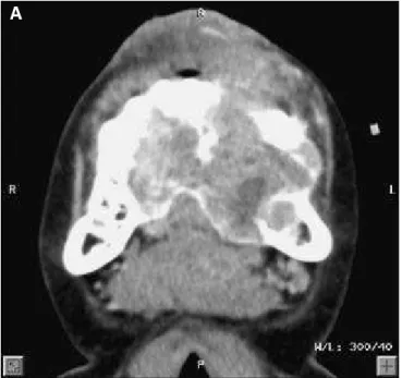
This soft wa re al lows for si mul ta ne ous dis play of 3D and MPR ima ges in three pla nes. In this study, axi al, co ro nal, and sa git tal pla nes were used. This per mit ted the le si on and as so ci a ted ana tomy to be vi e wed from dif fe rent pers pec ti ves, si mul ta ne ously (Fi gu re 1).
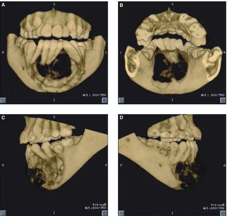
The 3D ima ges were vi e wed using ro ta ti on, trans la ti on, and a flythrough mode, which al lo -wed for the vi su a li za ti on of the le si on in re la ti on to re le vant ana to mi cal struc tu res. The soft wa re to ols also per mit ted vi su a li za ti on through the tu mor mass, pro vi ding de pic ti on of bone des truc ti on in the base of the man di ble, as well as os se ous pro li -fe ra ti on. The 3D-CT exa mi na ti on re ve a led bone des truc ti on along the an te ri or re gi on ex ten ding to the body of the man di ble, bi la te rally, in clu ding an -te ri or -te eth and pre mo lars, and cle arly de pic -ted the le si on wit hin the body of the man di ble (Fi gu re 2).
The 3D vo lu meren de ring soft wa re pac ka ge of -fers a wide cho i ce of set tings that apply dif fe rent
co lor ta bles and trans pa rency func ti ons to the CT data, ba sed on CT den sity. Using the vas cu lar set -ting, which pre fe ren ti ally co lo ri zed the tu mor due to the in cre a sed vas cu la rity and con trast enhan ce -ment around the pe rip hery of the le si on, it was pos si ble to lo ca li ze the con tour of the tu mor, vi su -a li ze -all of its bor ders in re l-a ti on to the m-an di ble, and per form li ne ar me a su re ments of the ne o plasm. Fron tal, la te ral, axi al, and in ter nal 3D vas
-cu lar set ting vi ews cle arly sho wed that the mass in vol ved the in fe ri or as pect of the man di ble (Fi gu -re 3).
RESULTS AND IMAGING
INTERPRETATION
The re sults reveal the num ber of le si ons that were de tec ted and in ter pre ted by two ra di o lo gists,
using dif fe rent pro to cols, and the cor re la ti on with the his to pat ho lo gi cal fin dings.
In 70% of the ca ses, it was pos si ble to de fi ne the fin dings through MPR ima ges. In 82% it was pos si -ble to de fi ne the fin dings through 3D-CT ima ges using the bone pro to col. In 85%, a very sa tis fac -tory as sess ment was pos si ble by me ans of the 3D-CT vas cu lar pro to col. Furt her mo re, in more than 90% of the ca ses, the com bi na ti on of both
3DCT pro to cols was fun da men tal for the di ag no -sis. We stu di ed ca ses of cen tral gi ant cell gra nu lo ma, os sif ying fi bro ma and fi brous dyspla -sia. All the ca ses of fi brous dyspla sia pre sen ted with poor vas cu la rity. The ca ses of cen tral gi ant cell gra nu lo ma and tho se of os sif ying fi bro ma pre sen ted with rich vas cu la rity, which was more pro -noun ced in the for mer. In re la ti on to the 3D-CT bone pro to col, the re sults ad ded va lu a ble in for ma
ti on sin ce the two ra di o lo gists no ti ced the in cre a -sed vo lu me of bone, wit hout des truc ti on, in fi brous dyspla si as; they also agre ed in de fi ning the tra be cu lar area of os sif ying fi bro mas, and the lar ge des -truc ti on of man di bu lar bone in the case of cen tral gi ant cell gra nu lo ma.
The MPR ima ges sho wed a high mul ti lo cu lar ra di o lu cency with nu me rous in ter nal sep ta. Con si -de ra ble ex pan si on of bone was evi -dent in both buc cal and lin gual as pects (Fi gu re 1). The 3D-CT bone pro to col de pic ted a lar ge des truc ti on of bone with dis lo ca ti on of the an te ri or te eth, also de mons tra ting the de gree of buc colin gual and in fe ri or ex -pan si on of the le si on in the body of the man di ble, as well as the in ter nal tra be cu lae (Fi gu re 2). The 3DCT vas cu lar pro to col de mons tra ted the di men -si ons of vas cu la rity (width from the pos te ri or view = 31.0 mm, and width from the la te ral view = 50.6 mm) and, sub se quently, the di men si -ons of the soft tis sue le si on (Fi gu re 3).
The 3D-CT vas cu lar pro to col sho wed a very good le vel of agre e ment with the his to pat ho lo gi cal exa mi na ti on, which was used as a gold stan dard. MPR sho wed some de gree of di sa gre e ment and, for the 3D-CT bone pro to col, the re was di sa gre e ment in only 2 ca ses.
DISCUSSION
3D-CT re cons truc ted ima ges of head and neck tu mors are an im por tant aid to cli ni ci ans in pre-and pos to pe ra ti ve as sess ment of le si ons12,9,10,11,14.
We car ri ed out our study ba sed on the use ful ness of 3D com pu ter grap hics in the de li ne a ti on of le si -ons using the vas cu lar set ting pro to col.
With the ad vent of sub se cond spi ral CT, the ran ge that can be co ve red in a gi ven pe ri od of time (e.g., du ring the cri ti cal pha se of vas cu lar enhan -ce ment) has in cre a sed from 25% to 30%15
. Now -adays, the mul tis li ce vo lu me tric CT scan ners greatly im pro ved both ima ge qua lity and pro duc ti -vity15
. In our study, we used the ne west ge ne ra ti on of spi ral CT, which per mits 0.5 sec for the thick -ness of each sli ce, with a high qua lity of MPR and 3D re cons truc ted ima ges.
One of the ad van ta ges of spi ral CT is the abi lity to re cons truct over lap ping sli ces at every ta ble po -si ti on wit hin the scan vo lu me. Thus, the qua lity of 3D re cons truc ted ima ges and co ro nal and sa git tal MPR ima ges is con si de rably im pro ved17,21
. Anot her ad van ta ge of spi ral CT is re la ted to the use of con trast me di um. For the cur rent study, con trast me di um was ne ces sary, in or der to per mit the de li ne
-a ti on of the ne o pl-asm using the v-as cu l-ar set ting. In the ir study, SPREER et al.21 (1995) found that
with spi ral CT they ne e ded one third less con trast me di um than with con ven ti o nal CT in or der to obtain suf fi ci ent tis sue con trast and soft tis su e enhan ce ment. The ho mo ge ne ity of con trast en -hance ment was sig ni fi cantly hig her when using the spi ral tech ni que.
BERTAND et al.3
(1993) sta ted that gi ant cell gra nu lo mas and os sif ying fi bro mas, but not fi -brous dyspla si as, pre sent at an ear li er age are more cli ni cally ag gres si ve and more vas cu la ri zed. We felt that, ba sed on this des crip ti on of in cre a sed vas cu la rity, the vas cu lar set ting pro to col that we have pre vi ously des cri bed would be use ful for de li -ne a ting the gi ant cell gra nu lo ma. This pro to col is ba sed upon the in cre a sed ac cu mu la ti on of con trast me di um in and around the le si on, due to tu morre la ted in cre a sed vas cu la tu re, re sul ting in in -cre a sed con trast. The vas cu lar set ting pro to col se lec ti vely co lo ri zes the are as of enhan ced con trast ac cu mu la ti on and, thus, de fi nes the li mits of the le si on.
EL-LABBAN13 (1997) per for med a study with the
pur po se of in ves ti ga ting the vas cu lar fin dings of the cen tral gi ant cell gra nu lo ma and dis cus sing the pat ho ge ne sis of this le si on. The aut hor con clu ded that the stro ma of the cen tral gi ant cell gra nu -lo ma has a strong vas cu la rity with he morr ha gic are as and di la ted blo od ves sels. Cen tral gi ant cell gra nu lo mas con sist of a rich con junc ti ve ma trix with a rich ca pil lar vas cu la rity23
; his to lo gi cally, tho se le si ons have highly vas cu lar growths2. The
dif fe ren ti al di ag no sis of ra di o lu cent le si ons can in -clu de vas cu lar le si ons. Most of the se le si ons are con di ti ons such as cen tral gi ant cell gra nu lo mas, he man gi o mas, tra u ma tic bone cysts and fi broos -se ous le si ons6
. Due to the thin-wal led vas cu lar spa ces, he morr ha ge can be very well seen. Intra -vas cu lar in va si on by gi ant cells can also be se en25
. He morr ha ge and di la ted vas cu lar chan nels are of ten clo sely as so ci a ted with cen tral gi ant cell gra -nu lo mas1,19
. The most in te res ting fin ding was the sta i ning pat tern of blo od ves sels in the cen tral gi -ant cell gra nu lo ma18.
here to lo ca li ze and me a su re a gi ant cell cen tral gra nu lo ma using a uni que com pu ter grap hics pro to col that had been pre vi ously va li da ted using ca -da ver he ads9
. In the cur rent pa per we show a cli ni -cal ap pli ca ti on of that ex pe ri ment, de mons tra ting the pos si bi lity of its cli ni cal use.
Ra di o grap hi cally, the cen tral gi ant cell gra nu lo -ma ap pe ars, with equal fre quency, as a uni lo cu lar ra di o lu cen ce or as mul ti lo cu lar ra di o lu cen ces. The bor der of the le si on may be well or po orly de fi ned, and the tu mor fre quently ex tends be yond the radiographic mar gins. The cli ni cal be ha vi or of the -se le si ons is qui te va ri a ble and dif fi cult to pre -dict4,24,27
. The re has been con si de ra ble in te rest in pre dic ting the ag gres si ve ness of the se le si ons; the per fo ra ti on of the cor ti cal bone, when pre sent, in -di ca tes more ag gres si ve le si ons4,24,27
.
In our study, MPR ima ges re ve a led des truc ti on, cor ti cal thin ning, lar ge le si ons and sep ta. Ho we -ver, 3D-CT ima ges were ne ces sary to con firm the ex tent of the le si on and of the des truc ti on. The cor -ti cal des truc -ti on was not very well seen in MPR ima ges but sa tis fac to rily seen in our 3D ima ges. The 3D vas cu lar ima ge sho wed the in va si on of the le si on wit hin the in te ri or as pects of the man di ble and its pro xi mity to ad ja cent struc tu res, de ter mi -ning the real to po graphy of the le si on. The se 3D pro to col vi ews cle arly sho wed both the os te o blas tic and os te oly tic as pects of the tu mor.
As sta ted abo ve, the abi lity of CT to pro du ce con trast enhan ce ment of a le si on is re la ted to se -lec ti ve ac cu mu la ti on of con trast me di um wit hin the ne o plasm. The le si ons tend to sti mu la te an gi o ge ne sis, so that the num ber of blo od ves sels as so -ci a ted with the tu mor in cre a ses, and the re is also an in cre a se in ex tra vas cu lar le a ka ge22
. That adds to the ac cu mu la ti on of con trast me di um around the le si on. Col lec ti vely, it le ads to in cre a sed ra di o pa city of the pe rip hery of the ne o plasm, of ten cal -led “blush”, which is com monly found on CT examinations20,22
. Con se quently, the at te nu a ti on co ef fi ci ent or CT num ber of the pe rip hery of the neoplasm is dif fe rent from that of the ne igh bor tis sue. Many aut hors have re por ted that this con -trast enhan ce ment can be used with 3D ima ges to pro du ce good vi su a li za ti on of le si ons16
. In our 3D ima ges, co lor as sign ment is ba sed on the at te nu a ti on co ef fi ci ent of tis su es as me a su red in Houns fi -eld units. Con trast me di um ac cu mu la ti on wit hin the tu mor due to the vas cu lar chan ges des cri bed abo ve al lows for se lec ti ve co lo ri za ti on of the ne o
-plasm in re la ti on to ad ja cent tis su es, using the vas cu lar set ting.
In a pa per that va li da ted li ne ar me a su re ments using the 3D vas cu lar set ting pro to col, CAVAL -CANTI et al.8 (2000) found no sta tis ti cally sig ni fi
-cant dif fe ren ces bet we en physi cal and ima ging me a su re ments using the 3D vas cu lar set ting pro to col. Using the same met ho do logy, we de mons tra -ted, in this pa per, a cli ni cal ap pli ca ti on in a pa ti ent with a gi ant cell cen tral gra nu lo ma in the man di ble. We lo ca li zed the le si on using the vas cu lar pro -to col, and per for med me a su re ments for tre at ment plan ning and eva lu a ti on. Also using the 3D bone pro to col we de mons tra ted some bony as pects of the le si on that were not well de fi ned on 2D-CT ima ges.
The ava i la ble com pu ter soft wa re al lows for the use of ima ge enhan ce ment al go rithms, 3D ren de -ring of gre at amount of data, vi e wing and sli cing of vo lu mes in any di rec ti on and pla ne, and iden ti fi ca ti on of dif fe rent sur fa ces and tis su es through co -lors8,26
. The pro cess of ac qui si ti on of data is im pro -ved using pro per ti es of the soft wa re such as co lor trans pa ren ce in the con trast are a8
. We think that this met ho do logy can dis tin guish some le si ons ba -sed on the ir vas cu la rity. We think that, with our met hods, 3DCT may play an im por tant role in es -ta blis hing tre at ment pro to cols sin ce it al lows for the de ter mi na ti on of the size, po si ti on and ex ten si -on of tu mor mas ses. Furt her re se ar ches must be de ve lo ped in or der to bet ter cla rify our hypot he sis.
CONCLUSIONS
We have shown that the 3D-CT vas cu lar set ting pro to col is a use ful ima ging mo da lity for the di ag no sis and tre at ment plan ning of ne o plas tic le si ons. For that, we de mons tra ted its cli ni cal ap pli ca -ti on in a case of ag gres si ve gi ant cell gra nu lo ma. The com pu ter soft wa re that per mits the uti li za ti on of pre set pro to cols ma kes it pos si ble to au to ma ti cally link vo lu me ren de ring to the scan ning pro cess, which ori gi na tes ap pro pri a te 3D re cons truc -ted ima ges with high ima ge qua lity. We have de mons tra ted the use ful ness of ima ging pro ces -sing with qua li ta ti ve and quan ti ta ti ve analy ses to im pro ve the di ag no sis and, thus, the tre at ment plan.
ACKNOWLEDGMENTS
CAVALCANTI, M. de G. P.; RUPRECHT, A.; VANNIER, M. W. Pro to co lo vas cu lar por meio da 3DTC uti li zan do a com pu -ta ção grá fi ca para ava li a ção de le sões ma xi lo-fa ci a is. Pes qui Odon tol Bras, v. 15, n. 3, p. 229-236, jul./set. 2001. Nes te tra ba lho, de mons tra mos os as pec tos de um gra nu lo ma cen tral de cé lu las gi gan tes por meio da to mo gra fia com -pu ta do ri za da (TC) em es pi ral ba se a da na re cons tru ção de ima gem em três di men sões (3D), uti li zan do a com -pu ta ção grá fi ca, e a im por tân cia do pro to co lo vas cu lar per mi tin do um me lhor di ag nós ti co, vi su a li za ção e ob ten do di men sões da le são. Fo ram ana li sa dos 21 pa ci en tes com le sões ma xi lofa ci a is de ori gens ne o plá si cas e pro li fe ra ti vas. Dois ra di o -lo gis tas ana li sa ram as ima gens. A uti li da de da in te ra ção da re cons tru ção da ima gem por meio da com pu ta ção grá fi ca, es pe ci al men te uti li zan do o pro to co lo vas cu lar para aná li ses qua li ta ti vas e quan ti ta ti vas para o di ag nós ti co, pla ne ja men to de tra ta men to e evo lu ção, as sim como para a lo ca li za ção da ex ten são da le são foi de mons trada. Isto é um im -por tan te ad jun to para a evo lu ção des tas le sões em re la ção a cor tes axi a is em TC e para ima gens -por meio de 3D-TC para es tru tu ras ós se as.
UNITERMOS: To mo gra fia com pu ta do ri za da por ra i os X; Pro ces sa men to de ima gem as sis ti do por com pu ta dor; Gra nu -lo ma de cé lu las gi gan tes; Man dí bu la.
BIBLIOGRAPHIC REFERENCES
1. ANDERSEN, L.; FEJERSKOV, O.; PHILIPSEN, H. P. Oral gi ant cell gra nu lo ma. A cli ni cal and his to lo gi cal study of 129 new ca ses. Acta Pathol Mi cro bi ol Scand, v. 81, p. 606-616, 1973.
2. ANDERSEN, L.; FEJERSKOV, O.; THEILADE, J. Oral gi ant cell gra nu lo ma. An ul tras truc tu ral study of ves sels.
Acta Pat hol Mi cro bi ol Scand, v. 83, p. 69-76, 1975. 3. BERTRAND, B.; ELOY, P.; CORNELIS, J. P. et al. Ju ve ni le
ag gres si ve ce men to-os sif ying fi bro ma: case re port and re vi ew of the li te ra tu re. Lary ngos co pe, v. 103, p. 1385-1390, 1993.
4. BORDNER, L.; BAR-ZIV, J. B. Ra di o grap hic fe a tu res of cen tral gi ant cell gra nu lo ma of the jaws in chil dren. Pe -di atr Ra -di ol, v. 26, p. 148-151, 1996.
5. BODNER, L.; BARZIV, J.; KAFFE, I. CT of cystic jaw le si -ons. J Com put Assist To mogr, v. 18, p. 22-26, 1994. 6. BURKES, E. F. Vas cu lar le i om yo ma of the man di ble: re port
of a case. J Oral Ma xil lo fac Surg, v. 53, p. 65-66, 1995. 7. CAVALCANTI, M. G. P.; RUPRECHT, A.; BONOMIE, J. M.;
VANNIER, M. W. Accu racy and pre ci si on of spi ral CT in the as sess ment of ne o plas tic le si ons as so ci a ted with the man di ble. Acad Ra di ol, v. 7, p. 94-99, 2000.
8. CAVALCANTI, M. G. P.; RUPRECHT, A.; BONOMIE, J. M.; VANNIER, M. W. The va li da ti on of 3D spi ral CT-ba sed me a su re ments of si mu la ted ma xil lo fa ci al ne o plasms.
Oral Surg Oral Med Oral Pat hol Oral Ra di ol Endod, v. 89. p. 753-758, 2000.
9. CAVALCANTI, M. G. P.; RUPRECHT, A.; QUETS, J. Eva lu a ti on of ma xil lo fa ci al fi bro sar co ma using com pu ter grap -hics and spi ral com pu ted to mo graphy. Den to ma xil lo -fac Ra di ol, v. 28, p. 145-151, 1999.
10. CAVALCANTI, M. G. P.; RUPRECHT, A.; QUETS, J. Pro -gres si on of squa mous cell car ci no ma eva lu a ted using com pu ter grap hics of spi ral com pu ted to mo graphy.
Den to ma xil lo fac Ra di ol, v. 28, p. 238-244, 1999. 11. CAVALCANTI, M. G. P.; RUPRECHT, A.; VANNIER, M. W.
Ima ging of cysts and tu mors using com pu ter grap hics form 2D-CT and 3D-CT re cons truc ted ima ges. Ra di o -logy, v. 207, p. 176-177, 2000.
12. CAVALCANTI, M. G. P.; VANNIER, M. W. Me a su re ment of the vo lu me of oral tu mours by three-di men si o nal spi ral com pu ted to mo graphy. Den to ma xil lo fac Ra di ol, v. 29, p. 35-40, 2000.
13. ELLABBAN, N. G. Intra vas cu lar fi brin throm bi and en dot -he li al cell da ma ge in cen tral gi ant cell gra nu lo ma. J Oral Pat hol Med, v. 26, p. 1-5, 1997.
14. FAGELMAN, D.; HUANG, A. B. Pros pec ti ve eva lu a ti on of le si ons of the man di ble and ma xil la: fin dings on mul ti pla -nar and three-di men si o nal CT. Am J Ro ent ge nol, v. 163, p. 693-698, 1994.
15. FOX, S. H.; TANENBAUM, L. N.; ACKELSBERG, S. Fu tu re di rec ti ons in CT tech no logy. Ne u ro i ma ging Clin N Am, v. 8, p. 497-513, 1998.
16. GHOLKAR, A.; ISHERWOOD, I. Threedi men si o nal to mo -grap hic re for ma ti ons of in tra cra ni al vas cu lar le si ons. Br J Ra di ol, v. 61, p. 258-262, 1988.
17. KALENDER, W. A.; POLACIN, A.; SUSS, C. A com pa ri son of con ven ti o nal and spi ral CT: an ex pe ri men tal study on the de tec ti on of sphe ri cal le si ons. J Com put Assist To -mogr, v. 18, p. 167-176, 1994.
18. LIM, L.; GIBBINS, J. R. Immu his to che mi cal and ul tras -truc tu ral evi den ce of a mo di fi ed mi cro vas cu la tu re in the gi ant cell gra nu lo ma of the jaws. Oral Surg Oral Med Oral Pat hol Oral Ra di ol Endod, v. 79, p. 190-198, 1995.
19. ODELL, E. W. Hybrid cen tral gi ant cell gra nu lo ma and cen -tral odon to ge nic fi bro ma-like le si ons of the jaws. His to -pat ho logy, v. 30, p.165-171, 1997.
20. SOM, P. M.; LANZIERI, S. F.; SACHER, M. et al. Extra cra ni -al tu mor vas cu la rity: de ter mi na ti on by dyna mic CT scan ning. Part I: con cepts and sig na tu re cur ves. Ra di o -logy, v. 154, p. 401-405, 1985.
21. SPREER, J.; KRAHE, T.; JUNG, G.; LACKNER, K. Spi ral
ver sus con ven ti o nal CT in rou ti ne exa mi na ti ons of the neck. J Com put Assist To mogr, v. 19, p. 905-910, 1995.
23. TIMOSCA, G.; GAVRILITA, L. Le gra nu lo me pé rip hé ri que à cel lu les géan tes des ma xil la i res. Étu de sur 173 cas. Re -vue de Sto ma to lo gie, v. 77, p. 587-597, 1976. 24. WALSTAD, R. W.; FIELDS, T.; SCHOW, S. R.; McKENNA, S.
J. Expan si le le si on of the an te ri or ma xil la. J Oral Ma xil -lo fac Surg, v. 57, p. 595-599, 1999.
25. WENIG, B. M.; MAFEE, M. F.; GHOSH, L. Fi bro-os so e us, os se ous, and car ti la gi nous le si ons of the or bit and pa ra
-or bi tal re gi on. Ra di ol Clin N Am, v. 36, p. 1241-1259, 1998.
26. WIEGAND, D. A.; PAGE, R. B.; CHANNIN, D. S. The sur gi cal work sta ti on: sur gi cal plan ning using ge ne ric soft wa -re. Oto lary ngol Head Neck Surg, v. 109, p. 434-440, 1993.
27. WHITAKER, S. B.; WALDRON, C. A. cen tral gi ant cell le si on of the jaws. Oral Surg Oral Med Oral Pat hol Oral Radiol Endod, v. 75, p. 199-208, 1993.