o
Journal of
Epilepsy and ClinicalNeurophysiology
J Epilepsy Clin Neurophysiol 2009;15(2):83-88
Incidence and Risk Factors for Seizures in
Central Nervous System Infections in Childhood
Paulo Breno Noronha Liberalesso*, Izabella Celidônio Bertoldo da Silva**, Karlin Fabianne Klagenberg***, Ari Leon Jurkiewicz***, Bianca Simone Zeigelboim***,
Victor Horácio Costa Júnior****
Departamento de Neurologia Infantil – Hospital Pequeno Príncipe, Curitiba, PR, Brasil
AbStRACt
Introduction: The infections of the central nervous system remain as a public health problem in several countries and there is a direct relation between poverty and underdevelopment with high mortality and morbidity rates. Seizures represents a complication related to infections of the central nervous system, are considered a clinical emergency and requiring neurological investigation. Objective: In this article, we propose to describe the incidence and risk factors for seizures in central nervous system infections in childhood. Methods: a retrospective study was performed between October 2007 and October 2008 and all patients who were hospitalized with the diagnosis of infections of the central nervous system were analyzed. Newborns were excluded. The patients were divided into GROUP 1 (without seizures) and GROUP 2 (with seizures). Results: 731 patients were included, 47.75% males, with average age of 15.7 years. GROUP 1 – with fever (652/92.35%), headache (580/82.15%), vomits (550/77.9%), and viral meningitis predominance (652/93.06%). GROUP 2 – with fever (25/100%), vomits (12/48), headache (6/24%), and viral encephalitis predominance (14/56%). Ten (40%) patients from the GROUP 2 presented EEG alterations. The incidence of seizures was 3.42% and a significant statistical difference was noticed related to mean age (p<0.000069), presence of headache (p<0.0000), vomits (p<0.0005), stiff neck (p<0.0105) and drowsiness (p<0.0265). Conclusions: the occurrence of seizures during the hospitalization is significantly more frequent in cases of viral encephalitis and bacterial meningitis compared to viral meningitis. The risk of seizures increases in early ages. Headache, vomits, stiff neck and drowsiness are more frequent symptoms in children with infection of the central nervous system who presented seizures during the hospitalization.
Key works: Seizures, central nervous system infection.
ReSuMO
Incidência e fatores de risco para crises epilépticas nas infecções do sistema nervoso central na infância
Introdução: a infecção do sistema nervoso central permanece como um problema de saúde pública em diversos países, havendo uma relação direta entre pobreza e subdesenvolvimento e taxas mais elevadas de morbidade e mortalidade. As crises convulsivas correspondem a uma das complicações das infecções centrais, representando uma emergência clínica e necessitando investigação neurológica. Objetivo: neste artigo nosso objetivo foi descrever a incidência e os fatores de risco para crises convulsivas em crianças com infecções do sistema nervoso central. Métodos: estudo retrospectivo realizado entre 01/10/2007 e 01/10/2008 sendo avaliados todos os pacientes internados com diagnóstico de infecção do sistema nervoso central. Foram excluídos os recém nascidos. Os pacientes foram divididos em GRUPO 1 (sem crise convulsiva) e GRUPO 2 (com crise convulsiva) para análise. Resultados: foram incluídos 731 paciente, 47,75% do sexo masculino, com idade média de 15,7 anos. GRUPO 1 – predomínio de febre (652/92,35%), cefaléia (580/82,15%), vômitos (550/77,90%) e do diagnóstico de meningite viral (657/93,06%). No GRUPO 2 - predomínio de febre (25/100%), vômitos (12/48%), cefaléia (6/24%) e do diagnóstico de encefalite viral (14/56%). Dez (40%) pacientes do GRUPO 2 apresentaram alteração no EEG. A incidência de crise convulsiva foi de 3,42% e houve diferença estatisticamente significativa quanto a
**** Departamento de Neurologia Infantil, Hospital Pequeno Príncipe, Curitiba, PR. Laboratório de EEG Digital do Hospital da Cruz Vermelha Brasileira, Curitiba, PR. Programa de Pós-Graduação em Distúrbios da Comunicação, Universidade Tuiuti do Paraná, Curitiba, PR.
**** Departamento de Neurologia Infantil, Hospital Pequeno Príncipe, Curitiba, PR.
**** Programa de Pós-Graduação em Distúrbios da Comunicação, Universidade Tuiuti do Paraná, Curitiba, PR. **** Departamento de Infectologia Infantil, Hospital Pequeno Príncipe, Curitiba, PR, Brasil.
INtRODuCtION
The infection of central nervous system (CNS) presents high prevalence in pediatric patients. The morbidity and mortality rates vary mainly according to the etiologic agent, although other factors such as age, immune condition, the early diagnosis and treatment also show great influence on prognosis.1,2 In pre-antibiotic’s epoch, infections in the CNS had high morbidity and mortality. The penicillin and sulfonamides represented a milestone in dealing with significant improvement of prognosis.3
The CNS infections can be classified as encephalitis and meningitis. The encephalitis is a disease characterized by inflammatory process of brain tissue due to the presence of an infectious agent, usually a virus. Meningitis is a disease characterized by inflammation of subarachnoid space and leptomeningeal membranes (pia mater and arachnoid), which may be caused by viruses, bacteria, protozoa and fungi. The etiologic agents can reach the CNS through hematogenous usually after pass over the upper airways mucous membrane, over a focus contiguous to the CNS (eg otitis, sinusitis) or direct way (eg craniocerebral trauma, neurosurgery).4 The encephalitis and meningitis pose a public health problem in several world places, especially in the poorest countries. Not only the incidence but also the risk of neurological complications is higher in poorer countries. In Brazil, states of lower socio-economic development are those with higher rates of encephalitis/ meningitis and also at greatest risk of complications.5
The high morbidity and mortality rates of these infections, particularly of bacterial meningitis in infants,6 justifies the importance of clinical and laboratoryaspects and the searching for factors that can predict the prognosis. The high incidence of seizures in the meningitis and encephalitis also justifies a detailed analysis of this association.
This study is aimed to identify the principal clinical and laboratory features of the CNS infections, evaluate their morbidity and mortality rates in childhood and seek factors that might help in determining the prognosis.
MetHODOLOGY
It was performed a retrospective study and has been reviewed the medical charts of 731 children hospitalized
at Hospital Pequeno Príncipe between 10/01/2007 and 10/01/2008. The inclusion criterion was the presence of clinical and laboratory diagnosis of CNS infection. The normal values of cerebrospinal fluid that were used for central infection diagnosis is in Table 1. After reviewing the medical records a clinical file was fulfilled containing age, gender, patient’s identity number, signs and symptoms that justified the cerebrospinal fluid gather, presence of seizures and the semiology of the events. The CNS infections were classified as viral meningitis, bacterial meningitis and encephalitis in conformity to clinical data and according to the cerebrospinal fluid analysis (number of leukocytes,
erythrocyte, glycorrhachia, protein, chloride, neutrophil percentage and polymorphonuclear and culture). All patients who had seizures or neurological signs were submitted to an electroencephalogram digital exam and a neuroimaging exam (CT scan or MRI). The clinical file also assessed the treatment (symptomatic, antibiotics or antiviral), the hospitalization period in infirmary and in the intensive care unit (ICU), presence of neurological sequel and use of antiepileptic drugs after discharge from hospital. All infants under 28 day-old (newborns) were excluded. There were excluded 24 medical records due to lack of data or incomplete data for analysis.
idade média (p<0,000069), presença de cefaléia (p<0,0000), vômitos (p<0,0005), rigidez de nuca (p<0,0105) e sonolência (p<0,0265). Conclusões: crise convulsiva durante o internamento é significativamente mais freqüente nos casos de encefalite viral e meningite bacteriana quando comparado à meningite viral. Quanto menor a idade da criança maior o risco de crise convulsiva durante o internamento. Cefaléia, vômitos, rigidez de nuca e sonolência são mais freqüentes nas crianças com infecção do sistema nervoso central que apresentam crise convulsiva no internamento.
unitermos: Crises convulsivas, infecção do sistema nervoso central.
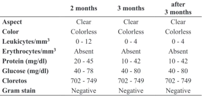
table 1. Normal values for cerebrospinal fluid according to age
2 months 3 months 3 monthsafter
Aspect Clear Clear Clear
Color Colorless Colorless Colorless
Leukicytes/mm3 0 - 12 0 - 4 0 - 4 Erythrocytes/mm3 Absent Absent Absent Protein (mg/dl) 20 - 45 10 - 42 10 - 42 Glucose (mg/dl) 40 - 78 40 - 80 40 - 80 Cloretos 702 - 749 702 - 749 702 - 749 Gram stain Negative Negative Negative
The research project was submitted, reviewed and approved by the Research Ethics Committee in Human Beings of the Hospital Pequeno Príncipe, Curitiba, PR, Brazil, in February 2008.
ReSuLtS
There were included 731 patients, 349 (47.75%) males and 382 (52.26%) females, with ages ranging between 0.1- and 2,88-year-old (average of 15.7-year-old, median 4.90-year- old, standard deviation of 3.41-year-old). The patients were divided into two groups for analysis: GROUP 1 (no seizures; 706/96.58%) and GROUP 2 (with seizures, 25/3.42%).
GROUP 1: 368 (52.13%) were female and 338 (47.88%) male, with ages ranging between 0.2- and 15.7-year-old (average of 5.55-year-old, median 4.9-year-old, standard deviation of 3.34-year-old). The most frequent clinical manifestations were fever (652/92.35%), headache (580/82.15%), vomits (550/77.90%), neck stiffness (262/37.11%), irritability (61/8.64%), drowsiness
(52/7.37%), neck pain (32/4.53%), petechiae (8/1.13%) and ataxia (7/1.00%). In this group, 657 (93.06%) patients were diagnosed with viral meningitis, 33 (4.67%) viral encephalitis and 32 (4.53%) bacterial meningitis.
GROUP 2: 14 (56%) patients were female and 11 (44%) male, with ages ranging between 0.1- and 12.9-year-old (average of 2.88 year-12.9-year-old, median 0.9-year-12.9-year-old; standard
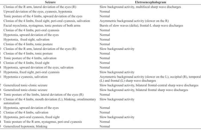
deviation of 4.25-year-old). The most frequent clinical manifestations were fever (25/100%), vomits (12/48%), headache (6/24%), drowsiness (6/24%), neck stiffness (3/12%) and irritability (3/12%). In this group, 14 (56%) patients were diagnosed with viral encephalitis, 7 (28%) viral meningitis and 4 (16%) bacterial meningitis. The seizures presented alternate semiology (Table 2). All patients in Group 2 were subjected to electroencephalography and imaging examinations (CT scan and/or MRI), 10 of them (40%) showed electrographic changes (Table 2) and 5 (20%) variation in neuroimaging (all suggestive of ischemic lesions).
Although no patients in this study received hospital discharge presenting neurological damage, 7 (28% of group 2) were discharged from hospital using antiepileptic drugs and also there was one decease in Group 2. The seizures incidence in children with central nervous system infection was 3.42%. The average age between the groups 1 and 2 showed high statistical significant difference (p<0.000069). The presence of headache (p<0.0000), vomits (p<0.0005), sniff neck (p<0.0105), drowsiness (p<0.0265) showed statistical significant difference (p<0.000069). Patients diagnosed with viral encephalitis (p<0.0000) and with diagnosis of bacterial meningitis (p<0.0094) had significant higher risk for seizures during the hospitalization. In Table 3 we have a statistical comparison of absolute values, percentages of the levels of significance between groups 1 and 2.
table 2. Seizures semiology e electroencephalogram findings
Seizure Eletroencephalogram
1 Clonias of the R arm, lateral deviation of the eyes (R) Slow background activity, multifocal sharp wave discharges 2 Upward deviation of the eyes, cyanosis, hypotonia Normal
3 Tonic posture of the 4 limbs, upward deviation of the eyes Normal
4 Clonias of the 4 limbs, ixed sight, peri-oral cyanosis, salivation Asymmetric background activity (slower on the R)
5 Facial myoclonia, nystagmus, tonic posture of both arms Bursts of slow waves (delta), frontal-L sharp wave discharges 6 Clonias of the 4 limbs, peri-oral cyanosis Normal
7 Hypotonia, upward deviation of the eyes Normal
8 Hypotonia, ixed sight, salivation Normal
9 Clonias of the 4 limbs, tonic posture Normal
10 Clonias of the R arm, lateral deviation of the eyes (R) Slow background activity 11 Clonias of the 4 limbs, tonic posture Normal
12 Tonic posture of the 4 limbs, salivation Normal
13 Clonias of the 4 limbs, ixed sight Normal
14 Hypotonia, upward deviation of the eyes, salivation Normal
15 Hypotonia, ixed sight, peri-oral cyanosis Slow background activity
16 Hypotonia e cyanosis, salivation Asymmetric background activity (slower on the L), occipital (R), temporal (L) and frontal (L) sharp wave discharges
17 Generalized tonic-clonic seizure Slow background activity, bilateral frontal-central sharp wave discharges 18 Generalized tonic-clonic seizure Slow background activity, bilateral frontal sharp wave discharges 19 Tonic posture of the limbs, lateral deviation of the eyes (R) Normal
20 Clonias of the 4 limbs, mouth deviation (L), blinking, oroalimentary automatism
Slow background activity
21 Hypotonia, upward deviation of the eyes Normal 22 Clonias of the 4 limbs, salivation Normal
23 Hypotonia, peri-oral cyanosis, ixed sight Slow background activity
24 Tonic posture of the R arm, nystagmus, peri-oral cyanosis Normal 25 Generalized hypotonia, blinking Normal
table 3. Comparison of variables – absolute value, percentages and significance levels
Variables GROUP 1
(n / %)
GROUP 2
(n / %) p
female 368 / 52,13 14 / 56 0,6941
male 338 / 47,88 11 / 44 0,7028
fever 652 / 92,35 25 / 100 0,1511
headache 580 / 82,15 6 / 24 0,0000 **
vomiting 550 / 77,9 12 / 48 0,0005 **
stiff neck 262 / 37,11 3 / 12 0,0105 *
irritability 61 / 8,64 3 / 12 0,5593
drowsiness 52 / 7,37 6 / 24 0,0265 *
neck pain 32 / 4,53 0 / 0 0,2768
petechiae 8 / 1,13 0 / 0 0,5663
ataxia 7 / 1 0 / 0 0,6552
viral meningitis 657 / 93,06 7 / 28 0,0000 **
viral encephalitis 33 / 4,67 14 / 56 0,0000 **
bacterial meningitis 32 / 4,53 4 / 16 0,0094 **
** significant statistical values (p<0,05). ** very significant statistical values (p<0,01).
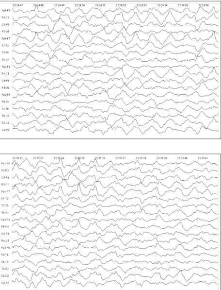
Figure 1. Electroencephalogram showing slow background activity in patient with seizure (clonias of the right arm and lateral deviation of the eyes – patient 10).
DISCuSSION
The CNS infections are related to several complications, such as increased intracranial pressure, cerebral abscesses, ventriculitis, hydrocephalus and cranial nerves injuries. Seizures are the most frequent complication, furthermore, their presence increases the morbidity and mortality rates.1,2
The differentiation between the viral meningitis
symptoms, viral encephalitis and meningitis bacteria is carried out through analysis of laboratory tests (especially the blood count, cerebrospinal fluid and blood glucose) and the clinical aspects lodged by the patient. Children with viral meningitis generally present feverish symptoms, cephalalgia, nausea and vomits without significant involvement of the general condition. Viral encephalitis in children usually have more acute neurological impairment, and there may be lowering of the level of consciousness (ranging from drowsiness to coma), acute ataxia, tremors, focal seizures and signs of focal neurological deficits.7
Lucena et al.6 studied 116 children aged between 28-days-old and 2-years-old with bacterial meningitis and demonstrated that the most frequent signs and symptoms were fever (113/97.4%), vomits (88/75.9%), neck stiffness (84/72.4%), decreased level of consciousness (42/36.2%), seizures (40/34.5%), Brudzinski sign (34/29.3%), upper motor neuron syndrome (32/27.6%), Kernig sign (11/9.5%), alteration in cranial nerves (10/8.6%), cerebellar syndrome (9/7.8%), intracranial hypertension syndrome (5/4,3%), Lasègue sign (4/3.4%) and extrapyramidal syndrome (2/1.7%). In our study fever, cephalalgia and vomits were the most frequent signs and symptoms of central nervous system infection in groups 1 and 2.
Seizures can occur in children with CNS infection mainly due to vasculitis, thrombosis, ischemia, cerebral abscess formation and subdural collections.8 In severe cases of central infection, where an association between vasculitis and cerebral edema occur, brain perfusion is intensively committed resulting in an increased risk of ischemic brain injury and seizures.9 Vieira5 evaluated 202 patients with bacterial meningitis aged between 0 and 12-year-old showing that 41.1% had neurological complications, and the most common problems were seizures, occurring in 38 patients (18.8%).
In a recent study published, Davenport et al.10 evaluated 81 children with bacterial meningitis with an average age of 7.5-year-old and showed that 18.5% evolved with neurological complications, the most frequent were seizures (8/9.9%), cerebral ischemia (5/6.2%), hydrocephalus (4/4.9%) and syndrome of inappropriate secretion of antidiuretic hormone (3/3.7%). The average values of cellularity, cerebrospinal fluid (CSF) glucose concentrations, proteinorachia, positive culture in blood and
cerebrospinal fluid were also evaluated and the authors concluded that the three major risk factors for neurological complications were the positive blood culture, high proteinorachia value and low age. The neurological sequelae related to bacterial meningitis are strongly dependent of the child’s age. According to Bresolin,11 the most common injuries in children younger than 2 months old are delayed neuropsychomotor development, hydrocephalus and seizures, while in children over 18 months seizures and deafness are the most frequent complications.
In our study, the seizures incidence (25/3.42%) was lower than reported in the international literature. The results obtained in our study, as demonstrated in the literature, showed that there is an inverse correlation between age and seizures risk in children with central nervous system infection and also that both viral encephalitis and bacterial meningitis have a significant higher risk of seizure compared to cases of viral meningitis.
Analyzing the signs and symptoms that the patients showed in hospital admission moment we verified that cephalalgia, vomiting, neck stiffness and drowsiness were significantly more frequent in children who subsequently develop seizures during the hospitalization. Moreover, fever, irritability, neck pain, petechiae and ataxia were not more frequent in children with central nervous system infection who developed seizures. We also evidenced that EEG is a medical exam that demonstrates a sensitivity of 40% for abnormality and should be performed in all children with central nervous system infection who present seizures during the hospitalization.
ACKNOWLeDGMeNt
We would like to thank Prof. Dr. Jair Marques Mendes, Professor of the Discipline of Advanced Scientific Methodology of Master and PhD’s degrees in Communication Disorders at the Universidade Tuiuti do Paraná by the support in the study and the statistical calculations.
ReFeReNCeS
Dawson KG, Emerson JC, Burns JL. Fifteen years of experience with 1.
bacterial meningitis. Pediatr Infect Dis J 1999; 18 (9): 816-22. Bryan JP, Silva HR, Tavares A, Rocha H, Scheld WM. Etiology and 2.
mortality of bacterial meningitis in Northeastern Brazil. Rev Inf Dis 1990; 12:128-35.
Radetsky M. Duration of treatment in bacterial meningitis: a historical 3.
inquiry. Pediatr Infect Dis J. 1990; 9:2-9.
Tunkel AR, Scheld WM. Acute meningitis. In: Mandell GL, Benentt 4.
JE, Dolin R. (ed) Mandell, Douglas and Bennett’s Principles and Practice of Infectious Diseases, 5. ed. New York: Churchill Livingstone, p. 959, 2000.
Vieira JFS. Incidência de meningite em pacientes de 0 – 12 anos no 5.
Lucena R, Gomes R, Cardoso E, Goes J, Nunes L, Cardoso A, 6.
Rodrigues B, Souza M, Novaes MA, Melo A. Aspectos clínicos e laboratoriais de meningite piogênica em lactentes. Arq. Neuro-Psiquiatr 2002: 60(2A): 281-4.
Diament A. Neuroviroses. In: Diament A, Cypel S. (ed) Neurologia 7.
Infantil, 4. ed. São paulo: Editora Atheneu, p. 983, 2005.
Pomeroy SL, Holmes SJ, Dodge FR, Feigin RD. Seizures and other 8.
neurologic sequelae of bacterial meningitis in children. N Engl Med 1990;336: 709.
Smith DH, Ingram DL, Smith AL, Gilles F, Bresnan MJ. Bacterial 9.
meningitis. Pediatrics 1973; 52(4):586-600.
Davenport MC, Del Valle MP, Gallegos P, Kannemann AL, Bokser 10.
VS. Meningitis bacteriana: factores de riesgo para el desarrollo de complicaciones agudas. Arch Pediatr 2007; 105(5): 405-410. Bresolin AU. Meningites bacterianas agudas e abscesso cerebral 11.
bacteriano. In: Diament A, Cypel S. (ed) Neurologia Infantil, 4. ed. São Paulo: Ed. Atheneu, p. 941, 2005.
endereço para correspondência:
Paulo Breno Noronha Liberalesso
Departamento de Neurologia Infantil – Hospital Pequeno Príncipe Curitiba, PR, Brasil