HFE ge ne m utatio ns in co ro nary
athe ro thro m bo tic dise ase
1Departamento de Clínica Médica, Faculdade de Medicina de Ribeirão Preto,
Universidade de São Paulo, Ribeirão Preto, SP, Brasil
2Fundação Hemocentro de Ribeirão Preto, Ribeirão Preto, SP, Brasil
R.T. Calado1, R.F. Franco1,2,
A. Pazin-Filho1,
M.V. Simões1,
J.A. Marin-Neto1
and M.A. Zago1,2
Abstract
Although iron can catalyze the production of free radicals involved in LDL lipid peroxidation, the contribution of iron overload to athero-sclerosis remains controversial. The description of two mutations in the HFE gene (Cys282Tyr and His63Asp) related to hereditary hemo-chromatosis provides an opportunity to address the question of the association between iron overload and atherosclerosis. We investi-gated the prevalence of HFE mutations in 160 survivors of myocardial infarction with angiographically demonstrated severe coronary ath-erosclerotic disease, and in 160 age-, gender- and race-matched healthy control subjects. PCR amplification of genomic DNA followed by
RsaI and BclI restriction enzyme digestion was used to determine the
genotypes. The frequency of the mutant Cys282Tyr allele was identi-cal among patients and controls (0.022; carrier frequency, 4.4%), whereas the mutant His63Asp allele had a frequency of 0.143 (carrier frequency, 27.5%) in controls and of 0.134 (carrier frequency, 24.5%) in patients. Compound heterozygotes were found in 2 of 160 (1.2%) controls and in 1 of 160 (0.6%) patients. The finding of a similar prevalence of Cys282Tyr and His63Asp mutations in the HFE gene among controls and patients with coronary atherothrombotic disease, indirectly questions the possibility of an association between heredi-tary hemochromatosis and atherosclerosis.
Co rre spo nde nce
R.F. Franco
Laboratório de Hematologia Departamento de Clínica Médica FMRP, USP
Av. Bandeirantes, 3900 14048-900 Ribeirão Preto, SP Brasil
Fax: + 55-16-633-1144 E-mail: rffranco@ netsite.com.br Research partially supported by FAPESP (No. 98/02821-0) and FUNDHERP.
Received April 9, 1999 Accepted January 18, 2000
Ke y wo rds
·Hemochromatosis ·HFE gene ·Atherosclerosis ·Myocardial infarction ·Risk factor
Intro ductio n
Based on the lower rate of coronary ath-erosclerotic disease (CAD) observed in pre-menopausal women compared to men and postmenopausal women and on the differ-ence in CAD inciddiffer-ence between developed and underdeveloped countries, in 1981, Sullivan (1) hypothesized that higher iron stores could be a risk factor for CAD. In this sense, many investigators have studied the contribution of iron overload to the patho-genesis of atherosclerosis. In addition, with
studies (8-11) have found a positive associa-tion between CAD and clinical markers of iron overload, other reports have found ei-ther a negative association (12) or none at all (13,14). Thus, the iron hypothesis remains controversial.
Hereditary hemochromatosis (HH) is an autosomal recessive genetic disease of iron regulation associated with iron overload and failure of several organs (15,16). The molec-ular bases of HH were elucidated in 1996 after the positional cloning of the involved gene, currently designated HFE (formerly HLA-H) (17). The HFE gene has been se-quenced, and two missense point mutations have been identified: a substitution of ty-rosine for cysteine at amino acid position 282 of the HFE protein (Cys282Tyr), and a substitution of aspartic acid for histidine at amino acid position 63 (His63Asp) (17). Cys282Tyr is strongly related to the disease, but the clinical significance of the His63Asp mutant in HH remains to be determined, although it seems to increase the risk for HH in the compound heterozygous state with Cys282Tyr (18). In addition, carriers of these mutations tend to present higher iron depos-its (19).
The analysis of HFE mutations in pa-tients with atherosclerotic disease represents a valuable opportunity to determine the spe-cific role of HH in atherosclerosis. In this respect, it should be emphasized that al-though HH is an autosomal recessive disor-der, heterozygotes usually exhibit evidence of iron accumulation that might result in increased predisposition to atherosclerosis. If indeed hemochromatosis contributes to atherogenesis, a higher prevalence of HH-related mutations in patients suffering from atherosclerotic disease in comparison to healthy individuals is predicted. Hence, the prevalence of HFE mutations in selected patients with objective diagnosis of coro-nary atherothrombosis in comparison to healthy controls was evaluated in the present study.
Subje cts and Me tho ds
Patie nts and co ntro ls
A total of 160 unrelated and relatively young individuals (124 men; mean age, 42 years; range, 25-55 years; and 36 women; mean age, 46 years; range, 30-55 years) with a diagnosis of acute myocardial infarction (MI) and angiographically proven CAD con-stituted the patient study group. MI was di-agnosed on the basis of clinical, enzymatic and electrocardiographic criteria. At least two of the following criteria were necessary to arrive at this diagnosis: typical chest pain (longer than 30 min), an increase in creatine kinase of more than twice the baseline level, and characteristic electrocardiographic changes in two or more adjacent leads. In-cluded were patients submitted to coronary angiography, which demonstrated stenosis of 50% or higher in a major artery. While the samples from patients were being collected, 160 unrelated subjects (mostly blood donor candidates at the local Blood Center) with-out a personal history of arterial disease or MI were selected as controls. Controls were enrolled before the first blood donation. Each case was matched to a control for gender, age (± 4 years) and race. Both patients and controls came from the same geographic region, i.e., the city of Ribeirão Preto, State of São Paulo, Southeastern Brazil. All indi-viduals enrolled gave their consent to par-ticipate in this study, which was approved by the Institutional Ethics Committee. Data re-garding classical risk factors for CAD and MI were obtained by reviewing records and interviewing the subjects included in the study. Subjects were classified as Whites, Blacks or Mulattoes on the basis of pheno-type characteristics.
Mutatio n analysis
leukocytes by conventional methods (20). For identification of the Cys282Tyr mutation, the following primers were used: 5' -CAA GTG CCT CCT TTG GTG AAG GTG ACA CAT - 3' and 5' - CTC AGG CAC TCC TCT CAA CC - 3'. For identification of the His63Asp mutation, the following primers were used: 5' - ACA TGG TTA AGG CCT GTT GC - 3' and 5' - CTT GCT GTG GTT GTG ATT TTC C - 3' (21). RsaI (Cys282Tyr
analysis) and BclI (His63Asp analysis)
re-striction enzyme digestion was employed after PCR genomic amplification to deter-mine the HFE genotypes.
Statistical analysis
Differences of allele frequencies of each mutation in controls and patients were ana-lyzed for statistical significance by the c2 test. Odds ratios (OR) and 95% confidence intervals (CI 95) were calculated using stan-dard methods (22).
Re sults
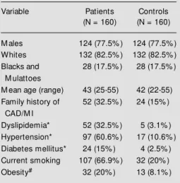
Table 1 lists relevant general characteris-tics of the patient and control groups. As expected, major risk factors for atheroscle-rotic disease were present in most cases and were rarer among subjects from the control group.
The mutant Cys282Tyr allele was found in the heterozygous state in 7 of 160 controls (allele frequency, 0.022; carrier frequency, 4.4%) and in 7 of 160 patients with MI (allele frequency, 0.022; carrier frequency, 4.4%; Table 2). These data yielded an OR for MI related to the Cys282Tyr mutation of 1.0 (CI 95: 0.34-2.91). No homozygotes for the Cys282Tyr mutation were found in con-trols or patients (Table 3). The His63Asp mutation was detected in 44 of 160 controls (allele frequency, 0.143; carrier frequency, 27.5%) and in 39 patients (allele frequency, 0.134; carrier frequency, 24.5%; Table 2). These data yielded an OR for MI related to
Table 1 - General characteristics of myocardial infarction (M I) survivors w ith angiographically demonstrated severe atherosclerosis and of con-trol subjects.
* Physician’s diagnosis and/or drug-treatment.
#Body mass index ³ 30 kg/m2. CAD, Coronary
atherosclerotic disease.
Variable Patients Controls
(N = 160) (N = 160)
M ales 124 (77.5% ) 124 (77.5% )
Whites 132 (82.5% ) 132 (82.5% )
Blacks and 28 (17.5% ) 28 (17.5% ) M ulattoes
M ean age (range) 43 (25-55) 42 (22-55) Family history of 52 (32.5% ) 24 (15% )
CAD/M I
Dyslipidemia* 52 (32.5% ) 5 (3.1% ) Hypertension* 97 (60.6% ) 17 (10.6% ) Diabetes mellitus* 24 (15% ) 4 (2.5% ) Current smoking 107 (66.9% ) 32 (20% )
Obesity# 32 (20% ) 13 (8.1% )
Table 2 - Allele frequencies of HFE mutations in controls and in patients w ith myocar-dial infarction (M I).
Number of mutated chromosomes/number of chromosomes analyzed; allele frequen-cies are given in parentheses. None of the differences reached statistical significance (P>0.05). OR: Odds ratio for M I; CI 95: 95% confidence interval.
M utation Patients Controls OR (CI 95)
Cys282Tyr 7/320 (0.022) 7/320 (0.022) 1.0 (0.34-2.91)
His63Asp 43/320 (0.134) 46/320 (0.143) 0.8 (0.51-1.40)
Table 3 - Hereditary hemochromatosis genotypes for controls and for myocardial infarction patients.
C282Y and H63D indicate Cys282Tyr and His63Asp, respectively. None of the differ-ences show n in this table w as statistically significant (P>0.05).
Genotype C282Y +/+ C282Y +/- C282Y -/- C282Y -/- C282Y +/- C282Y -/-H63D -/- H63D -/- H63D +/+ H63D +/- H63D +/- H63D
-/-Controls 0 (0) 5 (3.1% ) 2 (1.2% ) 40 (25% ) 2 (1.2% ) 111 (69.4% ) Patients 0 (0) 6 (3.7% ) 4 (2.5% ) 34 (21.3% ) 1 (0.6% ) 115 (71.9% )
de-ians (carrier frequency, 27.5%), with a fre-quency similar to that observed among white Europeans. In fact, the His63Asp mutation has been identified in populations of differ-ent ethnic origins and probably is an older mutation (21). Hence, the finding of a high prevalence of this mutation in the Brazilian population is also reasonable, since its preva-lence is less affected by racial admixture. Of note, since ethnicity may influence the com-parability of patients and controls in case-control studies, the case-control and the disease groups of this study were matched taking into account also the ethnic background of the subjects.
The mutant Cys282Tyr and His63Asp alleles had an identical frequency in controls and CAD patients, suggesting that mutations of the HFE gene have no significant effect on atherosclerosis. Although previous reports on the association between iron stores and atherosclerosis have yielded discrepant re-sults, it should be noted that epidemiological studies may be subjected to biases from dif-ferent environmental factors (24,25). For instance, the observation that postmeno-pausal women receiving estrogen replace-ment therapy have a risk for CAD similar to that of premenopausal women (27) suggested that the determining factor for increased risk after menopause is a reduction in estrogen levels, rather than iron overload, as it had been previously supposed (1). In fact, in several of the epidemiological studies de-signed to assess the possible relation be-tween body iron stores and atherosclerotic disease, no association was found, and even an inverse association was suggested in one of them (12). Moreover, supporting our find-ings is the recent demonstration that, in rab-bits, iron overload decreases plasma choles-terol levels and the formation of an aortic arch lesion, suggesting that excess iron does not contribute to atherosclerosis (7). It has also been observed that, although iron can mediate oxidant injury to endothelial cells, the damage may be attenuated by iron chela-tected among the patients (Table 3).
Com-pound heterozygosity for both mutations was observed in 2 controls (1.2%), whereas only one patient carried both mutations (0.6%). Separate analysis for White subjects (who corresponded to the majority of the subjects) did not alter the results (data not shown in tables).
D iscussio n
Since Sullivan (1) raised the hypothesis that iron overload could be a risk factor for CAD, many investigators have tested this possibility, but no definitive conclusion has been reached due to conflicting results (3,4,6-14,23-25). Since the occurrence of HFE gene mutations associated with HH provides a valuable opportunity to test indi-rectly whether iron overload plays a role in atherosclerosis, the prevalence of these two mutations in patients with coronary athero-thrombosis and in healthy controls was de-termined in the present study.
Brazil-tors such as ferritin (28).
Recently, we also analyzed the preva-lence of HH-related mutations in patients with premature atherosclerotic disease from the Netherlands. The results for the Dutch population were similar to those obtained in the present study insofar as they did not support genetic hemochromatosis as a major risk factor for atherosclerosis (29). In this respect, it should be noted that the recently described polymorphism in intron 4 does not affect our results (30), since we did not find a mutant homozygous for the Cys282Tyr mutation. However, it should be emphasized that, although a higher prevalence of the HFE mutations was not observed among CAD patients in comparison with healthy subjects in the present study, the possibility that iron may contribute to atherosclerosis cannot be completely ruled out. Firstly, we analyzed a population for which parameters of evaluation of body iron stores (such as transferrin saturation or serum ferritin lev-els) were not available, and in fact such analysis is beyond the scope of our study. Therefore, our conclusions are based on the absence of a significant association between specific gene variations and atherosclerosis, and do not take into account the possibility that a certain degree of iron deficiency may have diminished iron accumulation among carriers of the HH-related mutations and its impact as a putative atherogenic factor. In fact, our data do not exclude the possibility of an association between HH and athero-sclerosis in other populations. Secondly, our study deals with the analysis of HFE muta-tions in women and men, and the former are known to have increased iron loss (due to
menstruation and pregnancy) which can also minimize the effect of the HFE mutations on iron accumulation. On the other hand, women were only a minority of the individuals ana-lyzed, cases and controls were matched for gender, and separate analysis of data among males did not alter our results (data not shown). In addition, we investigated only individuals less than 55 years of age, and our results should not be extrapolated to older HFE carriers, in whom a more evident iron accumulation may be present. Moreover, an association with recurrent cardiovascular events was not explored in our study. Fi-nally, it could also be hypothesized that iron is mainly stored in parenchymal tissues in HH, and therefore iron deposits in the mac-rophage system (which could contribute to foam cell formation and atherosclerosis pro-gression) are not increased in carriers of the HFE mutations. Under any of these circum-stances, this study suggests that screening of HH mutations among patients with CAD and MI does not seem to be justified in our population.
To sum up, we found a similar preva-lence of the HFE gene mutations in rela-tively young patients with CAD and con-trols. We conclude that HFE mutations re-lated to HH are unlikely to be major genetic risk factors for coronary atherothrombotic disease in the Brazilian population.
Ackno wle dgm e nts
The authors are indebted to Mrs. Marli H. Tavella and Mrs. Amélia G. Araújo for ex-cellent technical assistance.
Re fe re nce s
1. Sullivan JL (1981). Iron and the sex differ-ence in the heart disease risk. Lancet, 307: 1293-1294.
2. M cCord JM (1998). Iron, free radicals, and oxidative injury. Seminars in Hematology, 35: 5-12.
3. Corti M C, Gaziano M & Hennekens CH (1997). Iron status and risk of cardiovascu-lar disease. Annals of Epidemiology, 7: 62-68.
4. M eyers DG (1996). The iron hypothesis -does iron cause atherosclerosis? Clinical
Cardiology, 19: 925-929.
5. Diaz M N, Frei B, Vita JA & Keaney Jr JF (1997). Antioxidants and atherosclerotic heart disease. New England Journal of M edicine, 337: 408-417.
V, Romano M , Bracho M , M ontano RF & Cardier J (1995). Iron overload augments the development of atherosclerotic le-sions in rabbits. Arteriosclerosis, Throm-bosis, and Vascular Biology, 15: 1172-1180.
7. Dabbagh AJ, Shw aery GT, Keaney Jr JF & Frei B (1997). Effect of iron overload and iron deficiency on atherosclerosis in the hypercholesterolemic rabbit. Arterioscle-rosis, Thrombosis, and Vascular Biology, 17: 2638-2645.
8. Lauffer RB (1990). Iron stores and the international variation in the mortality from coronary artery disease. M edical Hypoth-eses, 35: 96-102.
9. Salonen JT, Nyyssönen K, Korpela H, Tuomilehto J, Seppänen R & Salonen R (1992). High stored iron levels are associ-ated w ith excess risk of myocardial infarc-tion in eastern Finnish men. Circulation, 86: 803-811.
10. M orrison HI, Semenciw RM , M ao Y & Wigle DT (1994). Serum iron and risk of fatal acute myocardial infarction. Epidemi-ology, 5: 243-246.
11. Kiechl S, Willeit J, Egger G, Poew e W & Oberhollenzer F (1997). Body iron stores and the risk of carotid atherosclerosis. Cir-culation, 96: 3300-3307.
12. Sempos CT, Looker AC, Gillium RF & M aruk DM (1994). Body iron stores and the risk of coronary heart disease. New England Journal of M edicine, 330: 1119-1124.
13. Stampfer M J, Grodstein F, Rosemberg I, Willett W & Hennekens C (1993). A pro-spective study of plasma ferritin and risk of myocardial infarction in US physicians.
Circulation, 87: 688 (Abstract).
14. Reunanen A, Takkunen H, Knekt P, Seppänen R & Aromaa A (1995). Body iron states, dietary iron intake and coro-nary heart disease mortality. Journal of Internal M edicine, 238: 223-230.
15. Bothw ell TH & M acPhail P (1998). Heredi-tary hemochromatosis: etiologic, patho-logic, and clinical aspects. Seminars in Hematology, 35: 55-71.
16. Andrew s NC & Levy JE (1998). Iron is hot: an update on the pathophysiology of hemochromatosis. Blood, 92: 1845-1851. 17. Feder JN, Gnirke A, Thom as W , Tsuchihashi Z, Ruddy DA, Basava A, Dormishian F, Domingo Jr R, Ellis M C, Fullan A, Hinton LM , Jones NL, Kimmel BE, Kronmal GS, Lauer P, Lee VK, Loeb DB, M apa FA, M cClelland E, M eyer NC, M intier GA, M oeller N, M oore T, M orikang E, Prass CE, Quintana L, Starnes SM , Schatzman RC, Brunke KN, Drayna DT, Risch NJ, Bacon BR & Wolff RK (1996). A novel M HC class I-like gene is mutated in patients w ith hereditary haemochromato-sis. Nature Genetics, 13: 399-407. 18. Beutler E, Gelbart T, West C, Lee P,
Adams M , Blackstone R, Pockros P, Kosty M , Venditti CP, Phatak PD, Seese NK, Chorney KA, Ten Elshof AE, Gerhard GS & Chorney M (1996). M utation analysis in hereditary hemochromatosis. Blood Cells, M olecules, and Diseases, 22: 187-194. 19. Garry PJ, M ontoya GD, Baumgartner RN,
Liang HC, Williams TM & Brodie SG (1997). Impact of HLA-H mutations on iron stores in healthy elderly men and w omen.
Blood Cells, M olecules, and Diseases, 23: 277-287.
20. Saiki RK, Gelfand DH, Stoffel S, Scharf SJ, Higushi R, Horn GT, M ullis KB & Erlich HA (1988). Primer-directed enzymatic amplifi-cation of DNA w ith a thermostable DNA polymerase. Science, 239: 487-491. 21. M erryw eather-Clarke AT, Pointon JJ,
Shearman JD & Robson KJH (1997). Glo-bal prevalence of putative haemochroma-tosis mutations. Journal of M edical Ge-netics, 34: 275-278.
22. Woolf B (1955). On estimating the rela-tion betw een blood group and disease.
American Journal of Human Genetics, 19: 251.
23. Gillum RF (1997). Body iron stores and atherosclerosis. Circulation, 96: 3261-3263.
24. Burt M J, Halliday JW & Pow ell LW (1993). Iron and coronary heart disease. British M edical Journal, 307: 575-576.
25. Ascherio A & Willett WC (1994). Are body iron stores related to the risk of coronary heart disease? New England Journal of M edicine, 330: 1152-1153.
26. Annichino-Bizzachi JM , Saad STO, Arruda VR, Siqueira LH, Chiaparini LC & M ansur AP (1997). M utação CYS282TYR relacio-nada à hemocromatose em pacientes com infarto do miocárdio. Anais do XVI Congresso Nacional do Colégio Brasileiro de Hematologia, Belo Horizonte, P148 (Abstract).
27. Stampfer M J, Colditz GA, Willett WC, M anson JE, Rosner B, Speizer FE & Hennekens CH (1991). Postmenopausal estrogen therapy and cardiovascular dis-ease. Ten-year follow -up from the Nurses’ Health Study. New England Journal of M edicine, 325: 756-762.
28. Juckett M B, Balla J, Balla G, Jessurun J, Jacob HS & Vercellotti GM (1995). Ferritin protects endothelial cells from oxidized low density lipoprotein in vitro. American Journal of Pathology, 147: 782-789. 29. Franco RF, Zago M A, Trip M D, ten Cate
H, van den Ende A, Prins M H, Kastelein JJP & Reitsma PH (1998). Prevalence of hereditary haemochromatosis in prema-ture atherosclerotic vascular disease. Brit-ish Journal of Haematology, 102: 1172-1175.