HFE
gene mutations in Brazilian
thalassemic patients
1Departamento de Biologia, Instituto de Biociências, Letras e Ciências Exatas,
Universidade do Estado de São Paulo, São José do Rio Preto, SP, Brasil 2Departamento de Microbiologia, Instituto de Ciências Biomédicas,
Universidade de São Paulo, São Paulo, SP, Brasil
3Departamento de Gastroenterologia, Faculdade de Medicina,
Universidade de São Paulo, São Paulo, SP, Brasil
4Departamento de Saúde Coletiva e Epidemiologia, Faculdade de Medicina de
São Jose do Rio Preto, São José do Rio Preto, SP, Brasil
5Departamento de Patologia Clínica, Hospital Israelita Albert Einstein,
São Paulo, SP, Brasil T.M. Oliveira1, F.P. Souza2,
A.C.G. Jardim1, J.A. Cordeiro4, J.R.R. Pinho3,5, R. Sitnik5, I.F. Estevão1, C.R. Bonini-Domingos1 and P. Rahal1
Abstract
Hereditary hemochromatosis is a disorder of iron metabolism charac-terized by increased iron intake and progressive storage and is related to mutations in the HFE gene. Interactions between thalassemia and
hemochromatosis may further increase iron overload. The ethnic background of the Brazilian population is heterogeneous and studies analyzing the simultaneous presence of HFE and thalassemia-related mutations have not been carried out. The aim of this study was to evaluate the prevalence of the H63D, S65C and C282Y mutations in the HFE gene among 102 individuals with alpha-thalassemia and 168
beta-thalassemia heterozygotes and to compare them with 173 control individuals without hemoglobinopathies. The allelic frequencies found in these three groups were 0.98, 2.38, and 0.29% for the C282Y mutation, 13.72, 13.70, and 9.54% for the H63D mutation, and 0, 0.60, and 0.87% for the S65C mutation, respectively. The chi-square test for multiple independent individuals indicated a significant differ-ence among groups for the C282Y mutation, which was shown to be significant between the beta-thalassemia heterozygote and the control group by the Fisher exact test (P value = 0.009). The higher frequency of inheritance of the C282Y mutation in the HFE gene among
beta-thalassemic patients may contribute to worsen the clinical picture of these individuals. In view of the characteristics of the Brazilian population, the present results emphasize the need to screen for HFE
mutations in beta-thalassemia carriers.
Correspondence P. Rahal
Departamento de Biologia Instituto de Biociências, Letras e Ciências Exatas, UNESP 15054-000 São José do Rio Preto, SP Brasil
Fax: +55-17-221-2390 E-mail: [email protected]
Research supported by CNPq and FAPESP.
Received March 24, 2006 Accepted September 26, 2006
Key words
•HFE •H63D
•S65C
•C282Y
•Thalassemia
Introduction
Iron overload disease can be primary (he-reditary) or secondary (inborn or acquired). The latter disorders have in common the fact that the patient is anemic. Primary causes of hemochromatosis usually stem from inher-ited abnormalities of proteins implicated in
chromosome 6 and considered to be a candi-date for the gene bearing the primary defect responsible for hemochromatosis (3). How-ever, the phenotypic expression of mutated alleles seems to be highly variable and is possibly related to other co-inherited genetic modifiers, including genes related to heredi-tary anemia (4,5). HH can be the result of defects in the HFE gene (type 1) or to defects not associated with the gene (types 2A, 2B, 3 and 4). There are 5 major forms of HH caused by sequence variations in different genes. Classic hemochromatosis is due to variations in the HFE gene.
HH is most commonly found in Northern European descendants, affecting 1 in 300 persons from this ethnic group (6,7). Three missense mutations in the HFE gene are more frequently associated with HH and with an autosomal recessive pattern. About 85% of the patients with HH carry a cys-teine-to-tyrosine substitution at amino acid position 282 in the HFE gene (C282Y muta-tion) (6). This mutation is responsible for 60% of the HH cases in the Mediterranean population (8), with a North to South de-crease in frequency (9,10). An aspartic acid-to-histidine conversion at amino acid 63 (H63D) is widely spread among many popu-lations (8,11-13) and has a frequency of 15-20% in HH cases (7). The third mutation results from a serine-to-cysteine conversion in amino acid position 65 (S65C), with an allelic frequency of 1.4% (14). The last two mutations are associated with milder forms of HH (5,15), but the compound heterozy-gous state of one of them and the C282Y defect increase the risk to develop iron over-lap especially in men (15,16).
Genetic factors and acquired conditions are likely to modulate the expression of HFE hemochromatosis. Acquired factors, such as dietary habits, blood donation, gender-re-lated events (pregnancy, menopause and mode of contraception), and associated dis-orders, such as digestive malabsorption or blood loss, have been anecdotally reported
to influence phenotypic expression in hemo-chromatotic subjects but have not been ex-tensively studied. Excess body mass is com-monly associated with the lack of pheno-typic expression in detected C282Y homozy-gotes (17).
The thalassemias are characterized by ineffective erythropoiesis that could induce excess iron absorption and ultimately lead to iron overload (17,18). The thalassemias are a heterogeneous group of hereditary alter-ations caused by defects in the synthesis of one or more of hemoglobin’s polypeptide chains, which modify the normal ratio be-tween the globin subunits, impairing eryth-ropoiesis and, consequently, causing a dis-turbance in the hemoglobinization of the erythroblasts. The most frequent and de-fined types of thalassemia are alpha and beta, but other types exist, such as delta-beta-delta and gamma-delta-beta-thalassemia. When anemia is accompanied by increased erythroid activity and/or ineffective erythro-poieses there is a concomitant increase in the absorption of iron from the diet because of higher iron needs for hemoglobin synthesis. These patients develop iron overload even without erythrocyte transfusions. If transfu-sions are needed, they will add to the body iron excess. The interaction of the HH muta-tions with the thalassemias may have a syn-ergistic effect, increasing the iron intake and storage (19,20).
The aim of this study was to determine the prevalence of the C282Y, H63D and S65C mutations in the HFE gene of indi-viduals with alpha-thalassemia and beta-thalassemia heterozygotes and compare it to individuals without hemoglobinopathies.
Material and Methods
men). All individuals were of Caucasian origin. Blood samples (5 mL) were collected from individuals from different Brazilian regions (50% from the Southeast, 25% from the North, Northeast and West, and 25% from the South). Screenings for hemoglo-binopathies were carried out by the Hemo-globin and Hematological Diseases Labora-tory of the University of São Paulo State/ IBILCE, in São José do Rio Preto, SP, Bra-zil, by HPLC (BioRad, Rio de Janeiro, RJ, Brazil) and by electrophoretic procedures. Ferritin values were in accordance with the expected mean for the alpha-thalassemia group and the control group and slightly increased for the beta-thalassemia group. The hematologic values, indicating hypo-chromic microcytic anemia in beta-thalas-semics, were obtained with automatic equip-ment and agreed with the clinical evalua-tion.
The institutional Ethics Committee ap-proved the study and informed written con-sent was obtained from all patients.
Genomic DNA was extracted from pe-ripheral leukocytes by the phenol-chloro-form method (21). Two primer sets were used for DNA amplification. The first, 5'ACATGGTTAAGGCCTGTTGC3' and 5'GCCACATCTGGCTTGAAATT3', gener-ates a 207-bp fragment that comprises the H63D and S65C mutation sites. The second, 5'GGGTATTTCCTTCCTCCAACC3' and 5'CTCAGGCACTCCTCTCAACC3', gener-ates a 441-bp fragment for C282Y analysis. The amplified PCR products were di-gested with the restriction endonucleases BclI (H63D), HinfI (S65C) and RsaI
(C282Y). The H63D mutation abolishes the BclIrecognition site in the 207-bp PCR
prod-uct: while normal DNA is cut into two frag-ments (69 and 138 bp), the mutated DNA is not cut. The S65C mutation abolishes the HinfI recognition site in the 207-bp PCR
product: while normal DNA is cut into two fragments of 60 and 147 bp, the mutated DNA is not cut. The C282Y mutation
cre-ates a new RsaI site. The digested PCR product is cut into two fragments (145 and 296 bp) in the normal allele, while in the mutated DNA three fragments (29, 116, and 296 bp) are generated after digestion. The digestion products of the two first mutations were analyzed on 2.0% agarose gel stained with ethidium bromide, while the product of the digestion of the C282Y mutation was analyzed on 2.5% agarose gel also stained with ethidium bromide.
Statistical analysis
Data were analyzed by the chi-square test for multiple independent samples to iden-tify the relationship between the mutations and the groups studied. Fisher’s exact test was used to compare results between the two groups. A P value <0.05 was considered significant.
Results
Allelic frequencies in the alpha-thalas-semic group were 13.72% for the H63D mutation, 0% for the S65C mutation and 0.98% for the C282Y mutation. In the beta-thalassemic group, mutation frequencies were 13.70% for H63D, 0.60% for S65C and 2.38% for C282Y. Finally, in the control group, frequencies were 9.54, 0.87, and 0.29% for the H63D, S65C and C282Y mu-tations, respectively (Table 1).
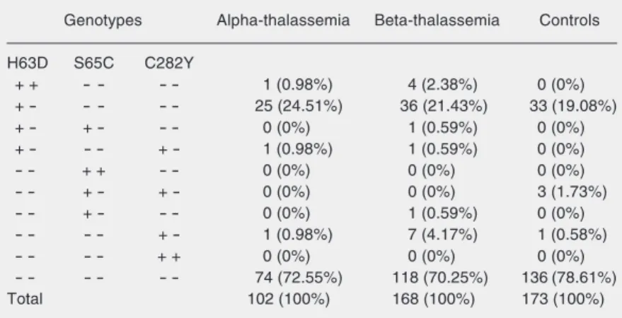
The genotypes found in each group are shown in Table 2. One beta-thalassemic case was a heterozygote for the H63D and S65C
Table 1. Allelic frequencies of the H63D, S65C and C282Y mutations.
mutations, one beta-thalassemic case and one alpha-thalassemic case were heterozy-gotes for the H63D and C282Y mutations, and three control cases were heterozygotes for the S65C and C282Y mutations. Ho-mozygote mutations were only found for H63D, including 1 alpha-thalassemic and 4 beta-thalassemic patients.
Statistical analysis using the chi-square test for multiple independent samples identi-fied a significant difference for the C282Y mutation among groups (P = 0.043). Fisher’s exact test indicated a statistically significant difference between the beta-thalassemic and control groups (P = 0.009). The H63D and S65C mutations did not show statistically significant differences among the groups studied (P > 0.05).
Discussion
HH is considered to be the most common inherited disorder in Caucasians and pre-sents a variable prevalence among different ethnic groups (2,22). Originally regarded as a rare affliction notable for its distinctive evolution to “bronze diabetes”, HH is now recognized as the most common genetic dis-order in populations of European ancestry. Recent advances in the understanding of
iron metabolism, the identification of the gene responsible for hemochromatosis, and extensive epidemiologic studies have changed the diagnostic approach to patients with HH and other forms of iron overload (23-25). Its phenotypic expression seems to be highly variable and possibly related to several fac-tors, such as availability of iron in the diet, loss of blood associated with menstruation or pregnancy, blood donation, hepatitis, and hemolytic anemia (9,23).
The C282Y missense mutation is respon-sible for about 80% of the cases of HH at least in Northern European populations. In other areas, e.g., Italy, its frequency is lower and decreases from North to South (9). In Brazil, H63D and C282Y screening in 4 specific populations (Caucasians, African descendants, Parakanã Indians, and a ra-cially mixed group) indicated a low preva-lence of the C282Y mutation (allelic fre-quency ranging from 0% for Parakanã Indi-ans to 1.4% for Caucasian descendants), while the H63D mutation varied from 0% in Parakanã Indians to 16.3% in Caucasian descendants (16). The allelic frequencies obtained in the present study for Caucasian individuals were 13.72, 13.70, and 9.54% for the H63D mutation in the alpha-thalas-semia, beta-thalassemia and control groups, respectively. For the C282Y mutation the frequencies observed were 0.98, 2.38, and 0.29%, respectively, differing from previ-ously published results from Brazil (16).
No data about the S65C mutation have been previously reported in Brazil. The low allelic frequencies observed here (0.0, 0.60, and 0.87% for the alpha-thalassemia, beta-thalassemia and control groups, respective-ly) agree with data about populations from other geographic areas (14,24,25). A 4.0% allelic frequency of the S65C mutation was detected in the Ecuadorian population, one of the highest observed until now (10).
In the present study, some cases of com-pound heterozygotes were found: one H63D/ C282Y patient in the alpha-thalassemic
Table 2. Genotypic frequencies of the H63D, S65C and C282Y mutations. Genotypes Alpha-thalassemia Beta-thalassemia Controls H63D S65C C282Y
group, two cases in the beta-thalassemic group (one of them H63D/S65C and the other H63D/C282Y), and three individuals with S65C/C282Y in the control group. Com-pound heterozygosis for C282Y and H63D seems to predispose to disease expression (5). The clinical significance of the other forms of compound heterozygosis, such as C282Y and S65C or H63D and S65C, is still controversial (5). However, some of these patients have thalassemia as another factor involving their iron status, which may be responsible for increasing the metal levels.
The clinical observation of a discordant course between individuals with the same hematological alteration (thalassemia) is very common and may be related to the different inherited mutations. Furthermore, the co-inheritance of iron metabolism disturbances can lead to an additional overload and an increase of oxidative stress in blood cells, influencing the phenotype expression and the manifestation of additional pathologies. Our findings suggest the need for an early screening for this alteration, especially in Brazil, with its multiethnic characteristics.
In the present study, a high incidence of the C282Y mutation was observed in the beta-thalassemia group, leading to a concern about the levels of iron deposition in the organism of these patients. Both diseases (hemochromatosis and thalassemia) affect iron metabolism and the meaning of co-inheritance of the two mutations is not well understood.
Screening for hemochromatosis muta-tions in beta-thalassemia minor patients from Iran indicated significant differences in the frequencies of C282Y and H63D mutants in relation to control individuals (20) but these differences were not observed in Portugal or in India (26,27). Two independent pathways have been proposed for iron metabolism, the erythroid regulator, which modulates intes-tinal iron absorption in response to the needs of the erythron, and the storage regulator, which controls iron accumulation (28-30).
There are suggestions that the erythroid regu-lator (beta-thalassemia) seems to be more pronounced than the storage regulator (the mutated HFE gene) in determining the de-gree of iron absorption (28,29). This hypo-thesis indicates that beta-thalassemia carri-ers might exhibit an advantage in balancing iron storage in their organisms. Neverthe-less, published data indicate more severe hemochromatosis symptoms when heterozy-gosis for the C282Y mutation is associated with beta-thalassemia (19), and when higher serum iron levels in beta-thalassemia co-in-heritance are associated with heterozygosis and homozygosis for H63D (4,26). However, these results remain controversial (29,31).
Alpha-thalassemia is a hereditary ane-mia that results from a defective synthesis of alpha-globin. There are insufficient produc-tion of normal hemoglobin and formaproduc-tion of unstable tetramers of γ4 (Hb Bart’s) or ß4 (Hb H) with accelerated destruction of the red blood cells. Alpha-thalassemia can be inherited or acquired and is originated by defects or deletions in one or more genes of the four alpha-globin genes. All the current knowledge about alpha-thalassemia, includ-ing its molecular biology understandinclud-ing, was obtained by clinical and laboratory observa-tions. Hypochromic and microcytic cells characterize alpha-thalassemia (32). The Hb H and Hb Bart’s are more stable than the alpha-chain aggregates observed in beta-thalassemia, and do not cause hemolysis. They precipitate in the red blood cells pro-voking cell damage and premature cell re-moval from the blood stream by endothelial reticulum systems, reducing the lifetime of the cells. In a study carried out in Hong Kong, iron overload in alpha-thalassemia was not related to hemochromatosis muta-tions (33). However, more data about the role of HFE mutations in alpha-thalassemic patients are still necessary. Nevertheless, HFE mutations were not found at a higher
The Brazilian population is a mixture of different ethnic groups and HFE mutations were observed at a higher frequency in beta thalassemic carriers. Hemoglobin chain syn-thesis disorders and HFE mutations may lead to severely increased iron storage and
worsen the clinical picture of hemochroma-tosis, reinforcing the need for this screening in thalassemic patients in Brazil. This ap-proach may improve a normal life expect-ancy and the response to specific anemia treatments.
References
1. Griffiths W, Cox T. Haemochromatosis: novel gene discovery and the molecular pathophysiology of iron metabolism. Hum Mol Genet
2000; 9: 2377-2382.
2. Hash RB. Hereditary hemochromatosis. J Am Board Fam Pract
2001; 14: 266-273.
3. Swinkels DW, Janssen MC, Bergmans J, Marx JJ. Hereditary hemo-chromatosis: genetic complexity and new diagnostic approaches.
Clin Chem 2006; 52: 950-968.
4. Melis MA, Cau M, Deidda F, Barella S, Cao A, Galanello R. H63D mutation in the HFE gene increases iron overload in beta-thalas-semia carriers. Haematologica 2002; 87: 242-245.
5. Pietrangelo A. Hereditary hemochromatosis - a new look at an old disease. N Engl J Med 2004; 350: 2383-2397.
6. Brissot P, Guyader D, Loreal O, Laine F, Guillygomarc’h A, Moirand R, et al. Clinical aspects of hemochromatosis. Transfus Sci 2000; 23: 193-200.
7. Vautier G, Olynyk JK. Porphyria cutanea tarda in the HFE-gene and hepatitis C virus era. Am J Gastroenterol 2000; 95: 3350-3352. 8. Candore G, Mantovani V, Balistreri CR, Lio D, Colonna-Romano G,
Cerreta V, et al. Frequency of the HFE gene mutations in five Italian populations. Blood Cells Mol Dis 2002; 29: 267-273.
9. Bomford A. Genetics of haemochromatosis. Lancet 2002; 360: 1673-1681.
10. Leone PE, Gimenez P, Collantes JC, Mino C. Analysis of HFE gene mutations (C282Y, H63D, and S65C) in the Ecuadorian population.
Ann Hematol 2005; 84: 103-105.
11. Rochette J, Pointon JJ, Fisher CA, Perera G, Arambepola M, Arichchi DS, et al. Multicentric origin of hemochromatosis gene (HFE) mutations. Am J Hum Genet 1999; 64: 1056-1062.
12. Papanikolaou G, Politou M, Terpos E, Fourlemadis S, Sakellaropou-los N, LoukopouSakellaropou-los D. Hereditary hemochromatosis: HFE mutation analysis in Greeks reveals genetic heterogeneity. Blood Cells Mol Dis 2000; 26: 163-168.
13. Campo S, Restuccia T, Villari D, Raffa G, Cucinotta D, Squadrito G, et al. Analysis of haemochromatosis gene mutations in a population from the Mediterranean Basin. Liver 2001; 21: 233-236.
14. Mura C, Raguenes O, Ferec C. HFE mutations analysis in 711 hemochromatosis probands: evidence for S65C implication in mild form of hemochromatosis. Blood 1999; 93: 2502-2505.
15. Asberg A, Thorstensen K, Hveem K, Bjerve KS. Hereditary hemo-chromatosis: the clinical significance of the S65C mutation. Genet Test 2002; 6: 59-62.
16. Agostinho MF, Arruda VR, Basseres DS, Bordin S, Soares MC, Menezes RC, et al. Mutation analysis of the HFE gene in Brazilian populations. Blood Cells Mol Dis 1999; 25: 324-327.
17. Laine F, Jouannolle AM, Morcet J, Brigand A, Pouchard M, Lafraise B, et al. Phenotypic expression in detected C282Y homozygous women depends on body mass index. J Hepatol 2005; 43:
1055-1059.
18. Piperno A, Mariani R, Arosio C, Vergani A, Bosio S, Fargion S, et al. Haemochromatosis in patients with beta-thalassaemia trait. Br J Haematol 2000; 111: 908-914.
19. Rees DC, Singh BM, Luo LY, Wickramasinghe S, Thein SL. Nontransfusional iron overload in thalassemia. Association with he-reditary hemochromatosis. Ann N Y Acad Sci 1998; 850: 490-494. 20. Jazayeri M, Bakayev V, Adibi P, Haghighi RF, Zakeri H, Kalantar E,
et al. Frequency of HFE gene mutations in Iranian beta-thalassaemia minor patients. Eur J Haematol 2003; 71: 408-411.
21. Sambrook J, Fritsch EF, Maniats T. Molecular cloning: a laboratory manual. 2nd edn. New York: Cold Spring Harbor Laboratory Press; 1989.
22. Brissot P. Hemochromatosis at the intersection of classical medi-cine and molecular biology. C R Acad Sci III 2001; 324: 795-804. 23. Yen AW, Fancher TL, Bowlus CL. Revisiting hereditary
hemochro-matosis: current concepts and progress. Am J Med 2006; 119: 391-399.
24. Siah CW, Trinder D, Olynyk JK. Iron overload. Clin Chim Acta 2005; 358: 24-36.
25. Beutler E. Hemochromatosis: genetics and pathophysiology. Annu Rev Med 2006; 57: 331-347.
26. Alexander J, Kowdley KV. Hereditary hemochromatosis: genetics, pathogenesis, and clinical management. Ann Hepatol 2005; 4: 240-247.
27. Martins R, Picanco I, Fonseca A, Ferreira L, Rodrigues O, Coelho M, et al. The role of HFE mutations on iron metabolism in beta-thalassemia carriers. J Hum Genet 2004; 49: 651-655.
28. Levy JE, Montross LK, Andrews NC. Genes that modify the hemo-chromatosis phenotype in mice. J Clin Invest 2000; 105: 1209-1216. 29. Ajioka RS, Levy JE, Andrews NC, Kushner JP. Regulation of iron
absorption in Hfe mutant mice. Blood 2002; 100: 1465-1469. 30. Politou M, Kalotychou V, Pissia M, Rombos Y, Sakellaropoulos N,
Papanikolaou G. The impact of the mutations of the HFE gene and of the SLC11A3 gene on iron overload in Greek thalassemia inter-media and beta(s)/beta(thal) anemia patients. Haematologica 2004; 89: 490-492.
31. Cappellini MD, Fargion SR, Sampietro M, Graziadei G, Fiorelli G. Nontransfusional iron overload in thalassemia intermedia: role of the hemochromatosis allele. Blood 1998; 92: 4479-4480.
32. Higgs DR, Vickers MA, Wilkie AO, Pretorius IM, Jarman AP, Weatherall DJ. A review of the molecular genetics of the human alpha-globin gene cluster. Blood 1989; 73: 1081-1104.