Received on 09 July 2002; revised 30 September 2002. Address for correspondence: Dr. Terezinha de Jesus Teixeira Santos – Lab. de Neurologia Experimental e Neurofisiologia – Depto. de Fisiologia e Farmacologia – Universidade Federal do Ceará – Rua Cel. Nunes de Melo, 1127 – Rodolfo Teófilo – Fortaleza-CE – 60.430-270 – Fax: +55 85 288-8333 – E-mail: mcastro@ufc.br
The Brazilian Journal of Infectious Diseases 2003;7(3):202-209 © 2003 by The Brazilian Journal of Infectious Diseases and Contexto Publishing. All rights reserved.
Western Blot Seroindeterminate Individuals for Human T-lymphotropic Virus 1/2
(HTLV-1/2) in Fortaleza (Brazil): A Serological and Molecular Diagnostic and
Epidemiological Approach
Terezinha de Jesus Teixeira Santos, Department of Physiology and Pharmacology, Carlos Maurício de Castro Costa, Federal University of Ceará, Fortaleza, CE, Brazil; Patrick Goubau,Anne-Mieke Vandamme, Unit of Virology, Université Catholique de Louvain, Jan Desmyter, Sonia Van Dooren, Rosa M. S. Mota, Louvain, Belgium; Rega Institute for Medical Francine Bovy de Castro Costa, Ana C. S. Oliveira, Research, Katholieke Universiteit Leuven, Leuven, Vânia Barreto A.F. Gomes, Anna B. Carneiro-Proietti, Belgium; Department of Statistics and Applied Veralice Meireles Sales de Bruin, Francisca C. F. de Sousa Mathematics, Federal University of Ceará,
and Reinaldo Barreto Oriá Fortaleza, CE, Brazil; HEMOCE of Fortaleza,
Fortaleza, CE, Brazil; HEMOMINAS Foundation, Belo Horizonte, MG, Brazil
How to handle Western blot (WB) seroindeterminate individuals for Human T-lymphotropic Virus 1/2 (HTLV-1/2) constitutes a challenge for blood banks and families. We made a cross-sectional study of 191 enzyme linked immunoassay (EIA) reactive individuals from the hematological center (HEMOCE) of Fortaleza (Brazil), examining their serological (WB) and molecular (PCR) diagnosis, and demographic profiles, as well as a possible association of their condition with other infectious pathologies and risk factors. Ethical institutional approval and personal consent were obtained. Out of 191 EIA reactive individuals, 118 were WB seroindeterminate and 73 were seropositive for HTLV-1/2. In the PCR analysis of 41 WB seroindeterminate individuals, 9 (22%) were positive and 32 (78%) were negative for HTLV-1/2. The demographic analysis indicated a trend towards a predominance of males among the seroindeterminate individuals and females in the seropositive ones. The seroindeterminate individuals were younger than the seropositive ones. We did not find any association of these conditions with syphilis, Chagas disease or HIV or hepatitis, and with risk factors such as breast-feeding, blood transfusion, STD (syphilis) and IDU.
Key Words: Epidemiology, PCR, HTLV-1/2, seroindeterminate individuals, hepatitis, syphilis, blood transfusion.
The human T-lymphotropic virus (HTLV) belongs to the family Retroviridae and the subfamily Oncovirinae. It is divided into types 1 and 2, sharing around 60% similarity. Based on molecular epidemiology HTLV-1 is
divided into the 1a (cosmopolitan), 1b (Central Africa), 1c (Melanesia), 1d, 1e and 1f (Central Africa) subtypes, and HTLV-2 into the 2a, 2b and 2d subtypes. HTLV-1/ 2 is variably endemic in Central and West Africa, the Caribbean, South America, Japan and Melanesia. In Brazil, its prevalence is 0.46% [1]. This retrovirus is transmitted vertically (mother-to-child) and horizontally (sexual contact, blood transfusion and intravenous drug use). Tropical Spastic Paraparesis/HTLV-1-associated Myelopathy (TSP/HAM) and Adult T-cell Leukemia/Lymphoma (ATL) are the main associated pathologies [2,3].
particle agglutination), confirmatory (Western blot, IFA, RIPA, and more recently INNO-LIA) and molecular (PCR, NASBA and bDNA) tests. Serological tests for HTLV infection have been mandatory in some countries since 1986, and in Brazil since 1993. Screening and confirmatory results from blood banks are varied, with negative, positive and indeterminate profiles apparent.
Blood banks worldwide are thus faced with blood donor candidates exhibiting a seroindeterminate Western blot (WB) profile. This is mainly reported in tropical and in some temperate regions endemic for HTLV-1/2 infection, and less frequently in northern countries, where prevalence of HTLV-1/2 is very low [4,5].
Individuals seroindeterminate for HTLV-1/2 represent a challenge for blood banks, as well as in familial contexts, due to the risk they represent in spreading infections through blood donation and sexual contact, if truly infected, and because of the psychological burden it presents for the person with this undefined condition.
With the aim of delineating a profile of Western Blot (WB) seroindeterminate individuals for HTLV-1/2 from the hematological center (HEMOCE) of Fortaleza (Brazil), we carried out a cross-sectional study, taking into consideration their serological and molecular diagnosis, and demographic characteristics, as well as the possible association of this condition with other infectious pathologies and risk factors.
Materials and Methods
One hundred and ninety one EIA reactive candidates among blood donors at the HEMOCE were tested with confirmatory HTLV WB version 2.4 (WB2.4) from Genelabs Diagnostic. Seropositivity was interpreted according to the stringent criteria indicated by Genelabs Diagnostics. A WB test was scored as HTLV-1 positive only if bands for the gag proteins (p19 with or without p24) and two env proteins (GD21 and rgp46-1) were present. It was scored as HTLV-2 positive if bands for the gag proteins (p24 with or without p19) and two env proteins (GD21 and rgp46-2) were found. The test was considered as indeterminate if
specific bands for HTLV that did not fulfill the criteria of positivity for HTLV-1 and 2 were present. Those that did not exhibit bands specific for HTLV were considered negative.
For HTLV-1/2 molecular analysis, 41 out of the 118 samples from WB seroindeterminate individuals were tested with PCR at the Rega Institute for Medical Research and University Hospitals of the Katholieke Universiteit Leuven (Belgium).
For provirus detection in patient samples, 106 peripheral blood mononuclear cells were pelleted and lysed in PCR buffer (10 mM Tris-HCl, pH 8.3, 50 mM KCl Perkin-Elmer), containing 2 mM MgCl2, 0.5% Tween-20, 0.5% NP40, and 100 µg/ml proteinase K (Boehringer Mannheim, stabilized proteinase K solution), for 1 hr at 56ºC. The DNA was extracted using phenol/chloroform (Life Technologies), precipitated with ethanol, and dissolved in Milli-Q water (Millipore system). The Qiagen blood kit was used (Westburg) for extracting the DNA of the control cell lines.
DNA from 105 cells was used for amplification in a 50 µl reaction volume containing 10 mM Tris-HCl, pH 8.3, 50 mM KCl, 200 µM nucleotide triphosphates, 0.2 µM outer or 0.5 µM inner primers and 00.25 U/µl AmpliTaq (Perkin-Elmer), with 1.5 mM MgCl
HTLV-1 MT2 (kindly provided by Luc Montaigner, Institut Pasteur, Paris, France), SI-5 cells harboring HTLV-1 Mel5 (kindly provided by Richard Yanagihara, NIH, Bethesda, MD), 729 cells harboring HTLV-2 Mo (kindly provided by Helen Lee, Abbott Laboratories, North Chicago, IL) clone 19 cells harboring HTLV-2 (kindly provided by Dana Gallo, Viral and Rickettsial Disease Laboratory, Berkeley, CA), Cu cells harboring HTLV-2, Gu, PH969 cells harboring STLV-PH969, and PP1664 cells harboring STLV-PP1664. Negative controls for the PCR were Hut-78 cells (kindly provided by Institut Pasteur, Brussels, Belgium) and ACH-2 cells (kindly provided through the AIDS reagent project of the Medical Research Council, UK).
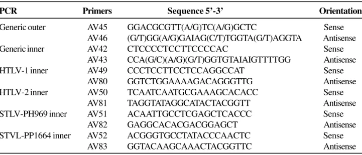
The PCR primers were designed using the tax gene alignment of all available HTLV and STLV strains. The primer sequences for the generic nested PCR and the four discriminatory inner PCRs are given in Table 1. Primers were developed and analyzed using the Oligo software (Medprobe, Oslo, Norway). Synthesis was carried out by Pharmacia Biotech and Perkin-Elmer/Applied Biosystems.
For the demographic study, age and gender were considered. Thus, 118 seroindeterminate and 73 seropositive individuals previously diagnosed by WB, and 41 out of 118 WB seroindeterminate individuals diagnosed by PCR, were included.
The analysis of co-infection and risk factors was based on previous information obtained from some of the individuals of our series. This explains the different numbers shown below.
The analysis of co-infection included laboratorial evidence for infections such as syphilis, Chagas disease, HIV, and hepatitis. Thus, 83 WB seroindeterminate and 27 seropositive individuals for HTLV-1/2, as well as 31 WB seroindeterminate individuals, diagnosed by PCR, were studied.
The risk factors, breast-feeding, blood transfusion, sexually transmitted diseases (STD) (syphilis) and intravenous drug use (IDU) were analyzed in 95 seroindeterminate and 65 seropositive individuals diagnosed by WB, and in 36 seroindeterminate ones diagnosed by PCR.
This research project was approved by the Committee for Ethics in Research of the Faculty of Medicine of the Federal University of Ceará (Brazil), with the participating individuals each signing a consent document.
Descriptive statistics and association tests for contingency tables (Fischer’s exact test) were used in the analysis. Levene’s and Student’s t-tests were used for analysis of variance and to determine if the means differed [7]. A level of significance of p≤0.05 was considered.
Results
The WB confirmatory test of our sample of 191 EIA reactive individuals showed that 73 were seropositive and 118 were seroindeterminate for HTLV-1/2.
The PCR analysis of the 41 WB seroindeterminate individuals revealed that 32 (78%) were negative and 9 (22%) were positive (7 for HTLV-1 and 2 for HTLV-2). The demographic analysis of the 118 WB seroindeterminate and 73 seropositive individuals showed that there was a significant (p=0.0420) association between sex and WB results, from which one could depict a trend for female predominance in the group of individuals seropositive for HTLV-1/2, and for male predominance in the group of seroindeterminate individuals (Table 2). The mean age of HTLV-1/2 seroindeterminate and seropositive individuals was 34.5 years (SD=9.6; Median=32) and 40.4 years (SD = 14.7; Median = 41.5), respectively. The mean age of seroindeterminate individuals was significantly lower (p=0.0029) than that of HTLV-1/2 seropositive individuals .
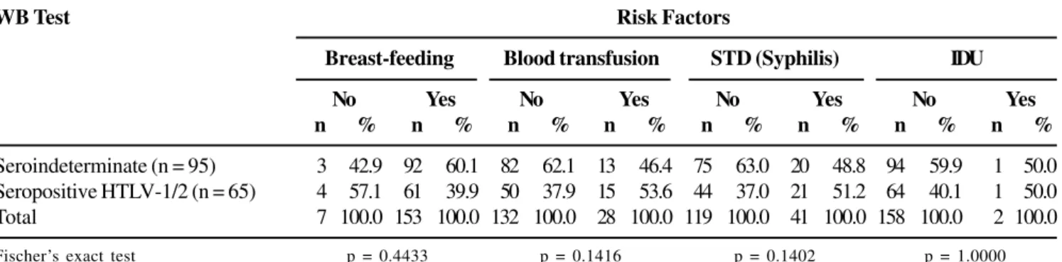
When we examined associated diseases, there was no significant correlation between the results of WB and PCR tests and co-infections (syphilis, Chagas disease, HIV, and hepatitis) (Tables 3 and 4).
Table 2. Distribution of WB seroindeterminate (n = 118) and seropositive (n = 73) individuals by gender
Table 1. Primers in the tax/rex gene used for the generic and discriminatory HTLV PCRs
Discussion
We found that 9 (22%) and 32 (78%) of the 41 WB seroindeterminate individuals were positive and negative for HTLV-1/2, respectively, as determined by PCR; consequently, this method could be useful for defining the condition of the WB seroindeterminate individuals for HTLV-1/2.
These findings are coherent with previous investigations. For example, Medrano et al. [8] studied 16 seroindeterminate individuals, and found HTLV-1/ 2 with PCR in 4 (25%). In addition, Garin et al. [9] studied 67 blood samples of seroindeterminate individuals in Zaire, and he found HTLV-1 (with PCR) in only 10 (15%) of them, whilst none were positive for HTLV-2. On the other hand, Caterino-de-Araújo
et al. [10] analyzed 553 samples from HIV positive individuals from São Paulo (Brazil), of which 24 (4.3%) were seroindeterminate for HTLV-1/2, 25% of them being positive for HTLV-2 by PCR. Delaporte et al. [11] carried out two studies in Gabon and found positivity by PCR in 16% and 31% of the WB seroindeterminate samples. However, Lipka et al. [12] analyzed 67 seroindeterminate samples and did not find positivity for HTLV-1/2 by PCR in any of them. Nerurkar et al. [13] studied 27 seroindeterminate individuals from Papua New Guinea and the Salomon Islands, and viral sequences of HTLV-1 were not found in any of them. Gessain et al. [14] reported a high prevalence of seroindeterminate patterns in the tropics, with PCR negative results. This may mean that there are additional etiologies for this condition.
WB Test Sex
Female Male Total
n % n % n %
Seroindeterminate (n = 118) 33 28.0 85 72.0 118 100.0 Seropositive (n = 73) 31 42.5 42 57.5 73 100.0 Fischer’s exact test p = 0.0420
PCR Primers Sequence 5’-3’ Orientation
Generic outer AV45 GGACGCGTT(A/G)TC(A/G)GCTC Sense
AV46 (G/T)GG(A/G)GAIAG(C/T)TGGTA(G/T)AGGTA Antisense
Generic inner AV42 CTCCCCTCCTTCCCCAC Sense
AV43 CCA(G/C)(A/G)(G/T)GGTGTAIAIGTTTTGG Antisense
HTLV-1 inner AV49 CCCTCCTTCCTCCAGGCCAT Sense
AV80 GGTCTGGAAAAGACAGGGTTG Antisense
HTLV-2 inner AV50 TCAATCAATGCGAAAGCACACC Sense
AV81 TAGGTATAGGCATACTACGGTT Antisense
STLV-PH969 inner AV51 ACAATTGCCTCGAGCTCACCC Sense
AV82 GAGGCACACGACGGAGCT Antisense
STVL-PP1664 inner AV52 ACGGGTGCCTATACCCAACTC Sense
Table 3. Distribution of WB seroindeterminate (n = 83) and seropositive (n = 27) individuals for HTLV-1/2 as a function of the Co-Infection test results
Table 4. Distribution of PCR negative (n = 25) and PCR/WB positive (n = 33) individuals for HTLV-1/2 as a function of the Co-Infection test results
PCR/WB Test Co-infection Test Results
Hepatitis B (HBsAg) Hepatitis B (HBc) Hepatitis C (HCV)
Negative Positive Negative Positive Negative Positive
n % n % n % n % n % n %
Negative (PCR) 23 42.6 2 50.0 21 47.7 4 26.7 22 44.9 3 30.0
Positive HTLV-1 18 33.3 1 25.0 13 29.5 6 40.0 13 26.5 6 60.0
(PCR/WB)
Positive HTLV-2 13 24.1 1 25.0 10 22.7 5 33.3 14 28.6 1 10.0
(PCR/WB)
Total 54 100.0 4 100.0 44 100.0 15 100.0 49 100.0 10 100.0
PCR/WB Test Co-Infection Test Results
Chagas Disease Syphilis HIV
Negative Positive Negative Positive Negative Positive
n % n % n % n % n % n %
Negative (PCR) 25 43.9 – – 25 43.1 – – 24 42.1 1 100.0
Positive HTLV-1 19 33.3 – – 19 32.8 – – 19 33.3 – –
(PCR/WB)
Positive HTLV-2 13 22.8 1 100.0 14 24.1 – – 14 24.6 – –
(PCR/WB)
Total 57 100.0 1 100.0 58 100.0 – – 57 100.0 1 100.0
WB Test Co-Infection Test Results
Chagas disease Syphilis HIV
Negative Positive Negative Positive Negative Positive
n % n % n % n % n % n %
Seroindeterminate 83 76.1 – – 83 75.5 – – 81 75.0 2 100.0 Seropositive 26 23.9 1 100.0 27 24.5 – – 27 25.0 – –
Total 109 100.0 1 100.0 110 100.0 – – 108 100.0 2 100.0
WB Test Co-Infection Test Results
Hepatitis B (HBsAg) Hepatitis B (HBc) Hepatitis C (HCV)
Negative Positive Negative Positive Negative Positive
n % n % n % n % n % n %
Seroindeterminate 78 75.7 5 71.4 71 78.9 12 57.1 78 77.2 5 50.0 Seropositive 25 24.3 2 28.6 19 21.1 9 42.9 23 22.8 5 50.0
Total 103 100.0 7 100.0 90 100.0 21 100.0 101 100.0 10 100.0
According to data (unpublished) from the hematological center of Fortaleza (HEMOCE-Ceará) from 1997 to 2000, the prevalence of seroindeterminate individuals was 0.16% in the general population of blood donors and 21.99% among the population of EIA reactive individuals. In another study, also in Ceará (Brazil), De Castro-Costa et al. [15] analyzed 1042 blood samples of non-neurological patients from Fortaleza (n=593) and Crato (n=449), of which 6 (1.0%) in Fortaleza and 2 (0.4%) in Crato exhibited a seroindeterminate profile.
There is however variation of prevalence depending on the geographic area and the population studied. While in Europe and USA the prevalence is low, in tropical areas it reaches higher values [4,5]. Lal et al. [16] analyzed 267,650 samples from blood donors, among which 379 (0.14%) were WB seroindeterminate. Césaire et al. [17] carried out an analysis of 9,759 blood donors from the West Indies and found 39 (0.4%) seropositive and 49 (0.5%) seroindeterminate individuals for HTLV-1. Mauclère et al. [18] studied 3783 individuals from the rural population of Cameroon and found a prevalence of 1.6% seroindeterminates. Garin et al. [9] studied the frequency of positivity for HTLV-1/2 in 98 samples of blood from individuals from Zaire and found that 28 (28.6%) were seropositive, 3 (3.0%) were seronegative and 67 (68.4%) were seroindeterminate. In Brazil, Caterino-de-Araújo et al. [10] investigated HTLV-1/2 infection in 553 samples of serum from HIV patients in São Paulo, of which 24 (4.3%) had an indeterminate profile.
Demographically, young men predominated among the seroindeterminate individuals, while older women predominated among the ones seropositive for HTLV-1/2. The difference in age between seroindeterminate and seropositive individuals could be due to tardive seroconversion; while the trend of women predominating among the seropositive individuals could be due to their greater exposure to infection with HTLV-1/2 [18,19]. Césaire et al. [17], however, did not find significant gender differences between groups seroindeterminate and seropositive for HTLV-1/2,
although consistent with our findings, they found that the seroindeterminate and seronegative individuals were younger than the seropositives.
Our investigation of HTLV-1/2 seroindeterminate and seropositive individuals with co-infections revealed seropositive individuals with anti-HBc (hepatitis B) and HCV (hepatitis C) antibodies, which is also shown in supported by previous reports from Brazil [20,21] and from other countries [17,19].
Other presumptive associations of indeterminate profiles have been reported with Plasmodium
falciparum [22-27], varicella zoster virus or herpes
simplex [28-31], and dengue virus [32]. Moreover, recent seroconversion to positivity for HTLV-1/2 [17], incomplete reactions to molecularly distinct variants of HTLV-1/2 or eventually to another exogenous or endogenous retrovirus [30], and transient infection by HTLV-1 [14], have also been reported. Other conditions are also supposedly correlated with a seroindeterminate profile. For example, Haynes et al. [33] and Palker et al. [34] analyzed the antigenic relationship between p19 and the thymic epithelium, possibly linked to immune conditions in HTLV-1 infected individuals. Seroindeterminate patterns have been found in Europe and the USA in a series of patients with multiple sclerosis, although at a very low frequency [35]. A seroindeterminate profile may also be associated with hematological abnormalities compatible with viral infection [36] or with a possible, as yet unidentified, genetic or environmental factor [37].
Acknowledgments
We thank the team of researchers and technicians of the Rega Institute of the Katholieke Universiteit Leuven (Belgium), the hematological centers of Fortaleza, CE (HEMOCE) and of Belo Horizonte, MG (HEMOMINAS), the Central Laboratory of the University Hospital of Fortaleza, CE (Brazil) for their collaboration, Dr. David Neil Criddle for the review of English style and Sílvio A Costa for the accurate electronic typewriting of this manuscript.
References
1. Galvão-Castro B., Loures L., Rodrigues L.G., et al. Distribution of human T-lymphotropic virus type 1 among blood donors: a nationwide Brazilian study. Transfusion 1997;37(2):242-3.
2. Gessain A., Barin F., Vernant J.C., et al. Antibodies to human T-lymphotropic virus type-1 in patients with tropical spastic paraparesis. Lancet 1985;2:407-10. 3. Osame M., Izuma S., Igata A., et al. Blood transfusion and
HTLV-1 associated myelopathy. Lancet 1986;2:104-5. 4. Lal R.B., Lipka J.J., Foung S.K., et al. Human T
lymphotropic virus type 1/2 in Lake Lindu Valley, Central Sulawesi, Indonesia. J Acquir Immune Defic Syndr Hum Retrovirol 1993;6:1067-8.
5. Mahieux R., Horal P., Mauclère P., et al. Human T-cell lymphotropic virus type 1 gag indeterminate Western blot patterns in Central Africa: relationship to Plasmodium falciparum infection. J Clin Microbiol 2000;38:4049-57. 6. Vandamme A.M., Fransen K., Debaisieux L., et al.
Standardization of primers and an algorithm for HIV-1 diagnostic PCR evaluated in patients harbouring strains of diverse geographical origin. The Belgian AIDS Reference Laboratories. J Virol Methods 1995;51:305-16.
7. Freeman J., Daniel H. Applied categorical data analysis. Marcel Dekker, 1987.
8. Medrano F.J., Soriano V., Calderón E.J., et al. Significance of indeterminate reactivity to human T-cell lymphotropic virus in Western blot analysis of individuals at risk. Eur J Clin Microbiol Infect Dis
1997;16:249-52.
9. Garin B., Gosselin S., De Thé G., Gessain A. HTLV-1/2 infection in a high viral endemic area of Zaire, Central Africa: comparative evaluation of serology, PCR, and significance of indeterminate Western blot pattern. J Med Virol 1994;44:104-9.
10. Caterino-de-Araújo A., de los Santos-Fortuna E., Meleiro M.C., et al. Sensitivity of two enzyme-linked immunosorbent assay tests in relation to Western blot in detecting human T-cell lymphotropic virus type 1 and 2 infection among HIV-1 infected patients from São Paulo, Brazil. Diagn Microbiol Infect Dis 1998;30:173-82. 11. Delaporte E., Monplaisir N., Louwagie J., et al. Prevalence
of HTLV-1 and HTLV-2 infection in Gabon, Africa: comparison of the serological and PCR results. Int J Cancer 1991;49:373-6.
12. Lipka J.J., Young K.K., Kwok S.Y., et al. Significance of human T-lymphotropic virus type 1 indeterminant serological findings among healthy individuals. Vox Sang 1991;61:171-6.
13. Nerurkar V.R., Miller M.A., Leon-Monzon M.E., et al. Failure to isolate human T cell lymphotropic virus type 1 and to detect variant-specific genomic sequences by polymerase chain reaction in Melanesians with indeterminate Western immunoblot. J Gen Virol 1992;73:1805-10.
14. Gessain A., Mahieux R., De Thé G. HTLV-1 “indeterminate” Western blot patterns observed in sera from tropical regions: the situation revisited (Letter). J Acquir Immune Defic Syndr Hum Retrovirol 1995;9(3):316-8.
15. De Castro-Costa C.M., Goubau P., Liu H.-F., et al. HTLV-negative and HTLV-positive tropical spastic paraparesis in Northeastern Brazil. AIDS Res Hum Retroviruses
1995;11:315-18.
Table 5. Distribution of WB seroindeterminate (n = 95) and seropositive (n = 69) individuals for HTLV-1/2 as a function of the Risk Factors
WB Test Risk Factors
Breast-feeding Blood transfusion STD (Syphilis) IDU
No Yes No Yes No Yes No Yes
n % n % n % n % n % n % n % n %
Seroindeterminate (n = 95) 3 42.9 92 60.1 82 62.1 13 46.4 75 63.0 20 48.8 94 59.9 1 50.0 Seropositive HTLV-1/2 (n = 65) 4 57.1 61 39.9 50 37.9 15 53.6 44 37.0 21 51.2 64 40.1 1 50.0 Total 7 100.0 153 100.0 132 100.0 28 100.0 119 100.0 41 100.0 158 100.0 2 100.0
16. Lal R.B., Rudolph D.L., Coligan J.E., et al. Failure to detect evidence of human T-lymphotropic virus (HTLV) type 1 and type 2 in blood donor with isolated gag antibodies to HTLV-1/2. Blood 1992;80:544-50.
17. Césaire R., Bera O., Maier H., et al. Seroindeterminate patterns and seroconversions to human T-lymphotropic virus type 1 positivity in blood donors from Martinique, French West Indies. Transfusion 1999;39:1145-9. 18. Mauclère P., Le Hesran J.Y., Mahieux R., et al.
Demographic, ethnic, and geographic differences between human T cell lymphotropic virus (HTLV) type-1-seropositive carriers and persons with HTLV-1 gag-indeterminate Western blots in Central Africa. J Infect Dis 1997;176:505-9.
19. Rouet F., Meertens L., Courouble G., et al. Serological, epidemiological, and molecular differences between human T-cell lymphotropic virus Type 1 (HTLV-1)-seropositive healthy carriers and persons with HTLV-1 gag indeterminate Western blot patterns from the Caribbean. J Clin Microbiol 2001;39:1247-53.
20. Nogueira C.M., Cavalcanti M., Schechter M., Ferreira O.C. Jr. Human T lymphotropic virus type 1 and 2 infections in healthy blood donors from Rio de Janeiro, Brazil (Letter). Vox Sang 1996;70:47-8.
21. Carneiro-Proietti A.B., Lima-Martins M.V., Passos V.M., et al. Presence of human immunodeficiency virus (HIV) and T-lymphotropic virus type 1 and 2 (HTLV-1/2) in a haemophiliac population in Belo Horizonte, Brazil, and correlation with additional serological results. Haemophilia 1998;4:47-50.
22. Biggar R.J., Gigase P.L., Melbye M., et al. ELISA HTLV retrovirus antibody reactivity associated with malaria and immune complexes in healthy Africans. Lancet
1985;2:520-3.
23. Yanagihara R., Jenkins C.L., Alexander S.S., et al. Human T-lymphotropic virus type 1 infection in Papua New Guinea: high prevalence among the Hagahai confirmed by Western analysis. J Infect Dis 1990;162:649-54. 24. Hayes C.G., Burans J.P., Oberst R.B. Antibodies to
human T lymphotropic virus type 1 in a population from the Philippines: evidence for cross-reactivity with Plasmodium falciparum. J Infect Dis
1991;163:257-62.
25. Lal R.B., Rudolph D., Alpers M.P., et al. Immunological cross-reactivity between structural protein of human T-cell lymphotropic virus type 1 and the blood stage of
Plasmodium falciparum. Clin Diagn Lab Immunol
1994;1:5-10.
26. Porter K.R., Liang L., Long G.W., et al. Evidence for
anti-Plasmodium falciparum antibodies that cross-react with human T-lymphotropic virus type 1 proteins in a population in Irian Jaya, Indonesia. Clin Diagn Lab Immunol 1994;1:11-5.
27. Porter K.R., Anthony R.L., Solihin A., Hayes C.G. Mapping of a human T-lymphotropic virus type 1 gag protein epitope that cross-reacts with anti-Plasmodium falciparum antibodies. J Med Virol 1995;45:469-74. 28. Sato A., Isaka Y., Morita F., et al. Human sera from
varicella-zoster virus (VZV) infections cross-react with human T-cell leukaemia virus type 1 (HTLV-1): common epitopes in VZV gene 22 protein and HTLV-1 p19 gag protein. J Gen Virol 1992; 73:2969-73.
29. Banki K., Maceda J., Hurley E., et al. Human T-cell lymphotropic virus (HTLV)-related endogenous sequences, HRES-1, encodes a 28-kDa protein: a possible autoantigen for HTLV-1 gag-reactive autoantibodies. Proc Natl Acad Sci USA 1992;89: 1939-43.
30. Miyakoshi H., Sugimoto M., Igarashi H., et al. Improvement of simultaneous detection of antibodies to gag and envelope antigens of human T-lymphotropic virus type 1 by Western immunoblot assay. J Clin Microbiol 1992;30:2555-59.
31. Segurado A.A., Malaque C.M.S., Sumita L.M., et al. Laboratory characterization of human T-cell lymphotropic virus types 1 (HTLV-1) and 2 (HTLV-2) infections in blood donors from São Paulo (Brazil). Am J Trop Med Hyg 1997;57(2):142-48.
32. Carvalho S.M.F., Pombo-de-Oliveira M.S., Thuler L.C.S., et al. Cross-reactivity between human T-cell leukemia/ lymphoma virus indeterminate Western blot and dengue virus in individuals from Rio de Janeiro, Brazil. J Acquir Immune Defic Syndr Hum Retrovirol 1999;20:4(PO94). 33. Haynes B.F., Robert-Guroff M., Metzgar R.S., et al. Monoclonal antibody against human T cell leukemia virus p19 defines a human thymic epithelial antigen acquired during ontogeny. J Exp Med 1983;157:907-20. 34. Palker T.J., Singer K.H., Vahlne A. Characterization of an antigen shared by human thymic epithelium and human T cell leukemia virus p19 gag protein. J Acquir Immune Defic Syndr Hum Retrovirol 1996;11:10-19.
35. Soldan S.S., Graf M.D., Waziri A., et al. HTLV-1/2 seroindeterminate Western blot reactivity in a cohort of patients with neurological disease. J Infect Dis
1999;180: 685-94.
36. Picchio G.R., Bare P., Savignano R., et al. HTLV-1/2 indeterminate serology and natural killer cell expansion. J Acquir Immune Defic Syndr Hum Retrovirol 1996; 12:428-31.