Experimental chronic entrapment of
the sciatic nerve in adult hamsters: an
ultrastructural and morphometric study
Departamentos de 1Anatomia and 2Histologia e Embriologia,
Universidade Federal do Rio de Janeiro, Rio de Janeiro, RJ, Brasil R.A.D. Prinz1,
M. Nakamura-Pereira1,
B. De-Ary-Pires1,
D.S. Fernandes1,
B.D.S.V. Fabião-Gomes1,
P.S. Bunn1,
A.M.B. Martinez2,
M.A. Pires-Neto1
and R. Ary-Pires1
Abstract
Entrapment neuropathy is a group of clinical disorders involving compression of a peripheral nerve and interference with nerve func-tion mostly through tracfunc-tion injury. We have investigated the chronic compression of peripheral nerves as an experimental procedure for detecting changes in ultrastructural nerve morphology. Adult ham-sters (Mesocricetus auratus, N = 30) were anesthetized with a 25%
pentobarbital solution and received a cuff around the right sciatic nerve. Left sciatic nerves were not operated (control group). Animals survived for varying times (up to 15 weeks), after which they were sacrificed and both sciatic nerves were immediately fixed with a paraformaldehyde solution. Experimental nerves were divided into segments based upon their distance from the site of compression (proximal, entrapment and distal). Semithin and ultrathin sections were obtained and examined by light and electron microscopy. Ultra-structural changes were qualitatively described and data from semi-thin sections were morphometrically analyzed both in control and in compressed nerves. We observed endoneurial edema along with both perineurial and endoneurial thickening and also the existence of whorled cell-sparse structures (Renaut bodies) in the subperineurial space of compressed sciatic nerves. Morphometric analyses of myeli-nated axons at the compression sites displayed a remarkable increase in the number of small axons (up to 60%) in comparison with the control axonal number. The distal segment of compressed nerves presented a distinct decrease in axon number (up to 40%) compara-tively to the control group. The present experimental model of nerve entrapment in adult hamsters was shown to promote consistent histo-pathologic alterations analogous to those found in chronic compres-sive neuropathies.
Correspondence
R. Ary-Pires
Departamento de Anatomia UFRJ, CCS, Bloco F 21941-590 Rio de Janeiro, RJ Brasil
Fax: +55-21-2553-6321 E-mail: [email protected]
Research supported by FAPERJ, CNPq, CAPES and TWAS.
Received October 2, 2002 Accepted May 9, 2003
Key words
Entrapment neuropathies are produced by existing anatomical arrangements that cause compression or constriction of a pe-ripheral nerve. The three most common en-trapment syndromes (the thoracic outlet syn-drome, the carpal tunnel synsyn-drome, and the cubital entrapment syndrome) are remark-able examples of the dramatic clinical fea-tures with motor and sensory symptoms of-ten associated with autonomic dysfunction. They occur at specific places where nerves are confined to narrow anatomic pathways and therefore are particularly susceptible to constricting pressures. In those neuropathies the neural lesion is directly attributable to local causes comprising disturbances to nerve vascular supply (1), axonal transport mechan-isms (2,3), degeneration of myelin sheaths (4), axonal damage (5), and/or connective tissue changes such as endoneurial edema (6). Nerves can be damaged in a number of ways such as: a) ischemia; b) physical agents such as traction, pressure stretching, cold or heat; c) infectious and inflammatory pro-cesses; d) ingestion of drugs and metals; e) infiltration by or pressure from tumors; f) the effects of systemic disease; g) compression and/or section. In the present study we inves-tigated by light and electron microscopy the ultrastructural changes of peripheral nerve lesions induced by chronic compression us-ing an experimental model for detectus-ing tis-sue changes promoted by entrapment.
All experimental procedures involving animal use were done in full agreement with the Animal Care and Use Committee of the Instituto de Biofísica, Universidade Federal do Rio de Janeiro. Adult hamsters ( Mesocri-cetus auratus, N = 30) were deeply
anesthe-tized with a 25% pentobarbital solution and each animal received a surgically applied small silicone cuff embracing the right sci-atic nerve. The left scisci-atic nerves were not operated and represented the control group. After surgery, animals were kept under vet-erinary care for variable survival intervals (2-15 weeks), after which they were
sacri-ficed by ether inhalation, being immediately perfused transcardially with 4% paraformal-dehyde. The nerves were then dissected and divided into segments based upon their dis-tance from the site of nerve compression (proximal, entrapment and distal). These seg-ments were then immersed in 2% glutaralde-hyde in 0.1 M cacodylate buffer, pH 7.4, for 2 h followed by 1% osmium tetroxide with 0.8% potassium ferricyanide and CaCl2 in 0.1 M cacodylate buffer, pH 7.4, for 1 h. Nerve blocks were subsequently dehydrated in graded acetone solutions and embedded in PolyBed 812. Semithin (500 nm) and ultrathin (60-70 nm) sections were stained with toluidine blue, uranyl acetate and lead citrate, respectively. Ultrathin sections were examined under a Zeiss EM-900 (Carl Zeiss, Oberkochen, Germany) electron microscope. Semithin sections of both control and com-pressed nerves were photographed with a light microscope (Zeiss-Axioscope) and then analyzed morphometrically (Image Pro Plus software package, Media Cybernetics, Carlsbad, CA, USA) and central nerve gions were compared to marginal nerve re-gions in each group. The central region of the sciatic nerve was defined as the inner circular zone corresponding to 50% of the nerve diameter, surrounded by the marginal region corresponding to 25% of the nerve diameter. Myelinated axons in each nerve region were systematically counted with the use of a uniform sampled area (3600 µm2 grid) and the data are respectively expressed as percentage of axon number (mean ± SEM). It was assumed that axon number in the central or marginal regions of the control nerves represented 100% of the normal axon population in each respective region. Statis-tical analysis was performed with a compu-terized package (SPSS 11.0 for Windows) and consisted of bivariate followed by uni-variate analysis of variance. The Duncan multiple range test was used to compare the experimental groups (P < 0.01).
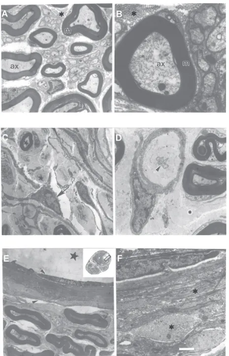
represent the first observations of experi-mental nerve entrapment in adult hamsters. After surgery, the main pathologic findings were already thoroughly established after a 5-week survival interval and most alterations followed a similar pattern of presentation in animals enduring longer compression (up to 15 weeks). Ultrastructural nerve morpholo-gy exhibited endoneurial edema, with thick-ening of both the endoneurium and perineu-rium, implying a distinct displacement of nerve fibers. The vasa nervorum pattern was also altered in compressed sciatic nerves compared to the control group. Intense my-elin sheath degeneration was also observed at the entrapment site and mainly in the distal segment with an influx of fibroblasts and Schwann cells to the lesioned regions (Figure 1). We also observed loosely tex-tured and whorled cell-sparse structures, ranging from 15 to 145 µm in diameter, found in the subperineurial space of the com-pressed sciatic nerve. In these areas we often observed isolated nerve fibers surrounded by collagen fibers. Some of these structures have a morphology suggestive of Renaut bodies (nerve cell-sparse structures with a predilection for sites of nerve compression and mechanical stress) (7,8).
The number of myelinated axons in each nerve segment - control, proximal, entrap-ment or distal - was analyzed according to axonal position either in the central or mar-ginal region of the nerve. Data are reported as percentage of the respective control re-gion (Figure 2). Interestingly, the results ob-served at the entrapment site displayed a remarkable increase in the number of small axons either in the central (26.36 ± 12.48%) or marginal (53.54 ± 8.92%) nerve region in comparison with the axonal number in the control group. In addition, the distal segment of the compressed nerve presented a distinct decrease in axon number in both the central (34.29 ± 4.44%) and marginal (24.04 ± 3.68%) entrapped nerve regions compara-tively to control nerves.
In the entrapment segment, we observed neural structures with a morphology highly suggestive of Renaut bodies. These bodies contained a loosely textured amorphous and fibrillar material (9-11). Renaut bodies ex-hibit mostly fibroblasts of perineurial and/or endoneurial origin with the extracellular matrix comprised of collagen fibrils, basal lamina material, and oxytalan filaments (10). Renaut bodies seem to be derived mainly from primary connective tissue elements of the endoneurium or from the degeneration of formed endoneurial structures such as capillaries. The latter possibility emerged from the study of human nerves showing a close association between thickened endo-neurial capillaries and Renaut bodies (12). We observed similar changes in the hamster sciatic nerve. Histologic characteristics that differentiate Renaut bodies from malignant neurotropic infiltration are: a) a cell-sparse mass, b) absence of nuclear atypia, c) a less
extensive than expected inflammatory infil-trate, and d) well-defined borders (10).
This model of experimental chronic en-trapment caused a remarkable increase of axons of small diameter (less than 3 µm) at the site of compression. This finding may be notably attributable to axonal sprouting in response to nerve compression. This bio-logical phenomenon was more evident in the marginal region, where it could be statisti-cally confirmed both in the proximal and in the entrapment nerve segment. In addition, we observed an important axonal degenera-tion occurring at the distal segment both in central and in marginal regions that may be considered as a probable direct consequence of mechanical stress upon the sciatic nerve affecting its normal physiology. The present study has proven the usefulness of this ex-perimental model in the production of clear-cut histopathologic findings observed in en-trapped sciatic nerves of adult hamsters. The entrapment procedure used in this investiga-tion produces the formainvestiga-tion of recognizable neuropathological signs (13,14) at a faster rate than previously described for the com-pressed sciatic nerve of the rat (15). In addi-tion, entrapped nerves exhibited Renaut bod-ies at the sites of nerve compression. Further investigations are necessary in order to clarify some of the neurobiological phenomena in-volved in the appearance of these abnormal structures in compressed nerves.
Acknowledgments
We thank Adiel Batista Nascimento, Antonia Lima Carvalho, Elizabeth Cunha Pena de Moraes and Jorge Luís da Silva for excellent technical assistance.
% Axon number (mean + SEM)
180 160 140 120 100 80 60 40 20 0 123 123 123 123 123 123 123 123 123 123 123 123 123 123 123 123 123 123 123 123 123 123 123 123 123 123 123 123 123 123 123 123 123 123 123 123 Central Marginal * ** Control nerve Proximal fragment 123 123 Entrapment site Distal fragment Figure 2. The number of
myeli-nated axons in each nerve seg-ment - control, proximal, entrap-ment or distal - was analyzed according to axonal position ei-ther in central or marginal re-gions of the nerve after 5 weeks of survival. Data are reported as percentage of the respective control region. The number of myelinated axons in the respec-tive regions of the control nerve was assumed to be 100%. Data are reported as means ± SEM. All symbols placed on top of the bars represent statistically differ-ent groups (P < 0.01) compared with respective control region (ANOVA followed by post hoc Duncan multiple range test).
References
1. Harrison MJG (1976). Pressure palsy of the ulnar nerve with pro-longed conduction block. Journal of Neurology, Neurosurgery and Psychiatry, 39: 96-99.
2. Rydevik B, McLean WG & Sjostrand J (1980). Blockage of axonal
transport induced by acute graded compression of the rabbit vagus nerve. Journal of Neurology, Neurosurgery and Psychiatry, 43: 690-698.
3. Dahlin LB, Rydevik B & McLean WG (1984). Changes in fast axonal #
+
transport during experimental nerve compression at low pressures. Experimental Neurology, 84: 29-36.
4. Jefferson D & Eames RA (1979). Subclinical entrapment of the lateral femural cutaneous nerve. Muscle and Nerve, 2: 145-154. 5. Thomas PK (1963). The connective tissue of peripheral nerve; an
electron microscope study. Journal of Anatomy, 97: 35-42. 6. Neary D, Ochoa J & Gilliatt RW (1975). Sub-clinical entrapment
neuropathy in man. Journal of the Neurological Sciences, 24: 283-298.
7. Renaut J (1881). Récherches sur quelque points particuliers de l’histologie des nerfs. Archives de Physiologie Normale et Pathologique, 8: 161-190.
8. Ortman JA, Sahenk Z & Mendell JR (1983). The experimental pro-duction of Renaut bodies. Journal of the Neurological Sciences, 62: 233-241.
9. Weis J, Alexianu ME, Heide G & Schroder JM (1993). Renaut bodies contain elastic fiber components. Journal of Neuropathology and Experimental Neurology, 52: 444-451.
10. Skidmore RA, Woosley JT & Tomsick RS (1996). Renaut bodies.
Benign disease process mimicking neurotropic tumor infiltration. Dermatologic Surgery, 22: 969-971.
11. Elcock LE, Stuart BP, Hoss HE, Crabb K, Millard DM, Bopp B, Mueller RE, Hastings TF & Lake SG (2001). Renaut bodies in the sciatic nerve of beagle dogs. Experimental and Toxicologic Patholo-gy, 53: 19-24.
12. Asbury AK (1973). Renaut bodies. A forgotten endoneurial struc-ture. Journal of Neuropathology and Experimental Neurology, 32: 334-343.
13. MacKinnon SE, Dellon AL, Hudson AR & Hunter DA (1986). Chronic human nerve compression - a histological assessment. Neuropa-thology and Applied Neurobiology, 12: 547-565.
14. Campbell WW (1997). Diagnosis and management of common compression and entrapment neuropathies. Neurologic Clinics, 15: 549-567.