Alte ratio ns in pro te ins o f bo ne m arro w
e xtrace llular m atrix in unde rno urishe d
m ice
Departamentos de 1Análises Clínicas, Centro de Ciências da Saúde and 2Biologia Celular, Embriologia e Genética, Centro de Ciências Biológicas,
Universidade Federal de Santa Catarina, Florianópolis, SC, Brasil
3Departamento de Análises Clínicas e Toxicológicas,
Faculdade de Ciências Farmacêuticas, Universidade de São Paulo, São Paulo, SP, Brasil
C.L. Vituri1,
M. Alvarez-Silva2,
A.G. Trentin2
and P. Borelli3
Abstract
The objective of the present study was to determine the effect of protein malnutrition on the glycoprotein content of bone marrow extracellular matrix (ECM). Two-month-old male Swiss mice were submitted to protein malnutrition with a low-protein diet containing 4% casein as compared to 20% casein in the control diet. When the experimental group had attained a 20% loss of their original body weight, we extracted the ECM proteins from bone marrow with PBS buffer, and analyzed ECM samples by SDS-PAGE (7.5%) and ECL Western blotting. Quantitative differences were observed between control and experimental groups. Bone marrow ECM from under-nourished mice had greater amounts of extractable fibronectin (1.6-fold increase) and laminin (4.8-(1.6-fold increase) when compared to the control group. These results suggest an association between fluctua-tions in the composition of the hematopoietic microenvironment and altered hematopoiesis observed in undernourished mice.
Co rre spo nde nce
M. Alvarez-Silva Divisão de Biologia Celular Centro de Ciências Biológicas, UFSC 88040-900 Florianópolis, SC
Brasil
Fax: + 55-48-331-9672 E-mail: malvarez@ ccb.ufsc.br
Presented at the 5th Brazilian Symposium on Extracellular Matrix - SIMEC, Angra dos Reis,
RJ, Brasil, September 7-10, 1998.
Research supported by CNPq, FUNPESQ UISA 97, CAPES and FAPESP (No. 13347-4/97).
Received January 22, 1999 Accepted April 10, 2000
Ke y wo rds
·Fibronectin ·Bone marrow ·Extracellular matrix ·Malnutrition
Intro ductio n
Protein-calorie malnutrition decreases blood cell production and interferes with both innate and adaptive immunity, as it affects both phagocytosis and the immune response (1-3). Blood cells arise in the bone marrow from stem cells able to undergo pro-cesses of proliferation and differentiation in the hematopoietic microenvironment. The bone marrow microenvironment is highly organized and regulates the location and phys-iology of the stem cells as well as the produc-tion and release of leukocytes and
erythro-cytes from bone marrow to peripheral blood. The hematopoietic microenvironment is com-posed of stromal cells (fibroblasts, macro-phages, endothelial cells, adipocytes), ac-cessory cells (T lymphocytes, monocytes), and their products (extracellular matrix (ECM) proteins and cytokines), which influ-ence the self-renewal, proliferation and dif-ferentiation of hematopoietic stem and pro-genitor cells (4).
and de-adhesion, binding and presentation of various cytokines, as well as regulation of cell growth (5,6).
Fibronectin is a ubiquitous molecule, consisting of two similar subunits joined by disulfide bonds, and can be synthesized by bone marrow stromal cells. Due to alterna-tive splicing this matrix component exists in a variety of isoforms (7). Fibronectin is pres-ent in soluble form in plasma, and in in-soluble form both in the ECM of connective tissue-forming cells and in basement mem-branes (8). Fibronectin is involved in the adhesion and maturation of the erythrocyte lineage (5). Adhesion of multipotent hemato-poietic progenitor cells to fibronectin via the
a5ß1 integrin may control the growth of hematopoietic cells, inhibiting apoptosis of primitive cells (9) or negatively controlling hematopoiesis. Negative control of hemato-poiesis may inhibit cytokine-induced prolif-eration of myeloid cell lineages (5).
Laminins are also important glycopro-teins from the bone marrow ECM. Laminins are heterotrimeric glycoproteins. Laminin-1, the prototype of the laminin family, origi-nally purified from the murine Engelbreth-Holm-Swarm (EHS) tumor, consists of three polypeptide chains, a1 (400 kDa), ß1 (200 kDa) and g1 (200 kDa). A variety of biologi-cal activities has been ascribed to laminin. These activities include interaction with type IV collagen, entactin, and heparan sulfate proteoglycan, binding to cells and regulation of their development and differentiation (5,10).
In a previous report we observed lympho-hematopoietic modifications, such as bone marrow myeloid hypoplasia in experimental models of malnutrition (11). The objective of the present study was to evaluate the effects of protein malnutrition on the glyco-protein components of the ECM in bone marrow, focusing on fibronectin and lami-nin accumulation. The experimental model consisted of mice submitted to a low-protein diet. We observed many alterations in
elec-trophoretic profile of ECM proteins, with accumulation of fibronectin and laminin in undernourished mice compared to controls. These data suggest that profound changes in the bone marrow microenvironment leading to changes in hematopoiesis occur in under-nourished mice.
Mate rial and Me tho ds
Expe rim e ntal die t
Two-month-old male Swiss mice obtained from Universidade Federal de Santa Catarina were housed in individual metabolic cages. The undernourished group received a diet containing 4% casein (low-protein diet) and the control group received 20% casein (con-trol diet) for 15 days. The diet contained fibers, saline and balanced vitamin mixtures, supplemented with 0.2% choline and 0.15% methionine (11). The two groups were main-tained on a light/dark cycle of 12 h, with water and food supplied ad libitum. Body weight was monitored every 48 h and the animals were submitted to experimental as-says when the undernourished group attained a 20% loss of their original body weight. The differences between control and undernour-ished mice were analyzed by the unpaired Student t-test.
Extrace llular m atrix pro te in e xtractio n
Bone marrow ECM was obtained from the femurs of the mice as described by Peters et al. (12). Marrow was aspirated into PBS (80 mM NaH2PO4, 20 mM Na2HPO4, 100
mM NaCl), pH 7.3, containing 2 mM dithiothreitol, 100 mM 6-aminohexanoic acid, 1 mM benzamidine-HCl, and 1 mM phenyl-methylsulfonyl fluoride (all from Sigma Chemical Co., St. Louis, MO, USA) at 4o
C and allowed to stand for 30 min. The samples were then pooled and centrifuged at 2,500 g
D ialysis and pro te in de te rm inatio n
The ECM proteins from bone marrow were dialyzed exhaustively (cut-off 12 kDa; Gibco BRL, Grand Island, NY, USA) for 48 h against distilled water. The protein con-centration recovered after dialysis was de-termined using the method of Bradford (13). Bovine serum albumin was used as standard. The ECM extracts from bone marrow were pooled and maintained at -20oC.
Po lyacrylam ide ge l e le ctro pho re sis
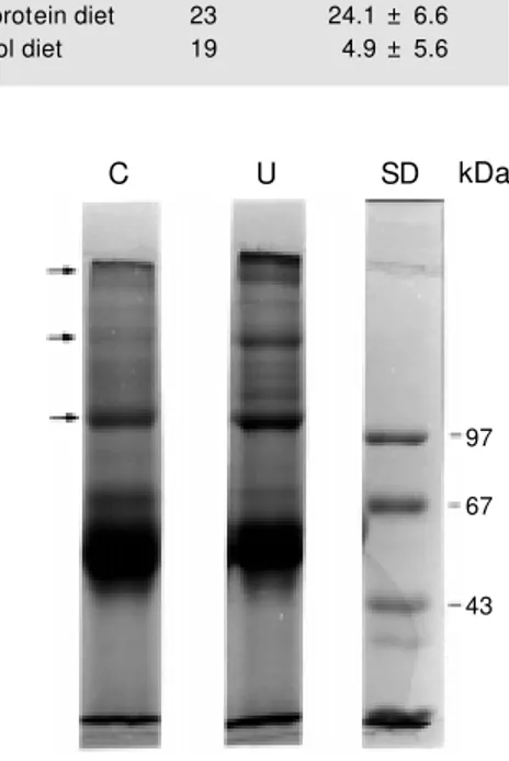
Denaturing SDS-polyacrylamide gel elec-trophoresis (SDS-PAGE) was performed on a 4% stacking gel and a 7% separating acry-lamide gel according to the discontinuous system of Laemmli (14) under reducing con-ditions (2 mM dithiothreitol added to the sample buffer). Gels were stained with 0.2% Coomassie blue R-250 (Sigma), 50% metha-nol and 10% acetic acid and destained over-night in 50% methanol and 10% acetic acid. Apparent molecular mass was determined using calibration kits (Pharmacia Biotech, São Paulo, SP, Brazil). The wells were loaded with 45 µg of protein obtained from bone marrow ECM.
We ste rn blo tting
Electrophoretic transfer of proteins from SDS-PAGE gels to nitrocellulose membranes was performed with a mini-V8 system, ac-cording to manufacturer instructions (Gibco BRL). Transfer was performed with 45 µg of protein/well run on an SDS-PAGE slab. The molecular mass calibration kit used for blot-ting was BenchMark Prestained Protein Lad-der (Gibco BRL). The nitrocellulose mem-brane was removed and incubated for 1 h at room temperature in 5% dry skim milk in PBS, pH 7.5. The following primary anti-bodies were used: rabbit anti-human fibro-nectin (A0245; Dako, Carpinteria, CA, USA) or rabbit anti-human laminin (a gift from Dr.
V. Moura Neto, Departamento de Anatomia, UFRJ). Biotinylated anti-rabbit IgG (B-7389; Sigma) and streptavidin-horseradish peroxi-dase (E-2886; Sigma) were incubated at room temperature for 1 h. Immunodetection was performed by the luminescence method (ECL, Amersham Life Science, Amersham, Buckinghamshire, UK). The reagents were incubated for 1 min on the nitrocellulose membrane, which was then exposed to an X-Omat film (Sigma) for 30 s. Alternatively the membranes were rinsed with PBS and re-acted with DAB (Sigma).
D e nsito m e tric analysis
Densitometric analysis was performed with scanned images of SDS-PAGE gels or with X-ray films from Western blot using a Microsoft Photo Editor Scanner. Alterna-tively, we scanned the membranes reacted with DAB. Images were analyzed and band area was determined using the Scion Image program of the National Institutes of Health, modified for Windows. We constructed lin-ear standard curves with 0.5-5 µg of albumin (fraction V, purchased from Sigma), fibro-nectin (1.5 µg, purchased from Gibco) and laminin (1 µg, purchased from Gibco) and the protein concentration obtained by densi-tometric analysis was calculated using the GraphPad Prism program, with 95% confi-dence intervals.
Re sults
Mice treated with the low-protein diet displayed a reduction in body weight (about 24%), while mice treated with the control diet displayed a small reduction in body weight of about 5% (Table 1).
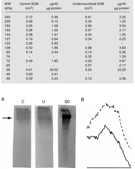
differ-(Table 2), and were absent in undernour-ished mice. Quantitative differences were observed for the similar bands with molecu-lar mass of 240, 220, 182, 150, 144, 127, 108, 85, 72, 56 and 46 kDa from both groups. Western blots for fibronectin obtained from mouse bone marrow ECM are depicted in Figure 2A. We observed differences in fibronectin expression in control and under-nourished samples, as demonstrated by den-sitometry. Measurement of the fibronectin Western blot demonstrated that the amount of fibronectin was 0.63 µg in the undernour-ished sample and 0.39 µg in the control sample (95% confidence interval). The un-dernourished group showed higher expres-sion of fibronectin compared to control groups, with a larger peak area also demon-strated by densitometry (Figure 2B).
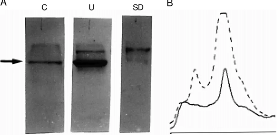
As observed for fibronectin, expression of laminin was modified in undernourished mice (Figure 3A and B). Measurement of laminin blots demonstrated that the amount of laminin was 2.44 µg in the undernour-ished mouse sample and 0.51 µg in control samples (95% confidence interval).
D iscussio n
The main finding of this study is that protein malnutrition induced an increase in the amount of extractable fibronectin and laminin from bone marrow ECM. It is not clear whether this increase is due to an in-crease in laminin and fibronectin synthesis rate or to an altered pattern of deposition of these molecules in the supramolecular struc-ture of the matrix. Regardless of the mechan-ism involved, it is clear that qualitative dif-ferences in bone marrow ECM composition and/or organization followed malnutrition.
Protein malnutrition modifies both spe-cific and nonspespe-cific resistance of organ-isms to infectious agents and impaired he-matopoiesis and/or lymphopoiesis follows modifications in cellularity and structure of hemato- and lymphopoietic tissues (1,15).
Table 1 - Effects of the low -protein and control diet on mouse body w eight.
Experimental mice w ere treated w ith a low -pro-tein and a control diet as described in M aterial and M ethods. Body w eight w as monitored every 48 h. The results are reported as percent (mean ± SD) w eight loss after 15 days of treatment. The differences in body w eight at the end of treat-ment among the experitreat-mental groups w ere sig-nificant (P<0.0001).
Treatment No. of animals Weight loss (% )
Low -protein diet 23 24.1 ± 6.6
Control diet 19 4.9 ± 5.6
kDa
97
67
43
C U SD
Figure 1 - SDS-PAGE profile of ECM pro-teins obtained from control (C) and under-nourished (U) mice. Each SDS-PAGE lane w as loaded w ith 45 µg of protein obtained from bone marrow ECM (control or under-nourished). After running, the bands w ere stained and measured and molecular mass determined. We observed differences in both groups. A similar profile w as obtained in five different experiments done w ith the same ECM pool (control and undernour-ished). We tested three different groups of control and undernourished mice w ith simi-lar results. SD, Standard molecusimi-lar mass.
The precise mechanisms underlying these effects are not clear. It was demonstrated that the mobilization and production of my-eloid cells during inflammatory reactions in mice are affected in protein malnutrition (11). The complete maturation and release of myeloid cell lineages is delayed due to specific modifications of the stimulatory fac-tors that control proliferation, differentia-tion and release of leukocytes in bone mar-row. Over the last years, many lines of evi-dence have indicated that ECM proteins play crucial roles in hematopoiesis. In normal adults, myelopoiesis and partial lymphopoie-sis occur in bone marrow. Cells of the med-ullary stroma locally produce cytokines and ECM proteins. These cells and their prod-ucts spatially organized in the ECM form the hematopoietic bone marrow microenviron-ment (16). The production of cytokines de-pends on the organization of the bone mar-row microenvironment determined by ad-equate levels of ECM proteins and cytokines (5). Based on these findings, modifications in proteins that compose bone marrow ECM may compromise the microenvironment and modify cytokine synthesis. The final result may be a change in cell production and/or differentiation in bone marrow.
In this study we demonstrated that fibro-nectin and laminin were expressed in both control and undernourished mice, although the undernourished group showed higher expression of these glycoproteins than the control group. This difference in expression may dramatically modify the bone marrow microenvironment, since fibronectin and la-minin are central proteins involved in the regulation of hematopoietic cell prolifera-tion and/or differentiaprolifera-tion (4,5).
Fibronectin and laminin are synthesized and deposited on the extracellular network by bone marrow stromal cells. Fibronectin is involved in the adhesion and maturation of the myeloid lineage (17,18) and lymphoid precursor cells (19). More recent work has demonstrated that multipotent
hematopoi-Table 2 - Comparative profile of bone marrow protein bands in control and undernour-ished mice.
These data w ere obtained by SDS-PAGE. The bands w ere measured and molecular mass w as determined by densitometry. Results are reported as band areas (cm2), and albumin (0.5-5 µg) w as used as standard. Results are reported as concentration (µg) per 45 µg of protein applied to each SDS-PAGE lane. Absence of bands is indicated by the (-) symbol. Proteins of less than 30 kDa are not show n. Control ECM indicates the ECM proteins obtained from the bone marrow of control mice. Undernourished ECM indicates the ECM proteins obtained from the bone marrow of undernourished mice.
M W Control ECM µg/45 Undernourished ECM µg/45
(kDa) (cm2) µg protein (cm2) µg protein
240 0.12 0.48 0.61 2.33
220 0.06 0.12 0.34 1.25
182 0.35 1.28 0.90 3.54
150 0.28 1.00 0.57 2.17
144 0.38 1.41 0.34 1.25
127 0.19 0.64 0.34 0.25
123 0.26 0.93 -
-108 0.50 1.89 0.98 3.83
85 0.14 0.44 0.12 0.36
76 - - 0.35 1.29
72 0.49 1.85 0.23 0.87
60 - - 0.57 2.17
56 5.01 20.02 5.55 22.20
49 0.63 2.41 -
-46 0.09 0.24 0.12 0.36
C U SD
A B
etic progenitor cells adhere to fibronectin in bone marrow (20). Bone marrow precursor cells may attach to fibronectin through a5ß1 and/or a4ß1 integrins or through membrane-bound proteoglycans (20,21). It has been demonstrated that the a4ß1 and a5ß1 inte-grin receptors are differentially expressed during myeloid maturation (21). Cell adhe-sion to fibronectin may be an important step in the control of hematopoiesis. It has been demonstrated that the interaction of fibro-nectin with hematopoietic precursor cells through a5ß1 integrin may prevent cell pro-liferation in the presence of hematopoietic mitogenic growth factors (22). The role of laminin in bone marrow has been less stud-ied. However its importance for the homing of precursor cells to bone marrow has been demonstrated (4,5). Modification in laminin level may alter the differentiation route of cells in bone marrow, as well as the release of differentiated hematopoietic cells from
bone marrow to blood.
The correlation between malnutrition and bone marrow ECM has not been investi-gated. However, ECM has been studied in other tissues, such as liver and thymus, in malnutrition. Perinatal food restriction re-duces the content but not the concentration of liver ECM in rats (23). A dense thymic ECM positively correlated with the degree of thymocyte depletion has been shown to occur in human malnutrition (24). The au-thors suggested that these effects may repre-sent a cause-effect relationship in which the contact of thymocytes with abnormally high amounts of thymic ECM triggers and/or en-hances programmed cell death.
ECM not only functions as the structural supporting element for cells and tissues but plays a major role in modulating the biology of the cell and its response to growth factors, hormones and vitamins. The composition of the matrix, however, is not static, but changes both during normal development and tissue repair and regeneration (10,24). On the basis of these considerations, we suggest that pro-tein malnutrition might be altering bone mar-row ECM structure (maybe due to altered synthesis of its components), thus modifying the hematopoietic microenvironment and/or hematopoiesis. Whether malnutrition modi-fies other components of bone marrow ECM, such as collagens, thrombospondin and pro-teoglycans, is currently under investigation in our laboratory.
Ackno wle dgm e nts
Prof. Dr. Vera Lucia Cardoso Tramonte (Departamento de Nutrição, UFSC) is grate-fully acknowledged for permitting the use of metabolic cages and full access to the Labo-ratório de Nutrição Experimental.
C U SD
A B
Re fe re nce s
1. Fried W, Shapiro S, Barone J & Anagnos-tou A (1978). Effect of protein deprivation on hematopoietic cells and on peripheral blood counts. Journal of Laboratory and Clinical M edicine, 92: 303-400.
2. Chandra RK (1991). Nutrition and immuni-ty: lessons from the past and new in-sights into future. American Journal of Clinical Nutrition, 53: 1083-1094. 3. Keusch RL & Farthing M JG (1986).
Nutri-tion and infecNutri-tion. Annual Review of Nu-trition, 6: 131-154.
4. M ayani H, Guilbert LJ & Janow sa-Wieczorek A (1992). Biology of the he-matopoietic microenvironment. European Journal of Haematology, 49: 225-233. 5. Klein G (1995). The extracellular matrix of
the hematopoietic microenvironment. Ex-perientia, 51: 914-926.
6. Alvarez-Silva M & Borojevic R (1996). Granulocyte-macrophage colony-stimulat-ing factor (GM -CSF) and interleukin-3 (IL-3) activities in schistosomal liver granulo-mas are controlled by stroma-associated heparan sulfate proteoglycans. Journal of Leukocyte Biology, 59: 435-441. 7. French-Constant C (1995). Alternative
splicing of fibronectin - many different pro-teins but few different functions. Experi-mental Cell Research, 221: 261-271. 8. Hynes RO (1990). Fibronectins. Rich A
(Editor), Springer Series in M olecular Biol-ogy. Springer Verlag, New York. 9. Yoshikaw a H & Sakihama Y (1996).
Co-stimulation of fibronectin receptor pro-motes Fc gamma R-mediated rescue of IL-3-dependent bone m arrow -derived cells from apoptosis. Journal of
Immunol-ogy, 156: 1832-1840.
10. Yurchenco PD & O’Rear J (1994). Basal lamina assembly. Current Opinion in Cell Biology, 6: 674-681.
11. Borelli P, M ariano M & Borojevic R (1995). Protein malnutrition: effect on myeloid cell production and mobilization into in-flammatory reactions in mice. Nutrition Research, 15: 1477-1485.
12. Peters C, Budde CL, Breon TA, Kuper A & Kim J (1995). Ovine bone marrow extra-cellular matrix and soluble protein extrac-tion: fetuin amino terminus microhetero-geneity. American Journal of the M edical Sciences, 309: 285-294.
13. Bradford M M (1977). A rapid and sensi-tive method for the quantitation of micro-gram quantities of protein utilizing the principle of protein-dye binding. Analytical Biochemistry, 72: 248-254.
14. Laemmli UK (1970). Cleavage of struc-tural proteins during the assembly of the bacteriophage T4. Nature, 277: 680-685. 15. Gross RL & New berne M P (1980). Role of
nutrition in immunologic function. Physi-ological Review s, 60: 188-302.
16. Gordon M Y (1988). Extracellular matrix of the marrow microenvironment. British Journal of Haematology, 70: 1-4. 17. Patel VP & Lodish HF (1984). Loss of
ad-hesion of murine erythroleukemia cells to fibronectin during erythroid differentia-tion. Science, 224: 996-998.
18. Patel VP & Lodish HF (1987). Fibronectin matrix is required for differentiation of murine erythroleukemia cells into reticu-locytes. Journal of Cell Biology, 105: 3105-3118.
19. Bernard P, Patel VP & Lodish HF (1987). Lymphoid precursor cells adhere to tw o different sites on fibronectin. Journal of Cell Biology, 105: 489-498.
20. Verfaillie CM , M cCarthy JB & M cGlave PB (1991). Differentiation of primitive hu-man multipotent hematopoietic progeni-tors into single lineage clonogenic pro-genitors is accompanied by alterations in their interaction w ith fibronectin. Journal of Experimental M edicine, 174: 693-703. 21. Kerst JM , Sanders JB, Slaper-Cortenbach IC, Doorakers M C, Hooibrink B, van Oers RH, von der Born AE & van der Schoot CE (1993). Alpha 4 beta 1 and alpha 5 beta 1 are differentially expressed during my-elopoiesis and mediate the adherence of human CD34+ cells to fibronectin in an activation-dependent w ay. Blood, 81: 344-351.
22. Sugahara H, Kanakura Y, Furit su T, Ishikaw ama J, Hashimoto K, Kanayama Y & M atsuzama Y (1994). Induction of pro-grammed cell death in human hematopoi-etic cell lines by fibronectin via its interac-tion w ith very late antigen 5. Journal of Experimental M edicine, 179: 1757-1766. 23. Reif S, Lu R, Tano M , Terranova V, Young C, Fisher J, Petell J & Lebenthal E (1993). Perinatal food restriction in rats reduces the content but not concentration of liver extracellular matrix proteins. Journal of Nutrition, 123: 811-816.