ISSN 0100-879X
BIOMEDICAL SCIENCES
www.bjournal.com.br
www.bjournal.com.br
Volume 45 (4) 291-375 April 2012
Braz J Med Biol Res, April 2012, Volume 45(4) 314-320
doi: 10.1590/S0100-879X2012007500044
Estrous cycle and stress: influence of progesterone on the female
brain
T.A. Lovick
Institutional Sponsors
The Brazilian Journal of Medical and Biological Research is partially financed by
Faculdade de Medicina de Ribeirão Preto Campus
Ribeirão Preto
Explore High - Performance MS Orbitrap Technology In Proteomics & Metabolomics
Estrous cycle and stress: influence of
progesterone on the female brain
T.A. Lovick
School of Clinical and Experimental Medicine, University of Birmingham, Birmingham, UK
Abstract
The female brain operates in a constantly changing chemical milieu caused by cyclical changes in gonadal hormones during the estrous cycle (menstrual cycle in women). Such hormones are highly lipophilic and pass readily from the plasma to the brain
where they can influence neuronal function. It is becoming clear that the rapid reduction in peripheral circulating progesterone,
which occurs during the late diestrous phase of the cycle, can trigger a withdrawal-like response, in which changes in GABAA receptor expression render hyper-responsive certain brain areas involved in processing responses to stressful stimuli. The periaqueductal gray matter (PAG) is recognised as an important region for integrating anxiety/defence responses. Withdrawal from progesterone, via actions of its neuroactive metabolite allopregnanolone, triggers up-regulation of extrasynaptic GABAA receptors on GABAergic neurons in the PAG. As a consequence, ongoing GABAergic tone on the output cells decreases, leading to an increase in functional excitability of the circuitry and enhanced responsiveness to stressful stimuli during the late diestrous phase. These changes during late diestrus could be prevented by short-term neurosteroid administration, timed to produce a more gradual fall in the peripheral concentration of allopregnanolone than the rapid decrease that occurs naturally, thus removing the trigger for the central withdrawal response.
Key words: Female; Estrous cycle; Stress; Anxiety; Progesterone; Neurosteroid replacement
Correspondence: T.A. Lovick, School of Clinical and Experimental Medicine, University of Birmingham, Birmingham B15 2TT, UK. E-mail: [email protected]
Presented at the III Fórum em Neurobiologia do Estresse, Araraquara, SP, Brazil, September 8-10, 2011.
Received November 26, 2011. Accepted March 13, 2012. Available online March 30, 2012. Published April 9, 2012.
Responsiveness to psychological stress in
females
Adverse responsiveness to psychological stress is a major health hazard, which is more prevalent in women than in men (1). The female brain, unlike its male coun-terpart, operates in a changing chemical milieu caused by cyclical changes in production of female sex hormones in the periphery during the menstrual cycle (estrous cycle in animals). Many of these compounds are neuroactive and their lipophilic nature enables them to pass readily through
the blood brain barrier and produce significant influences
on brain function through actions at both membrane-bound and nuclear receptors (2). In large numbers of women, the late luteal phase of the menstrual cycle is characterised by the development of premenstrual syndrome (PMS): a constellation of adverse psychological symptoms that include worsening of mood and episodes of aggressive behaviour, often triggered by relatively minor stressors related to everyday life (3). PMS and its more severe coun-terpart premenstrual dysphoric disorder (PMDD) interferes
significantly with the woman’s quality of life, impairing work,
family relationships and other social activities. Interestingly, PMS does not occur in anovulatory cycles (4,5), suggesting that cyclical changes in sex hormones, rather than absolute levels may be the trigger for the syndrome. The late luteal phase of the menstrual cycle is characterised by a rapid decline in secretion of the sex hormones progesterone and estrogen, suggesting that one or both of these hormones may be linked to the development of symptoms. This article will focus on the recent evidence obtained in rodents that indicates that the fall in progesterone secretion, which occurs during the late diestrous phase in rats, leads to a change
in gamma-aminobutyric acid (GABAA) receptor expression
in the brain that is manifested behaviourally as enhanced responsiveness of the animal to mild anxiogenic stress. A consideration of the effect of estrogen on brain function and behaviour is beyond the scope of this article and has recently been reviewed elsewhere (6).
Responsiveness to stress varies during the
estrous cycle in rodents
Stress and the female brain 315
changes in responsiveness to stressful situations. In gen-eral, higher levels of anxiety and responsiveness to mild anxiogenic stress have been reported in the non-receptive phase of the cycle (diestrus) compared to the receptive phase (proestrus and estrus) in both rats and mice (7-13, but see Ref. 14 for an opposite view). Unlike women in whom the late luteal (premenstrual) phase is character-ised by falling levels of both estrogen and progesterone, the latter part of diestrus in rats, when responsiveness to stress is increased, is associated with a rapid fall only in plasma concentration of progesterone, whilst estrogen secretion remains at a stable low level (15). Since
proges-terone passes the blood brain barrier readily, fluctuations in peripheral secretion will be reflected in the brain. Falling
concentration of progesterone in the brain could therefore be the key factor that triggers increased responsiveness to acute stress during late diestrus in rats and, by analogy, during the late luteal phase in women. In support of this idea, behavioural changes similar to those seen in late diestrus can be induced in rats by withdrawal from long-term dosing with exogenous progesterone (16-19). The behaviour of spontaneously cycling rats in late diestrus therefore appears to offer a useful model in which to study the role of progesterone in the development of premenstrual syndrome in women.
Falling progesterone leads to changes in
neuronal excitability
There are several indications that rapid fluctuations in
endogenous steroid concentrations result in altered GABAA
receptor kinetics, which impacts on neuronal excitability. In rats, withdrawal from progesterone, either during the late diestrus phase in cycling animals or following an exog-enous progesterone withdrawal dosing regimen, has been
shown to evoke up-regulation of α4, β1 and δ subunits of
the GABAA receptor in several brain structures (7,19-23).
The effect was due not to progesterone itself but to the fall in concentration of its neuroactive metabolite allopreg-nanolone (ALLO), which triggers enhanced transcription of
the genes encoding α4, β1 and δ subunits of the GABAA
receptor (16,17,23). Alpha4, β1 and δ subunits are able to assemble into functional receptors. Recombinant α4β1δ
GABAA receptors show electrophysiological properties
characteristic of other extrasynaptic receptors containing
δ subunits. Typically, these act tonically to modify the level
of ongoing GABAergic inhibitory activity that regulates the excitability of neuronal ensembles (22,24-26).
Functional consequences of progesterone
withdrawal
There is now compelling evidence that a reduction in brain concentration of progesterone enhances stress-induced anxiety-linked behaviours. Whether stress-induced
experi-mentally by withdrawal from an exogenous progesterone dosing regimen (17,22,23,27,28) or occurring naturally as
a response to natural fluctuations during the estrous cycle
(10-12,18), falling progesterone is anxiogenic in a variety of behavioural tests.
The periaqueductal gray matter (PAG) is a midbrain region recognised as key to mediating behavioural and autonomic components of responses to stressful chal-lenge. As such, it has the potential to provide a useful brain locus for investigating the mechanisms underlying female hormone-induced links between transcriptional changes at the receptor level and changes in neuronal excitability and behaviour. As with other brain regions, the vast majority of studies on this region have been carried out in males. However, as the results from more studies on females be-gin to emerge, it is becoming clear not only that the PAG is involved in processing stress-evoked behaviour in the female sex, as in males, but also that the excitability of its
circuitry fluctuates during the estrous cycle. In females in
late diestrus, the fall in brain concentration of progester-one, and hence ALLO, that occurs during this stage,
trig-gers up-regulation of extrasynaptic α4βδ GABAA receptor
expression in the PAG, which is confined principally to the
GABAergic neuronal subpopulation (Figure 1). This tran-scriptional change correlates with an increase in functional excitability of the intrinsic PAG circuitry that translates in the conscious animal to raised anxiety levels and increased behavioural responsiveness to acute psychogenic stress (Figure 2A). At the cellular level electrophysiological studies
in spontaneously cycling rats in vivo have demonstrated
that GABAergic tone on output neurons within the PAG is reduced during the late diestrous phase (29), which renders the circuitry intrinsically more excitable. Direct evidence for an increase in functional excitability of the PAG circuitry during late diestrus has been provided by the recent report of a lowered threshold for escape behaviour evoked by direct electrical stimulation in the PAG in late diestrus (30). Autonomic responsiveness to a pentagastrin, a panicogenic agent known to activate PAG circuitry following systemic administration, was also increased during the late diestrous phase (31). At this time, functional activation of PAG circuitry evoked by acute exposure to a mild anxiogenic stress is also
changed, as reflected by decreased regional expression of
the immediate early gene c-fos (32 and Figure 2B). At the
present time the phenotype of the Fos-positive population
in the PAG has not been determined. However, the find
-ings are consistent with a stress-induced decreased c-fos
expression in the GABAergic population, and at the same
time with an increase in c-fos expression in PAG output
neurons, which are likely to be less numerous than the GABAergic population.
Interestingly, in hippocampal tissue withdrawal from
progesterone evoked an increased α4 receptor expression
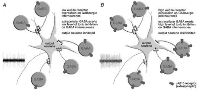
Figure 1. Schematic drawing showing the functional consequences of increased α4β1δ GABAA receptor expression in the
peri-aqueductal gray matter (PAG). A, When expression of α4β1δ receptors is low, spontaneous activity in GABAergic interneurones
in the PAG limits the excitability of the output neurones. B, Increased expression of α4β1δ receptors when progesterone levels
fall in late diestrous leads to an increase in tonic current carried by GABAergic cells, which limits their ongoing activity. The output neurones therefore become intrinsically more excitable, and the threshold for activation by stressful stimuli is lowered. Repro-duced from Ref. 41 with permission.
Figure 2. A, Change in latency of the tail flick reflex (s) following
exposure to 5-min anxiogenic vibration stress in rats at different stages of the estrous cycle. Data (mean ± SEM) are reported
as change in tail flick latency (TFL) from baseline in 6-min
post-vibration stress. B, Density of Fos-positive neurons in the periaq-ueductal gray matter (PAG) of rats exposed to anxiogenic stress
and tail flick testing at different stages of the estrous cycle. P = proestrus; E = estrus; ED = early diestrus; LD = late diestrus.
Figure drawn from data in Ref. 32 and reproduced with permis-sion. *P < 0.05, **P < 0.01 (post hoc Bonferroni test) stressed
group compared to the non-stressed group after significant (P <
0.05) one-way ANOVA. §§P < 0.01 LD stressed group compared
to stressed rats at all other stages of the estrous cycle (post hoc
Stress and the female brain 317
No effects were observed in cortex or cerebellum. Thus, there may be regional differences with respect to the effects triggered by withdrawal from progesterone. Moreover, at the cellular level the functional outcome of receptor upregulation appears to be dependent on the cell type that expresses
α4 subunit-containing GABAA receptors, e.g., interneurons
or principal neurons.
The kinetics of progesterone withdrawal
determines its anxiogenic effect
It is becoming clear that withdrawal from progester-one during the late diestrous phase induces a rebound
hyperexcitable state manifested by significant changes
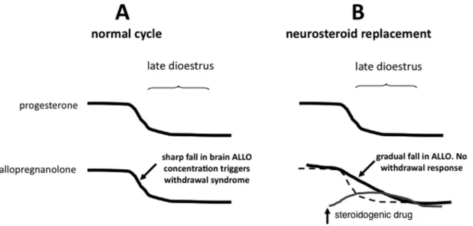
in responsiveness to anxiogenic stress. Interestingly, the critical event for inducing this effect may be related to the kinetics of the decline in steroid levels rather than the absolute magnitude of the fall. In a study on female rats chronically treated with estrogen and progesterone, a rapid withdrawal of these hormones precipitated increased startle and anxiety-like behaviour, which was not seen if the hormone levels were allowed to decline gradually (34,35). Since similar increases in anxiety behaviour can be pre-cipitated by abrupt withdrawal from progesterone alone (16-18), the trigger for anxiogenesis in the former study is likely to be the rapid decline in concentration in the brain of progesterone, and hence its neuroactive metabolite ALLO. Measures to produce a more gradually tapered fall in
pro-gesterone levels might therefore be effective in preventing the withdrawal effect.
Neurosteroid replacement in late diestrus
prevents the development of stress-induced
hyperalgesia
We hypothesized that short-term neurosteroid replace-ment, timed to blunt the rate of the natural fall in ALLO that occurs during the late diestrous phase in spontaneously cycling rats, should prevent the increased responsiveness to stress that normally occurs at this time (Figure 3). In males
the selective serotonin reuptake inhibitor (SSRI) fluoxetine
has been shown to produce rapid onset steroidogenic ef-fects following acute administration, by raising plasma and brain concentrations of ALLO (36-38). Importantly, these effects could be induced at doses below the threshold for effects on 5-hydroxytryptamine (5-HT) systems (37). We have shown that short-term administration of a low dose
of fluoxetine (1.75 mg/kg, ip) was also steroidogenic in females, producing a doubling of whole brain ALLO con-centration (39).
Compared to other cycle stages, rats in late diestrus normally show an exaggerated response to anxiogenic vibration stress, which is manifested as hyperalgesia (18).
Administering low-dose fluoxetine to female rats on the
evening of early diestrus, with a top up next morning when they had reached late diestrus, completely prevented the
development of stress-induced hyperalgesia (Figure 4A). Similarly, the functional deactivation within the PAG that is normally induced by exposure to the vibration stress in late diestrus was prevented as well (Figure 4B). Importantly,
the behavioural effects of fluoxetine could be reproduced
following dosing with ganaxolone (data not shown), a syn-thetic analogue of ALLO not reported to have effects on
5-HT systems (40). Thus, the effect of fluoxetine is likely
to be due to its steroidogenic action. The dramatic effects of short-term neurosteroid replacement in preventing the appearance of estrous cycle-linked anxiogenesis in rats may have relevance to the quest for more effective treatments of menstrual cycle-linked dysphoria in women.
In conclusion, the data summarized here show how cyclical changes in production of progesterone during the estrous cycle can have a powerful effect on functioning of the female brain. In rodents, this occurs over a remark-ably short time frame. The withdrawal effects evoked by the natural cyclical variation in secretion of progesterone during the estrous cycle occur over 12- to 24-h periods,
influencing anxiety levels and responsiveness to anxiogenic
stress. To date the overwhelming majority of the studies on
brain function and behaviour in animals has been confined
to males, a common argument for the single sex approach being that data obtained from females are more variable and
may be confounded by hormonal influences. Increasingly,
however, it is being recognized that sex differences do exist, both in terms of behaviour and responsiveness to certain
drugs. It is essential that the effects of possible influences
of the ovarian cycle on brain function and behaviour are incorporated into experimental designs in future studies using females. In a world in which 50% of the population is female, such differences are important and should no longer be overlooked.
References
1. Legato MJ. The skewed sex distribution in affective disor-ders - a diagnostic, social, or biological problem? Prog Brain Res 2010; 186: 159-166.
2. Paul SM, Purdy RH. Neuroactive steroids. FASEB J 1992; 6: 2311-2322.
3. O’Brien S, Rapkin A, Dennerstein L, Nevatte T. Diagnosis
and management of premenstrual disorders. BMJ 2011; 342: d2994.
4. Backstrom T, Andreen L, Birzniece V, Bjorn I, Johansson IM, Nordenstam-Haghjo M, et al. The role of hormones and hormonal treatments in premenstrual syndrome. CNS Drugs
2003; 17: 325-342.
5. Hammarback S, Ekholm UB, Backstrom T. Spontaneous anovulation causing disappearance of cyclical symptoms in women with the premenstrual syndrome. Acta Endocrinol
1991; 125: 132-137.
6. Handa RJ, Ogawa S, Wang JM, Herbison AE. Roles for oestrogen receptor beta in adult brain function. J
Neuroen-docrinol 2012; 24: 160-173.
7. Gangitano D, Salas R, Teng Y, Perez E, De Biasi M.
Pro-gesterone modulation of alpha5 nAChR subunits influences
anxiety-related behavior during estrus cycle. Genes Brain
Figure 4. A, Short-term dosing with fluoxetine (1.75 mg/kg) on
the evening of late diestrus, with a top up on the morning of late diestrus, prevents the development of stress-induced
Stress and the female brain 319
Behav 2009; 8: 398-406.
8. Gouveia A Jr, dos Santos UD, Felisbino FE, de Afonseca TL,
Antunes G, Morato S. Influence of the estrous cycle on the
behavior of rats in the elevated T-maze. Behav Processes
2004; 67: 167-171.
9. Ho HP, Olsson M, Westberg L, Melke J, Eriksson E. The
serotonin reuptake inhibitor fluoxetine reduces sex
steroid-related aggression in female rats: an animal model of pre-menstrual irritability? Neuropsychopharmacology 2001; 24: 502-510.
10. Koonce CJ, Walf AA, Frye CA. Type 1 5alpha-reductase may be required for estrous cycle changes in affective behaviors of female mice. Behav Brain Res 2012; 226: 376-380.
11. Marcondes FK, Miguel KJ, Melo LL, Spadari-Bratfisch RC. Estrous cycle influences the response of female rats in the
elevated plus-maze test. Physiol Behav 2001; 74: 435-440.
12. Mora S, Dussaubat N, Diaz-Veliz G. Effects of the estrous cycle and ovarian hormones on behavioral indices of anxiety in female rats. Psychoneuroendocrinology 1996; 21: 609-620.
13. Reddy DS, Kulkarni SK. Sex and estrous cycle-dependent changes in neurosteroid and benzodiazepine effects on food consumption and plus-maze learning behaviors in rats.
Pharmacol Biochem Behav 1999; 62: 53-60.
14. Hiroi R, Neumaier JF. Differential effects of ovarian steroids
on anxiety versus fear as measured by open field test and
fear-potentiated startle. Behav Brain Res 2006; 166: 93-100.
15. Butcher RL, Collins WE, Fugo NW. Plasma concentration of LH, FSH, prolactin, progesterone and estradiol-17beta throughout the 4-day estrous cycle of the rat. Endocrinology
1974; 94: 1704-1708.
16. Smith SS, Gong QH, Hsu FC, Markowitz RS, ffrench-Mullen JM, Li X. GABA(A) receptor alpha4 subunit suppression prevents withdrawal properties of an endogenous steroid.
Nature 1998; 392: 926-930.
17. Smith SS, Gong QH, Li X, Moran MH, Bitran D, Frye CA, et al. Withdrawal from 3alpha-OH-5alpha-pregnan-20-One using a pseudopregnancy model alters the kinetics of hip-pocampal GABAA-gated current and increases the GABAA receptor alpha4 subunit in association with increased anxi-ety. J Neurosci 1998; 18: 5275-5284.
18. Devall AJ, Liu ZW, Lovick TA. Hyperalgesia in the setting of anxiety: sex differences and effects of the oestrous cycle in Wistar rats. Psychoneuroendocrinology 2009; 34: 587-596. 19. Gangisetty O, Reddy DS. Neurosteroid withdrawal regulates
GABA-A receptor alpha4-subunit expression and seizure susceptibility by activation of progesterone receptor-inde-pendent early growth response factor-3 pathway. Neurosci-ence 2010; 170: 865-880.
20. Griffiths J, Lovick T. Withdrawal from progesterone increases
expression of alpha4, beta1, and delta GABA(A) receptor subunits in neurons in the periaqueductal gray matter in female Wistar rats. J Comp Neurol 2005; 486: 89-97.
21. Griffiths JL, Lovick TA. GABAergic neurones in the rat pe -riaqueductal grey matter express alpha4, beta1 and delta GABAA receptor subunits: plasticity of expression during the estrous cycle. Neuroscience 2005; 136: 457-466.
22. Lovick TA, Griffiths JL, Dunn SM, Martin IL. Changes in
GABA(A) receptor subunit expression in the midbrain during the oestrous cycle in Wistar rats. Neuroscience 2005; 131:
397-405.
23. Gulinello M, Orman R, Smith SS. Sex differences in anxiety, sensorimotor gating and expression of the alpha4 subunit of the GABAA receptor in the amygdala after progesterone withdrawal. Eur J Neurosci 2003; 17: 641-648.
24. Mody I. Aspects of the homeostatic plasticity of GABAA receptor-mediated inhibition. J Physiol 2005; 562: 37-46. 25. Smith SS, Gong QH. Neurosteroid administration and
with-drawal alter GABAA receptor kinetics in CA1 hippocampus of female rats. J Physiol 2005; 564: 421-436.
26. Wei W, Zhang N, Peng Z, Houser CR, Mody I. Perisynaptic localization of delta subunit-containing GABA(A) receptors and their activation by GABA spillover in the mouse dentate gyrus. J Neurosci 2003; 23: 10650-10661.
27. Gallo MA, Smith SS. Progesterone withdrawal decreases
latency to and increases duration of electrified prod burial:
a possible rat model of PMS anxiety. Pharmacol Biochem
Behav 1993; 46: 897-904.
28. Lofgren M, Johansson IM, Meyerson B, Lundgren P,
Back-strom T. Progesterone withdrawal effects in the open field
test can be predicted by elevated plus maze performance.
Horm Behav 2006; 50: 208-215.
29. Brack KE, Lovick TA. Neuronal excitability in the periaque-ductal grey matter during the estrous cycle in female Wistar rats. Neuroscience 2007; 144: 325-335.
30. Santos JM, Lovick TA, Brandão ML. Effects of acute
treat-ment with fluoxetine on the neural substrates of fear of the
periaqueductal gray matter in female rats. Neuroscience
2011; Abstract 281.02.
31. Brack KE, Jeffery SM, Lovick TA. Cardiovascular and respi-ratory responses to a panicogenic agent in anaesthetised female Wistar rats at different stages of the oestrous cycle.
Eur J Neurosci 2006; 23: 3309-3318.
32. Devall AJ, Santos JM, Lovick TA. Estrous cycle stage influ -ences on neuronal responsiveness to repeated anxiogenic stress in female rats. Behav Brain Res 2011; 225: 334-340.
33. Bitran D, Smith SS. Termination of pseudopregnancy in the rat produces an anxiogenic-like response that is associated with an increase in benzodiazepine receptor binding density
and a decrease in GABA-stimulated chloride influx in the
hippocampus. Brain Res Bull 2005; 64: 511-518.
34. Doornbos B, Fokkema DS, Molhoek M, Tanke MA, Postema F, Korf J. Abrupt rather than gradual hormonal changes in-duce postpartum blues-like behavior in rats. Life Sci 2009; 84: 69-74.
35. Saavedra M, Contreras CM, Azamar-Arizmendi G, Hernan-dez-Lozano M. Differential progesterone effects on defen-sive burying and forced swimming tests depending upon a gradual decrease or an abrupt suppression schedules.
Pharmacol Biochem Behav 2006; 83: 130-135.
36. Uzunov DP, Cooper TB, Costa E, Guidotti A. Fluoxetine-elicited changes in brain neurosteroid content measured by negative ion mass fragmentography. Proc Natl Acad Sci U S A 1996; 93: 12599-12604.
37. Pinna G, Costa E, Guidotti A. SSRIs act as selective brain steroidogenic stimulants (SBSSs) at low doses that are inactive on 5-HT reuptake. Curr Opin Pharmacol 2009; 9: 24-30.
38. Serra M, Pisu MG, Muggironi M, Parodo V, Papi G, Sari R, et al. Opposite effects of short- versus long-term administration
in rat plasma and brain. Psychopharmacology 2001; 158: 48-54.
39. Devall A, Fry JP, Honour JW, Lovick TA. Short term, low
dose fluoxetine treatment increases brain allopregnanolone
concentrations in female rats and abolishes estrous cycle-related stress-induced hyperalgesia? Proc Physiol Soc
2011; 22; PC11.
40. Nohria V, Giller E. Ganaxolone. Neurotherapeutics 2007; 4: 102-105.