Abstract
Objective: To determine the prevalence of celiac disease and to describe the histological alterations, clinical manifestations, and conditions associated with a group of irst-degree relatives of celiac disease patients in the municipality of Recife, Northeast Brazil.
Method: The study was conducted in outpatient clinics of pediatric gastroenterology located in Recife. We included in the study 174 irst-degree relatives who were screened for the anti-transglutaminase IgA antibody. Those relatives who had positive serological tests were invited to undergo a small intestine biopsy (classiied according to Marsh). They were also evaluated regarding weight, height, clinical symptoms and conditions associated with celiac disease. The chi-square test and Fisher’s exact test were used to assess the differences with a signiicance level of p < 0.05.
Results: The anti-transglutaminase IgA antibody was positive for 20.1% (34/174) of the relatives (95%CI 14.6-26.5). There was no difference in terms of positive serological tests regarding either degree of kinship or sex. Twenty-two patients underwent biopsy. Thirteen had histological alterations classiied as Marsh stage 3; seven had stage 1; and two had stage zero, with a probable prevalence of 11.5%. All patients, except for one, had symptoms; the only patient with no symptoms was short.
Conclusion: Celiac disease prevalence in this group of relatives was high. All new cases identiied were symptomatic or had associated conditions. In this group, there was a high frequency of individuals with positive serological tests, symptoms suggestive of celiac disease, and no evidence of villous atrophy in the intestinal mucosa.
J Pediatr (Rio J). 2010;86(4):331-336: Celiac disease, epidemiology, diagnosis, serology, pathology.
ORiginAl ARtiCle
Copyright © 2010 by Sociedade Brasileira de Pediatria331
introduction
Celiac disease (CD) is characterized by inlammatory and autoimmune changes triggered by the ingestion of gluten in genetically susceptible individuals.1 It affects from 0.5
to 1% of the world’s population, with treatment consisting of a gluten-free diet for life. In Brazil, CD is considered a relatively rare condition. However, recent serologic studies
have revealed that it is present in 0.15 to 1.75% of the general population.2-5 This frequency is similar to that found in most European countries.6
First-degree relatives of CD patients share genetic and environmental risk factors for CD. Therefore, they are at the most risk of developing the disease.7 The frequency
Celiac disease in irst-degree relatives of patients
Margarida M. Castro-Antunes,1 Roberta Magalhães,2 Josemar M. M. Nobre,3 Bruna P. Duarte,4 Giselia A. P. Silva5
1. Pós-Graduação, Saúde da Criança e do Adolescente, Centro de Saúde, Universidade Federal de Pernambuco (UFPE), Recife, PE, Brazil. Ambulatório de Gastroenterologia Infantil, Instituto de Medicina Integral Professor Fernando Figueira (IMIP), Recife, PE, Brazil.
2. Laboratório Marcelo Magalhães, Recife, PE, Brazil. 3. Laboratório de Imunologia, IMIP, Recife, PE, Brazil.
4. Acadêmica, Curso Médico, UFPE, Recife, PE, Brazil. Programa Institucional de Bolsas de Iniciação Científica (PIBIC), Conselho Nacional de Desenvolvimento Científico e Tecnológico (CNPq).
5. Pós-Graduação, Saúde da Criança e do Adolescente, Centro de Saúde, UFPE, Recife, PE, Brazil. Departamento Materno Infantil, Centro de Ciências da Saúde, UFPE, Recife, PE, Brazil.
Financial support: National Council for Scientific and Technological Development – CNPq (Process no.: 475120/2006-1).
No conflicts of interest declared concerning the publication of this article.
Suggested citation: Castro-Antunes MM, Magalhães R, Nobre JM, Duarte BP, Silva GAP. Celiac disease in first-degree relatives of patients. J Pediatr (Rio J). 2010;86(4):331-336.
of CD in these individuals is 10 to 20 times higher than in the general population. Because of that, there is consensus regarding the need for serologic screening in this population, even in individuals who claim to be asymptomatic.8,9
According to the European Society for Pediatric Gastroenterology, Hepatology and Nutrition (ESPGHAN) 1990 criteria, the diagnosis of CD is based on the presence of small intestinal biopsy with villous atrophy (Marsh stage 3) and presence of clinical symptoms and serological parameters of CD during gluten ingestion with relief of all symptoms with a gluten-free diet.10 However, these
parameters often lead to diagnostic uncertainty. In 2005, the North American Society for Pediatric Gastroenterology, Hepatology and Nutrition (NASPGHAN) revised the ESPGHAN criteria, and issued recommendations that also addressed diagnosis in special situations.8 These guidelines recommend serological testing in individuals at risk, even if asymptomatic, after 3 years of age.
A small intestine biopsy with villous atrophy is not always observed in relatives at risk with a positive serological test for CD. In addition, many of them have unspeciic symptoms or are asymptomatic.11 Currently, albeit minimal
alterations in inlammatory proile (Marsh stage 1) are still not suficient for diagnosing CD, some authors suggest that in the presence of positive serology and symptoms patients with evidence of alterations should be submitted to testing with a gluten-free diet and follow-up.12,13
Another aspect to be considered is that enteric infection and chronic malnutrition, which are highly frequent in Brazil and in other developing countries, can cause alterations in intestinal mucosa that are similar to those associated with celiac enteropathy.14,15 In addition, the racial mix in Brazil is
very different from that of Europe, where most studies on CD were carried out. Therefore, CD may be speciic clinical and histological characteristics in the Brazilian population.
Based on these assumptions, the objective of this study was to determine the prevalence of CD and to describe histological alterations, clinical symptoms and associated conditions in a group of irst-degree relatives of CD patients in the municipality of Recife, Northeast Brazil.
Method
Population, setting, and study design
The present study was conducted in outpatient clinics of pediatric gastroenterology in the municipality of Recife, state of Pernambuco, Northeast Brazil, from August 2007 to March 2009. Patients with previous diagnosis of CD were identiied as index cases and their irst-degree relatives, older than 18 months, were invited to participate in the study. We classiied as an index case the individual whose diagnosis had been established according to the ESPGHAN 1990 criteria,10 regardless of follow-up duration. Those
relatives who agreed to participate in the study underwent collection of 5 mL of blood, had their weight and height measured, and completed a questionnaire about symptoms. The relatives who had positive serological tests were invited to undergo intestinal biopsy.
Serological testing
Three mL of blood were placed in a dry tube for serological testing. Blood samples were centrifuged, and the serum obtained through this process was stored in Eppendorf tubes at -18 °C. The serological screeningfor anti-tissue transglutaminase (tTG) antibodies was performed by semi-quantitative ELISA using a kit available in the market (ImmuLisaTM, IMMCO Diagnostics Inc, Buffalo, NY,
USA). The test was performed in duplicate in two separate readers and the individuals whose two readings were higher than 25 IU/mL were considered positive, as deined by the manufacturer.
Intestinal biopsy
Four to six mucosa samples were collected from the duodenal bulb and distal duodenum through digestive endoscopy. The histopathological evaluation, performed after hematoxylin and eosin staining, used Marsh’s classiication for the diagnosis of CD.16
Clinical assessment
Weight and height were measured by the same investigator and categorized into percentiles according to the curve of the Centers for Disease Control and Prevention (CDC).17 Body mass index (BMI) was calculated
using the ratio of weight by the square of height. To investigate symptoms and associated conditions, we used a questionnaire of closed answers. Signs and symptoms were grouped into three categories: 1) weight loss, 2) gastrointestinal symptoms – abdominal pain, diarrhea, constipation, abdominal distension, latulence, fullness, and 3) non-gastrointestinal symptoms – oral ulcers, joint pain, fatigue, irritability, insomnia , alopecia, dermatitis. The morbid conditions assessed were: anemia, osteoporosis, dermatitis herpetiformis, thyroid, seizures, depression, rheumatic diseases, genetic syndromes, and chronic nephropathy.
Statistical analysis
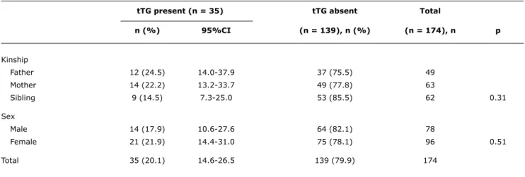
tTG present (n = 35) tTG absent Total
n (%) 95%CI (n = 139), n (%) (n = 174), n p
Kinship
Father 12 (24.5) 14.0-37.9 37 (75.5) 49
Mother 14 (22.2) 13.2-33.7 49 (77.8) 63
Sibling 9 (14.5) 7.3-25.0 53 (85.5) 62 0.31
Sex
Male 14 (17.9) 10.6-27.6 64 (82.1) 78
Female 21 (21.9) 14.4-31.0 75 (78.1) 96 0.51
Total 35 (20.1) 14.6-26.5 139 (79.9) 174
Table 1 - Frequency of anti-tissue transglutaminase antibodies by sex and degree of kinship with the index case
95%CI = 95% confidence interval; IgA = immunoglobulin A; tTG = human IgA anti-tissue transglutaminase antibody. Ethical aspects
A written consent form was obtained from all participants. The study was approved by the Research Ethics Committee of the Instituto de Medicina Integral Fernando Figueira (IMIP), Recife, state of Pernambuco, Brazil, and complied with the decisions of the Helsinki Declaration. Individuals who were diagnosed with CD or had positive serological tests were referred to follow-up in a specialized outpatient clinic.
Results
We identiied 199 relatives of 66 index cases of CD, and 174 (87.4%) agreed to participate in the study and were included. All families had only one individual with a prior diagnosis of CD. There were 78 men (44.8%) and 96 women (55.2%) among the relatives. Regarding the type of kinship with the index case, 49 were fathers, 63 mothers, and 62 brothers or sisters. Parents’ age ranged between 23 and 56 years, median of 38 years (P25-75% 33-42 years), and siblings’ age ranged from 20 months to 27 years, median of 13 years (P25-75% 8 -17 years ). There was a predominance of females in the study population (p = 0.05).
Serological testing
tTG was positive in 35/174 relatives, resulting in a seroprevalence of 20.1% (95%CI 14-26). The results of the serological tests, according to type of kinship and sex, are described in Table 1. No difference was found in the frequencies of tTG seropositivity either in terms of type of kinship with the index case or gender.
Histology of the small intestine
Of the relatives with positive serological tests, 22/35 (62.8%) agreed to undergo endoscopy combined with biopsy.
Among them, 20/22 (90.9%) had altered biopsies: seven were compatible with Marsh stage 1 and 13 were compatible with Marsh stage 3. Considering that those patients showing altered biopsy and symptoms had CD, we found a probable CD prevalence of 11.5% (95%CI 7.3-17.4). Figure 1 shows the sequence and distribution of the results of serological testing and biopsies.
Symptoms
One of two relatives with normal biopsy and positive tTG was asymptomatic, and the other one had abdominal distension and pain, diarrhea, fullness, flatulence, constipation, joint pain, fatigue, irritability, alopecia, and rheumatologic disease without a deined diagnosis. Table 2 describes age, kinship, tTG results, classiication of height, BMI, histological alterations, symptoms, and associated conditions in the 20 relatives who had an altered biopsy.
Discussion
In our study, we found a seroprevalence of 20.1% and an estimated prevalence of CD of 11.5% in relatives of CD patients. This frequency is high and similar to those of Europe and North America.13,18 The two Brazilian studies involving
relatives of CD patients that we are aware of so far were conducted in the Central South region of Brazil and found a maximum prevalence of 4.8%.Both used anti-endomysial antibody(EmA)for initial screening.19,20 We believe that the difference in seroprevalence between these studies and the present study was signiicant because, although the EmA testing is more speciic, it is less sensitive than tTG.21
Age
Patient (years) Kinship tTG pHgt pBMI Marsh Symptoms and morbid conditions
1 29 Father 32 2 96 1 Asymptomatic
2 36 Mother 30 39 58 1 Alopecia, autoimmune thyroiditis
3 33 Father 199 21 81 1 Joint pain, fatigue
4 11 Sister 32 32 16 1 Constipation, recurrent oral ulcers
5 46 Mother 28 8 67 1 Diarrhea, recurrent oral ulcers, joint pain
6 35 Mother 25 36 84 1 Abdominal pain and distension, diarrhea, fullness,
latulence, joint pain, fatigue, irritability, insomnia, anemia
7 29 Mother 29 1 80 1 Nausea, abdominal pain and distension, latulence,
constipation, recurrent oral ulcers, fatigue, insomnia, anemia
8 2 Brother 69 33 8 3 Weight loss, diarrhea, abdominal distension and
pain, vomiting, recurrent oral ulcers
9 14 Sister 207 73 56 3 Constipation, fatigue, irritability
10 33 Mother 27 18 89 3 Abdominal pain and distension, fullness, constipation,
recurrent oral ulcers, fatigue, joint pain
11 44 Mother 68 14 94 3 Abdominal pain and distension, vomiting, nausea,
fullness, constipation, recurrent oral ulcers, thyroiditis
12 42 Father 175 9 51 3 Abdominal pain, fullness, latulence, constipation,
fatigue
13 17 Sister 30 0 18 3 Weight loss, joint pain, fatigue, anemia, rheumatic
disease, osteoporosis
14 47 Father 45 18 64 3 Flatulence, irritability
15 45 Mother 81 3 79 3 Abdominal pain, fullness, abdominal distension,
constipation, joint pain, fatigue, alopecia, anemia
16 28 Mother 113 20 52 3 Weight loss, nausea, constipation, joint pain, fatigue,
irritability, depression
17 14 Sister 26 53 25 3 Weight loss, nausea, fullness, abdominal distension,
latulence, constipation, recurrent oral ulcers, fatigue, irritability
18 11 Sister 33 49 48 3 Constipation, recurrent oral ulcers, alopecia
19 36 Father 55 73 76 3 Abdominal pain and distension, diarrhea, fullness,
flatulence, recurrent oral ulcers, joint pain, insomnia
20 46 Mother 25 2 72 3 Abdominal pain and distension, joint pain, fatigue
Table 2 - Age, kinship with the index case, result of anti-transglutaminase antibody, percentiles of height and body mass index, histological classiication, and clinical characteristics of 20 irst-degree relatives with altered biopsy
pHgt = height classification in percentiles according to the 2000 CDC; pBMI = classification of body mass index in percentiles according to the 2000 CDC; tTG = human anti-tissue transglutaminase antibody.
Biopsy alterations classified according to Marsh.
IgA deiciency. In Brazil, IgA deiciency affects about 0.1% of the population, being one of the lowest rates reported in the world,22 and even considering the higher frequency among CD patients, this possibility is low in this group.
A major operational limitation of the study was the relatives’ dificulty to attend the follow-up visits because they lived in remote areas of the municipality of Recife.
Therefore, 13/34 (37%) of the seropositive patients did not undergo biopsy. It is likely that some of these patients have CD and that the prevalence of this group is higher than that we found.
IgA = immunoglobulin A; tTG = human anti-tissue transglutaminase antibody.
Figure 1 - Flowchart of the research with distribution of the results of the serological tests and biopsies, classiied according to Marsh.
References
1. Kagnoff MF. Celiac disease: pathogenesis of a model immunogenetic disease. J Clin Invest. 2007;117:41-9.
2. Gandoli L, Pratesi R, Cordoba JC, Tauil PL, Gasparin M, Catassi C. Prevalence of celiac disease among blood donors in Brazil. Am J Gastroenterol. 2000;95:689-92.
3. Crovella S, Brandão L, Guimarães R, Filho JL, Arraes LC, Ventura A, et al. Speeding up coeliac disease diagnosis in the developing countries. Dig Liver Dis. 2007;39:900-2.
4. Oliveira RP, Sdepanian VL, Barreto JÁ, Cortez AJ, Carvalho FO, Bordin JO, et al. High prevalence of celiac disease in Brazilian blood donor volunteers based on screening by IgA antitissue transglutaminase antibody. Eur J Gastroenterol Hepatol. 2007;19:43-9.
5. Melo SB, Fernandes MI, Peres LC, Troncon LE, Galvão LC. Prevalence and demograic characteristcs of celiac disease among blood donors in Ribeirão Preto, State of São Paulo, Brasil. Dig Dis Sci. 2006;51:1020-5.
6. Accomando S, Cataldo F. The global village of celiac disease. Dig Liver Dis. 2004;36:492-8.
7. Bonamico M, Ferri M, Mariani P, Nenna R, Thanasi E, Luparia RP, et al. Serologic and genetic markers of celiac disease: a sequential study in the screening of irst degree relatives.J Pediatr Gastroenterol Nutr. 2006;42:150-4.
8. Hill ID, Dirks MH, Liptak GS, Colletti RB, Fasano A, Guandalini S, et al. Guideline for the diagnosis and treatment of celiac disease in children: recommendations of the North American Society for Pediatric Gastroenterology, Hepatology and Nutrition. J Pediatr Gastroenterol Nutr. 2005;40:1-19.
9. NICE. National Institute for Health and Clinical Excellence - Clinical guideline 86. Coeliac disease: recognition and assessment of coeliac disease. May 2009. [online] http://www.nice.org.uk/nicemedia/ pdf/CG86CostReport.pdf.
10. Revised criteria for diagnosis of coeliac disease. Report of Working Group of European Society of Paediatric Gastroenterology and Nutrition. Arch Dis Child. 1990;65:909-11.
11. Esteve M, Rosinach M, Fernandes-Banares F, Farré C, Salas A, Alsina M, et al. Spectrum of gluten-sensitive enteropathy in irst-degree relatives of patients with coeliac disease: clinical relevance of lymphocytic enteritis. Gut. 2006;55:1739-45.
the following possibilities: a false-positive serological test, the sample collected was not obtained in an affected area of the small intestine, or CD was at an early stage and there was not villous atrophy. In this population, it is important to consider the differential diagnosis of environmental enteropathy, which is endemic in these regions, and may have clinical and histological symptoms similar to CD. Similar data were found in India, Egypt, and Turkey, countries where the population lives in conditions comparable to those of Brazil.14,23,24 In this context, serological testing
and follow-up of clinical response to the gluten-free diet are essential for diagnosis.
All patients diagnosed in the present study, except for one, were symptomatic, and the patient who reported to be asymptomatic was short. Possibly because their symptoms were less intense and less speciic, those patients were not motivated to seek a diagnosis. Although many studies have already demonstrated that a high proportion of patients with constipation, dyspepsia, and irritable bowel syndrome have undiagnosed CD, the diagnostic suspicion in such cases remains low.25,26
These data reinforce the need for active search using serological testing in relatives of CD patients, even in the non-European populations. In developing countries, serological testing and follow-up are essential to establish the diagnosis in relatives. We also suggest that, in cases with inconclusive biopsy that have some symptoms, tests with gluten-free diet and new biopsies for evaluation should be considered.
Acknowledgements
The authors would like to thank Sérgio Santos for collecting, transporting and processing the blood samples.
12. Tursi A, Brandimarte G. The symptomatic and histologic response to a gluten-free diet in patients with borderline enteropathy.J Clin Gastroenterol. 2003;36:6-7.
13. Fasano A, Berti I, Gerarduzzi T, Not T, Colletti RB, Drago S, et al. Prevalence of celiac disease in at-risk and not-at-risk groups in the United States: a large multicenter study.Arch Intern Med. 2003;163:286-92.
14. Groover R, Puri AS, Aggarwal N, Sakhuja P. Familial prevalence among irst-degree relatives of celiac disease in North India. Dig Liver Dis. 2007;39:903-7.
15. Cataldo F, Montalto G. Celiac disease in the developing countries: a new and challenging public health problem. World J Gastroenterol. 2007;13:2153-9.
16. Marsh MN. Gluten, major histocompatibility complex, and the small intestine. A molecular and immunobiologic approach to the spectrum of gluten sensitivity (‘celiac sprue’).Gastroenterology. 1992;102:330-54.
17. Kuczmarski RJ, Ogden CL, Guo SS, Grummer-Strawn LM, Flegal KM, Mei Z, et al. 2000 CDC growth charts for the United States: methods and development. National Center for Health Statistics. Vital Health Stat. 2002;246:1-190. http://www.cdc. gov/growthcharts.
18. Dube C, Rostom A, Sy R, Cranney A, Saloojee N, Garritty C, et al. The prevalence of celiac disease in average-risk and at-risk Western European populations: a systematic review. Gastroenterology. 2005;128:S57-67.
19. Kotze LM, Utiyama SR, Nishiara RM, Zeni MP, de Sena MG, Amarante HM. Antiendomysium antibodies in Brazilian patients with celiac disease and their irst-degree relatives. Arq Gastroenterol. 2001;38:94-103.
20. Almeida PL, Gandoli L, Modelli IC, Martins Rde C, Almeida RC, Pratesi R. Prevalence of celiac disease among irst degree relatives of Brazilian celiac patients. Arq Gastroenterol. 2008;45:69-72
Correspondence:
Margarida M Castro-Antunes
Av. Beira Rio, 240/2402, Ilha do Retiro CEP 50750-400 - Recife, PE - Brazil Tel.: +55 (81) 3446.8660
E-mail: [email protected]
21. Biagi F, Pezzimenti D, Campanella J, Vadacca GB, Corraza GR.
Endomysial and tissue transglutaminase antibodies in celiac sera: a comparison not inluenced by previous serological testing.Scand J Gastroenterol. 2001;36:955-8.
22. Carneiro-Sampaio MM, Carbonare SB, Rozentraub RB, de Araujo MN, Riberiro MA, Porto MH. Frequency of selective IgA deiciency among Brazilian blood donors and healthy pregnant women.
Allergol Immunopathol (Madr). 1989;17:213-6.
23. Bhatnagar S, Gupta SD, Mathur M, Phillips AD, Kumar R, Knutton S, et al. Celiac disease with mild to moderate histologic changes is a common cause of cronic diarrhea in Indian children. J Pediat Gastroenterol Nutr. 2005;41:204-209.
24. Abu-Zekry M, Kryszak D, Diab M, Catassi C, Fasano A. Prevalence of celiac disease in Egyptian children disputes the east-west agriculture-dependent spread of the disease.J Pediat Gastroenterol Nutr. 2008;47:136-40.
25. Usai P, Manca R, Cuomo R, Lai MA, Boi MF. Effect of gluten-free diet and co-morbidity of irritable bowel syndrome-type symptoms on health-related quality of life in adult coeliac patients. Dig Liver Dis. 2007;39:824-8.