238
Bayerl JS et al. Osteomyelitis of the wrist in a patient with paracoccidioidomycosis
Radiol Bras. 2012 Jul/Ago;45(4):238–240
Osteomyelitis of the wrist in a patient with disseminated
paracoccidioidomycosis: a rare presentation
*
Osteomielite de punho em paciente com paracoccidioidomicose disseminada: uma rara apresentação
Juliana Santos Bayerl1, André Ribeiro Nogueira de Oliveira2, Paulo Mendes Peçanha3, Aloísio Falqueto4
Paraccocidioidomycosis is the most frequently found endemic systemic mycosis in Brazil. No symptoms are observed in the early phases of the disease. As the disease progresses, the patient may present disseminated involvement, but bone involvement is extremely rare. The present report is aimed at evaluating bone changes found on imaging studies in a patient with osteomyelitis of the wrist as a result of disseminated paracoccidioidomycosis.
Keywords: Paracoccidioidomycosis; Endemic mycosis; Osteomyelitis.
Paracoccidioidomicose é a micose sistêmica endêmica mais frequente no Brasil. No início, o paciente não desenvolve sintomas. Com a progressão da doença, o indivíduo pode apresentar envolvimento disseminado, sendo que o acome-timento ósseo é extremamente raro. O objetivo deste artigo é avaliar as alterações ósseas encontradas em estudos de imagem em um paciente com osteomielite de punho decorrente de paracoccidioidomicose disseminada.
Unitermos: Paracoccidioidomicose; Micose endêmica; Osteomielite.
Abstract
Resumo
* Study developed at Hospital Universitário Cassiano Antônio de Moraes – Universidade Federal do Espírito Santo (UFES), Vi-tória, ES, Brazil.
1. MD, Resident in Radiology and Imaging Diagnosis, Univer-sidade Federal do Espírito Santo (UFES), Vitória, ES, Brazil.
2. Nuclear Physician, CMEN – Centro de Medicina Nuclear, Preceptor of Radiology and Imaging Diagnosis, Universidade Federal do Espírito Santo (UFES), Vitória, ES, Brazil.
3. Fellow Master Degree, Associate Professor, Universidade Federal do Espírito Santo (UFES), Vitória, ES, Brazil.
4. PhD, Associate Professor, Universidade Federal do Espírito Santo (UFES), Vitória, ES, Brazil.
Mailing Address: Dra. Juliana Santos Bayerl. Rua José Batista, 14, Bairro Recanto. Cachoeiro de Itapemirim, ES, 29303-012, Brazil. E-mail: [email protected]
Received January 4, 2012. Accepted after revision June 22, 2012.
Bayerl JS, Oliveira ARN, Peçanha PM, Falqueto A. Osteomyelitis of the wrist in a patient with disseminated paracoccidioidomycosis: a rare presentation. Radiol Bras. 2012 Jul/Ago;45(4):238–240.
0100-3984 © Colégio Brasileiro de Radiologia e Diagnóstico por Imagem CASE REPORT
Considering the radiographic findings, the patient was treated with antibiotics and referred to the center of infecto-parasitic diseases at Hospital Universitário Cassiano Antônio de Moraes. One month after the treatment, new radiographic images of the wrist and high resolution computed tomog-raphy (HRCT) of the chest were acquired in addition to fungal serology and cytology of the wrist secretion. The cytology re-vealed the presence of a high number of fungi compatible with Paracoccidioides brasiliensis.
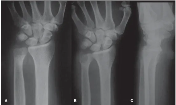
Radiography of the right wrist demon-strated a lytic area in the distal ulna (Fig-ures 1B and 1C). The patient was submit-ted to magnetic resonance imaging of the wrist, which demonstrated a significant involvement of the bone marrow in the dis-tal ulna, development of bone abscess and fistulous trajectory, with involvement of the muscle and adjacent subcutaneous tissues, with intense contrast-enhancement (Figure 2). At chest HRCT multiple consolidations were observed in the middle and lower thirds of the lungs, besides septal thicken-ing, cavitating nodules and bronchial walls thickening (Figure 3). Technetium and gal-lium scintigraphy was performed, confirm-ing the presence of active disease in the lungs and right wrist (Figure 4). Contrast The present report is aimed at
evaluat-ing bone changes observed on imagevaluat-ing studies of a patient with osteomyelitis of the wrist secondary to disseminated paracoccidioidomycosis, as a contribution to the early diagnosis and treatment of this disabling disease.
CASE REPORT
A male, 59-year old patient who as ag-ricultural worker and with a long history of smoking attended an emergency unit in the city of Vitória, ES, Brazil, reporting pro-gressive dyspnea for three months, ulcer-ative lesions on his skin and mucosas in association with pain and soft tissue en-largement in his right wrist, with serosan-guineous secretion. Findings at physical examination included cervical and axillary lymph nodes enlargement, ulcerative le-sions on the skin of his dorsal region and oral mucosa, besides a significant increase in volume of his right wrist.
Chest radiography demonstrated diffuse and confluent opacities in the middle thirds of the pulmonary fields and opacities cor-responding to fibrotic scars in the lung bases. Radiography of the right wrist dem-onstrated a subtle decrease in the bone den-sity of the distal ulna (Figure 1A). INTRODUCTION
239
Bayerl JS et al. Osteomyelitis of the wrist in a patient with paracoccidioidomycosis
Radiol Bras. 2012 Jul/Ago;45(4):238–240
uptake was not observed in other bones or in the central nervous system.
The patient was admitted and initially treated with Amphotericin B. After im-provement of the respiratory symptoms and decrease in volume of soft tissue compo-nents in his right wrist, the patient was dis-charged with sulfamethoxazole-trimetro-pim, remaining under regular follow-up on an outpatient basis.
DISCUSSION
Paracoccidioidomycosis affects prima-rily adult individuals in their most produc-tive phase, causing high social and eco-nomic impact. More than 90% of cases occur in male individuals(1,4), representing a relevant public health problem because of its high disabling potential and high mor-tality in cases of disseminated disease(4,5). Accurate epidemiological data on the dis-ease are not available in Brazil, since Bra-zilian regulations do not require mandatory notification(6).
Paracoccidioidomycosis may manifest in several organs, particularly in lungs, skin, mucosas and lymph nodes. Lungs are most commonly affected(7), with radio-graphic changes in 60% of acute cases and in up to 80% of chronic cases(8). Small opacities constitute the most common pul-monary findings, generally with bilateral and symmetric distribution of the le-Figure 2. MRI of right wrist. A,B: Coronal and axial T1-weighted images demonstrate intermediate signal
intensity in the distal ulna bone marrow, with cortical destruction and fistulous traject. C: Coronal, T2-weighted image reveals a significant bone marrow edema and extension of the infectious process to-wards soft tissues. D: Contrast-enhanced coronal T1-weighted image demonstrates enhancement of the bone marrow as well as of the muscle and adjacent subcutaneous tissues.
A
C
B
D
Figure 1. Plain radiograph of wrist. A: The first radiograph of the right wrist demonstrates subtle decrease in bone density of the distal ulna and preserved joint spaces. B,C: Antero-posterior and lateral radiographic images of wrist acquired one month later, clearly identify a lytic area in the distal ulna.
240
Bayerl JS et al. Osteomyelitis of the wrist in a patient with paracoccidioidomycosis
Radiol Bras. 2012 Jul/Ago;45(4):238–240 single bone involvement are reported in the literature.
In many cases, osteomyelitis secondary to paracoccidioidomycosis is a late diagno-sis, resulting in high morbidity and mortal-ity.
Imaging studies are useful to evaluate the disease extent. Suspicious areas must be radiologically investigated. Bone scin-tigraphy can assess the whole body. Gal-lium scintigraphy can detect inflammatory activity.
Therefore, the identification of imaging findings in cases of osteomyelitis second-ary to paracoccidioidomycosis is extremely relevant for early diagnosis and treatment, reducing morbidity.
REFERENCES
1. Shikanai-Yasuda MA, Telles Filho FQ, Mendes RP, et al. Consenso em paracoccidioidomicose. Rev Soc Bras Med Trop. 2006;39:297–310. 2. Souza AS Jr, Gasparetto EL, Davaus T, et al.
High-resolution CT findings of the 77 patients with untreated pulmonary paracoccidioidomycosis. AJR Am J Roentgenol. 2006;187:1248–52. 3. Pereira GH, Santos AQ, Park M, et al. Bone
mar-row involvement in a patient with paracocci-dioidomycosis: a rare presentation of juvenile form. Mycopathologia. 2010;170:259–61. 4. Moraes CS, Queiroz-Telles F, Marchiori E, et al.
Análise das alterações radiográficas pulmonares durante a terapêutica da paracoccidioidomicose. Radiol Bras. 2011;44:20–8.
5. Coutinho ZF, Silva D, Lazera M, et al. Paracocci-dioidomycosis mortality in Brazil (1980-1995). Cad Saúde Pública. 2002;18:1441–54. 6. Martinez R. Paracoccidioidomycosis: the
dimen-sion of the problem of a neglected disease. Rev Soc Bras Med Trop. 2010;43:480.
7. Muniz MAS, Marchiori E, Magnago M, et al. Paracoccidioidomicose pulmonar – aspectos na tomografia computadorizada de alta resolução. Radiol Bras. 2002;35:147–54.
8. Tobón AM, Agudelo CA, Osorio ML, et al. Re-sidual pulmonary abnormalities in adult patients with chronic paracoccidioidomycosis: prolonged follow-up after itraconazole therapy. Clin Infect Dis. 2003;37:898–904.
9. Fulciniti F, Troncone G, Fazioli F, et al. Osteomy-elitis by Paracoccidioides brasiliensis (South American blastomycosis): cytologic diagnosis on fine-needle aspiration biopsy smears: a case re-port. Diagn Cytopathol. 1996;15:442–6. 10. Krivoy S, Belfort EA, Mondolfi A, et al.
Para-coccidioidomycosis of the skull. Case report. J Neurosurg. 1978;49:429–33.
11. Nogueira SA, Guedes AL, Wanke B, et al. Osteo-myelitis caused by Paracoccidioides brasiliensis in a child from the metropolitan area of Rio de Janeiro. J Trop Pediatr. 2001;47:311–5.
Figure 4. Scintigraphy and cytology of right wrist. A,B: Cytology of the right wrist secretion demonstrates the presence of a high number of fungi compatible with Paracoccidioides brasiliensis. C: Significant ra-diopharmaceutical uptake in the distal wrist, indicating the presence of active disease.
sions(4). Bones involvement is extremely rare(3,9–11).
The typical finding is a well-defined lytic area, either with or without a sclerotic halo that may be observed in any bone, ei-ther with or without involvement of soft tis-sues. The disease is generally multifocal. Differential diagnoses include other infec-tious diseases such as chronic bacterial
os-teomyelitis, tuberculosis and primary or metastatic tumors such as lymphoma and osteosarcoma.