166 Rev Bras Hematol Hemoter. 2011;33(2):166
Acid phosphatase in blood smears of
Phrynops geoffroanus
(Testudines: Chelidae)
Images in Clinical Hematology
Universidade Estadual Paulista "Julio de Mesquita Filho" – UNESP, São José do Rio Preto, SP, Brazil
Maria Isabel Afonso da Silva Maria Tercília Vilela de Azeredo Oliveira
Claudia Regina Bonini-Domingos
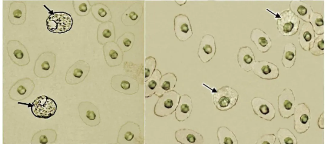
Acid phosphatases belong to the hydrolases class of enzymes; they act on organic esters, releasing phosphate ions in acidic conditions. These enzymes are found in lysosomes and secretory vacuoles. They are important for tissue autolysis and proliferation and differentiation and cell transformation processes, but also indicate possible tumors. Deficiency may limit leukocytes, resulting in recurrent infections.(1)Acid phosphatase staining was performed in blood smears of Phrynops geoffroanus using the lead phosphate method,(2) with eosinophils being strongly stained (Figure 1).
In human blood samples this cytochemical stains the neutrophils. In chelonians, there is a low number of neutrophils and a high number of circulating eosinophils which respond to parasitic infections.(3) Cytoplasmic staining suggests that eosinophils are rich in lysosomes which is probably
related to their mobilization as defense cells.
Conflict-of-interest disclosure: The authors declare no competing financial interest
Financial support: FAPESP, CNPq Submitted: 8/24/2010 Accepted: 9/3/2010
Corresponding author:
Maria Isabel Afonso da Silva
Universidade Estadual Paulista "Julio de Mesquita Filho"
Rua Cristóvão Colombo, 2265 – Jd. Nazareth 15054-000 – São José do Rio Preto, SP, Brazil
Phone: 55 17 3221-2392 [email protected] www.rbhh.org or www.scielo.br/rbhh DOI: 10.5581/1516-8484.20110041
Figure 1 – Acid phosphatase technique in blood smears of Phrynops geoffroanus. The image on the left shows detection of the enzyme in eosinophils and on the right is the control reaction performed on a sample from the same individual. The arrows identify eosinophils
References
1. Bull H, Murray PG, Thomas D, Fraser AM, Nelson PN. Acid phosphatases. Mol Pathol. 2002;55(2): 65-72.
2. Gomori G. An improved histochemical technique for acid phosphatase. Stain Technol. 1950;25(2): 81-5.
3. Goulart CE. Herpetologia, herpetocultura e medicina de répteis. Rio de Janeiro. LF Livros de Veterinária Ltda; 2004. p.21-56, 99-108, 131-144.