Current status of PET/CT in the diagnosis and follow up of lymphomas
Centro de Medicina Nuclear, Instituto de Radiologia, Hospital das Clínicas, Faculdade de Medicina, Universidade de São Paulo – USP, São Paulo, SP, Brazil
Instituto do Câncer do Estado de São Paulo – ICESP – Grupo Fleury, São Paulo, SP, Brazil
Carlos Alberto Buchpiguel Lymphomas are a heterogeneous group of malignancies that have a distinct biological behavior according to the subtype and degree of differentiation. Adequate staging, which has a direct impact on prognosis, is essential to properly plan therapy. Structural cross-sectional imaging, such as computed tomography, has been the standard imaging tool to stage and monitor patients with lymphoma. However, molecular imaging such as positron emission tomography has shown complementary diagnostic and prognostic values. This review discusses the current value of positron emission tomography imaging using 2-[fluorine-18]fluoro-2-deoxy-d-glucose in staging, restaging, monitoring and detecting relapse in Hodgkin's and non-Hodgkin lymphoma.
Keywords: Lymph Nodes/radionuclide imaging; Lymphoma/radionuclide imaging; Positron-emission
tomography; Fluorodeoxyglucose F18/diagnostic use; Lymphoma, Hodgkin’s disease; Lymphoma, non-Hodgkin
Introduction
Lymphomas consist of a heterogeneous group of malignancies that bear distinct biological behavior according to the subtype and the degree of differentiation. They are mainly characterized by enlargement of lymph nodes (nodal disease) although any organ in the body can be involved in different settings of the disease (extranodal disease). They account for approximately 5% of all cancers in the United States and for a US$ 4.6 billion annual healthcare cost.(1)
Lymphomas can be divided into two main groups: Hodgkin's (HL) and non-Hodgkin (NHL). The most common type is NHL which represents approximately 85% of all lymphomas. According to the biological behavior and consequently the prognosis, they can be grouped as indolent (low grade) and aggressive (intermediate or high grade) tumors.(2) The presence of extranodal disease has also prognostic implications as this may define a more advanced staging status (III or IV). However, patients with primary extranodal disease can still be categorized as stage I or II according to Ann Arbor system with the incorporation of the classification of bulky disease of the Cotswold consensus meeting held in 1989.(3)
The diagnosis of primary versussecondary lymphoma remains a challenge despite the great development in diagnostic tools. Part of this difficulty is related to the subtle changes frequently missed by conventional cross-sectional structural imaging. The limitations of computed tomography (CT) imaging in restaging, monitoring treatment and detecting relapse of lymphoma, especially with indolent lymphomas, are already well recognized.(4) Most of the limitations reside in the criterion of size, that is when to classify an enlarged lymph node as abnormal. It is not rare that normal-sized lymph nodes, according to the criteria, are involved by a tumor and enlarged lymph nodes are not infiltrated by lymphoma cells. Enlargement of lymph nodes can be seen in many non-malignant conditions such as inflammatory disorders.
The sensitivity to detect lung nodules has increased with current multi-detector CT scanners (MDCT) although the specificity is not that high.(5) The detection of gastric or small/large bowel involvement requires special techniques and much expertise in CT imaging.(6)
Therapeutic strategies for the management of lymphoma are constantly being refined to improve long-term survival with the lowest possible risk of toxicity to the patient. However, therapy planning is directly dependent on proper staging with definite prognostic implications in the various categories of lymphoma.
Positron emission tomography (PET) using 2-[fluorine-18]fluoro-2-deoxy-d-glucose (FDG) has already been validated to assess patients with different types of malignant
Conflict-of-interest disclosure: The authors declare no competing financial interest
Submitted: 2/13/2011 Accepted: 4/4/2011
Corresponding author:
Carlos Alberto Buchpiguel
Instituto de Radiologia, Hospital das Clínicas Av. Dr. Enéas de Carvalho Aguiar, 255 – Cerqueira César
05403-000 – São Paulo, SP, Brazil Phone: 55 11 3082-1015 [email protected]
www.rbhh.org or www.scielo.br/rbhh
tumors, including lymphomas. The principle of the imaging test is based on metabolic changes that reflect fundamental differences in the central metabolic pathways in malignant tissue. Most cancer cells exhibit elevated levels of glycolysis and this metabolic pathway seems to be related to a higher glucose uptake. Because of those changes, tumor cells produce lactate at higher levels compared to non-malignant tissue, even in the presence of oxygen, a phenomenon termed "aerobic glycolysis" or the "Warburg effect".(7) FDG-PET relies on this principle to detect foci of tumor proliferation.
This review comments on the value of adding molecular imaging information, as provided by FDG-PET, to the staging and monitoring of therapy of patients with HL and NHL.
Staging
FDG-PET has been widely used to stage and restage HL and NHL. Staging HL and NHL with FDG-PET is directly dependent on many different factors including biological and technical aspects of the lymphoma.
One of the most important factors is the histology. Normally, the most common subtypes of HL and the most aggressive NHL, such as diffuse large B cell lymphoma (DLBCL), show high levels of cell proliferation that reflect in a higher aerobic glycolysis rate (Figure 1).
Therefore, higher FDG uptake values are seen in aggressive tumors compared to more indolent ones that usually
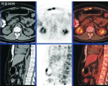
show faint or very low glucose concentrations. The level of FDG uptake can be evaluated semiquantitatively using the standardized uptake value (SUV). The SUV is the activity in the lesion measured as µCi/mL corrected for the patient's weight and the dose of FDG administered. Aggressive NHL lymphomas such as DLBCL and grade III follicular lymphoma (FL) have mean SUVs as high as 17.1 and 16.0, respectively. On the other hand, indolent lymphomas such as marginal zone lymphoma (MZL) and grade I/II FL can have SUVs as low as 9.0 and 7.0, respectively.(8) The sensitivity of PET in detecting the degree of FL may be as high as 98% but it drops to the 50% range when detecting MZL, peripheral T-cell lymphoma and small-T-cell lymphocytic lymphoma.(9) However, even though controversies still arise regarding the value of PET/CT in staging low-grade lymphomas, in special cases it seems to provide very reliable and important information for decisions on clinical management.(4) For example the limitations of PET/CT are well recognized in MZL of the mucosa-associated lymphoid tissue (MALT) subtype, since it has a tropism for gastrointestinal organs that usually show physiological FDG activity (Figure 2).
Figure 1 – High grade Hodgkin's lymphoma on PET/CT. First row shows the CT, second row shows the PET images and the third row shows the fused PET/CT images. Note the intense uptake do 2-[fluorine-18]fluoro-2-deoxy-d-glucose in the bulky mass and enlarged lymph nodes in the mediastinum
Figure 2 – Non-Hodgkin lymphoma MALT type involving the stomach. Note how difficult is to confirm and detect disease based only on the CT images
this kind of measurement might have a prognostic value in staging lymphoma.(10)
The molecular mechanisms involved in particular tumor micro-environments might also affect the performance of molecular imaging tools since a down-regulation or even a lack of expression of glucose transporter (GLUT) receptors (the proteins responsible to transport glucose from extracellular to intracellular moiety) on a tumor membrane would promote no FDG uptake by the tumor. However, the GLUT receptor is not the only mechanism that might increase the sensitivity of PET, since higher expressions of hexokinase-II (HK-II) have markedly increased FDG uptake, in particular in DLBCL.(11)
Benign tumors may show low FDG uptake and therefore this may reduce the specificity of FDG-PET. However, the integration of CT into the PET gantry, providing simultaneous structural and functional data, improves the interpretation and consequently the specificity of PET.
Another aspect that might impair specificity is the possibility that inflammatory cells trap glucose at a similar level to indolent or low-grade lymphomas. Hence, accurate correlation with CT findings and even with laboratory and clinical features may help improve the overall accuracy of the test.
A relatively recent meta-analysis evaluating the accuracy of FDG-PET on staging lymphoma showed a sensitivity of 90% and a specificity of 91%. The false-positive rate was 10.3% with a maximum accuracy of 88%.(12) Another systematic review showed very high sensitivity (97%) and specificity (100%) for staging HL and NHL.(13)
FDG-PET has also proved to be superior to a CT-alone strategy for staging. Upstaging can be up to 32% in HL(14) and 31% in NHL.(15)
PET/CT can detect any form of extranodal disease, including brain, head and neck, liver, spleen, muscle and skin involvement. However, in some locations special care in interpretation is recommended since physiological activity or inflammation after therapy can preclude an accurate staging or restaging. Physiological and symmetrical activity is commonly seen in the lymphatic structures of the head and neck however lymphoma involvement presents asymmetric activity and a more intense degree of FDG uptake that is often easily recognized (Figure 3).(16)
Staging lung involvement is feasible when the size of nodules is within the spatial resolution range of the modern PET/CT equipment (> 7-8 mm). However, specificity can be impaired due to the higher frequency of chemotherapy-associated pneumonitis, pneumonia and radiation-induced changes. Inflammatory processes such as sarcoidosis are one of the most common causes of false-positive results in evaluations of the mediastinum and lungs (Figure 4).(17)
Another area where interpretation requires special attention is the thymus, although thymus involvement does not change the staging in HL as it is considered a "nodal organ".(18) Enlargement of the thymus associated to mild/
Figure 3 – Patient with nasopharyngeal non-Hodgkin lymphoma showing marked asymmetrical 2-[fluorine-18]fluoro-2-deoxy-d-glucose uptake in the right pharyngeal tonsil
moderate diffuse FDG uptake, mainly after therapy, probably represents thymus hyperplasia (Figure 5).
However, distinction from a low grade thymoma can be challenging. Leptomeningeal metastasis can also be more difficult to detect by PET compared to MRI due to its spatial resolution limitations.
Detecting bone marrow involvement is also very important for a proper disease staging. Recent papers have shown a complimentary role of PET/CT on detecting bone marrow infiltration. PET/CT has higher sensitivity and comparable specificity in relation to bone marrow biopsy as it can detect disease in locations not usually covered by marrow biopsies (iliac crest or sternum) (Figure 6).(19)
Figure 6 – Patient with non-Hodgkin lymphoma showing multiple bone lesions seen only on PET/CT. A bone marrow biopsy of the iliac crest was negative. After reviewing the PET findings, a repeated marrow biopsy in the right femur confirmed bone marrow involvement
Figure 5 – FDG-PET showing increased uptake of 2-[fluorine-18]fluoro-2-deoxy-d-glucose in an enlarged thymus after therapy representing thymic hyperplasia
However, after chemotherapy, especially after using colony-stimulating factors, a diffuse bone marrow activity can normally be seen on PET/CT, which reflects bone marrow hyperplasia rather than infiltration. In contrast, a heterogeneous and irregular pattern of bone marrow FDG uptake suggests infiltration by the disease rather than a physiological reaction.
Another area that can be considered quite challenging for staging and restaging disease is the gastrointestinal tract. High gastric to large bowel activity is commonly observed since excretion and even lymphoid bowel elements might promote physiological concentrations of FDG without representing foci of disease activity. However, gastric and bowel involvement usually shows typical findings on CT with irregular or diffuse wall thickening when the proper diagnostic technique is applied. Therefore, the joint interpretation of PET together with CT helps to differentiate physiological uptake from pathological uptake in the gastrointestinal tract.
Another issue that has been widely discussed with the implementation of PET/CT is whether or not the use of iodine contrast in CT makes much difference in staging lymphomas. A recent paper showed that staging was almost similar using unenhanced low-dose PET/CT compared to enhanced full-dose PET/CT, although fewer indeterminate findings were seen with the enhanced technique and more extranodal lesions were detected.(21) Another recent study showed that a diagnostic CT did not have any incremental value on staging lymphoma when carried out concurrently with PET/CT.(22) Therefore the decision to use iodine contrast in CT depends on the imaging professional who is evaluating the patient, as this decision may change according to the PET findings, the availability of a recent enhanced MDCT exam and the probability of extranodal involvement among other things.
Therapy response evaluation
survival (OS) was 50% for PET positive versus92% for PET negative. A meta-analysis of FDG-PET in restaging HL and NHL included 20 studies and 854 subjects and showed a median sensitivity of 90.3% and a median specificity of 91.1%.(12)
A systematic review of 15 studies involving 705 patients with residual mass after therapy of HL and aggressive NHL showed that the pooled sensitivity and specificity for the detection of residual disease of HL by PET were 84% (95% CI: 71-92%) and 90% (95% CI: 84-94%), respectively. For NHL, the pooled sensitivity and specificity were 72% (95%: CI 61-82%) and 100% (95%: CI 97-100%), respectively.(24)
There are well known pretreatment prognostic factors that help to precisely predict the OS and the clinical stage status of patients with HL and NHL.(25,26) However, the tumor response is, in itself, an important prognostic factor that might not only tailor the therapy strategy, but also help to understand and apply the risk-adapted therapy strategy.
Structural imaging cannot always conclude whether therapy was successful in eliminating the tumor since residual masses are not rare after therapy, and might take time to shrink completely. It is difficult by CT to differentiate post-treatment fibrosis from active viable tumor in residual lesions seen in structural imaging. Even using standard criteria to quantify the size reduction on CT, limitations still exist in distinguishing active tumor from fibrosis.(27)
Molecular imaging such as PET/CT can be more effective in this task as it is more accurate at defining comple-te remission afcomple-ter therapy compared to the stacomple-te of the art structural imaging. The concept of its use is based on the fact that viable tumor cells still require glucose to maintain cell proliferation as opposed to fibrotic tissue where a very low level of activity or no activity at all is seen in the residual lesions after completing treatment. It is important to understand that PET/CT cannot be used, however, to identify microscopic malignant changes far below its threshold of spatial resolution.
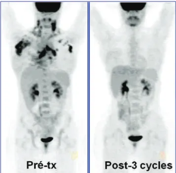
PET/CT can be used to evaluate treatment in two distinct situations with specific clinical purposes. The most common is to evaluate response after completion of therapy in order to verify incomplete remission that would require further therapy (Figures 7 & 8).
The early PET monitoring of lymphoma therapy (interim-PET) can be used to predict the relapse rate (prognosis) and in future may be used to tailor drug regimens and other treatment alternatives for refractory patients. There are groups that suggest that a negative interim-PET result might permit a reduced number of therapy cycles as this situation predicts a better outcome after treatment compared to a positive interim-PET result (Figure 9).
However, more prospective trials are required to prove that outcomes, including OS, are not impaired using that strategy.
In HL, the PFS directly reflects the PET/CT results. Retrospective and prospective studies have shown that
Figure 7 – Therapy monitoring with PET/CT. An example of complete response. Note that the multiple foci of 2-[fluorine-18]fluoro-2-deoxy-d-glucose uptake in abdominal lymph nodes seen on pre-therapy scan (left) show no more activity in the post-therapy study (right)
lower positive predictive value when compared to the ABVD regimen.(29) Another recent paper showed that interim-PET performed after 3 cycles of ABVD in HL patients is highly prognostic, since the 3-year event-free survival (EFS) was 53.4% for the PET-positive group compared to 90.2% for the PET-negative group.(30)
In NHL, especially with the aggressive subtypes, interim-PET can also group patients with high and low odds ratio for relapse. PFS ranges from 10-50% in the positive interim-PET group compared to 79-100% in the negative interim-PET group. In a large retrospective study, which included a cohort of 121 patients with high-grade NHL and a median follow-up of 28.5 months, 18F-FDG PET performed after 2 or 3 cycles of treatment strongly predicted PFS and OS. The estimated 5-year PFS was 89% for a negative interim-PET group and 16% for a positive-interim-PET group. Also an association was shown between early-PET results and OS.(31) In a prospective study, Haioun et al., performing PET after 2 cycles of chemotherapy in aggressive NHL patients, showed 2-year PFS and OS of 82% and 90%, respectively for a negative-PET group in contrast to 43% and 60%, respectively for a positive-PET group.(32) A recent study evaluating the prognostic value of interim-PET during R-CHOP therapy in aggressive NHL showed no differences in PFS and OS between the negative interim-PET and positive interim-PET groups. Only the end-therapy scan showed impact on PFS and OS.(33)
There is not enough evidence regarding the use of interim-PET in indolent low-grade lymphomas. Most data report small cohorts of patients, which precludes any definite
conclusions about its value.(34) However, more complex is the fact that indolent but advanced follicular NHL is quite different from HL and aggressive NHL. Current therapy is rarely curative, and the use of autologous stem cell transplantation (ASCT) or bone marrow transplantation for non-responding patients is feasible but the optimal timing of the procedure and its late effects remain questionable. Also, for indolent tumors it is possible to keep the disease under control for longer times using various therapy regimes but without improving the OS.
Adapting therapy according to the early PET response has been under investigation by many groups(35,36) however, so far there is not enough evidence to prove that changing to a more aggressive therapy or even ASCT to treat HL and NHL non-responders provides better OS and clinical outcomes.
Many studies have shown the impact of FDG-PET, performed at the end of therapy in HL and aggressive NHL, on the PFS and OS.(24,37-43) It is more accurate than conventional structural imaging, since the information is dependent on cellular activity rather than on the size of the involved structures. A recent systematic review and meta-analysis showed that the negative predictive value of PET in evaluating therapy response in HL ranged from 94.3% (95% CI, 92.8-95.7) to 100% (95% CI, 97.1-100).(44) In aggressive NHL, the result of PET at the end of treatment is also highly predictive for residual disease or relapse.(45)
In 2007 a group of experts brought together by the International Harmonization Project, developed new recommendations for response criteria using FDG-PET to monitor aggressive lymphomas during therapy.(46) This was very important to standardize the way PET is interpreted whether by visual or quantitative criteria. Relatively recent studies confirmed the superiority of the revised interpretation criteria to evaluate HL and NHL.(47) However, most of the evidence-based literature is related to studies performed using conventional treatment protocols. Very little data is available regarding the use of PET in tailoring therapy, even by abbreviating chemotherapy in early good-responders or by excluding radiotherapy as a complimentary therapy option in the end-of-treatment phase when the patient shows complete metabolic response at PET. The results of omitting radiotherapy at the end of chemotherapy in patients with residual mass but negative PET is controversial, since there are studies that confirm the high negative predictive value of PET (95%),(48) in contrast to others that show a higher relapse rate in the group of patients not randomized to radiotherapy in comparison to the group referred to radiotherapy.(49) However, a recent study showed that PET/CT detects occult visceral disease in the liver and spleen in HL which is not detected by conventional imaging, thereby identifying patients requiring consolidation radiation of the spleen. Moreover, the relapse rate was much lower in the PET/CT group compared to the conventional imaging staged group.(50)
Another potential role of PET/CT is predicting outcome (PFS and OS) in patients who are candidates for ASCT. Several studies have shown that a positive scan before ASCT is highly predictive of relapse. Studies in HL and NHL have shown that the 2-year PFS is up to 82% better in patients with a negative pre-ASCT scan.(51)
However, most of the available data is based on retrospective studies using heterogeneous criteria to interpret the histological and PET results. Therefore, more prospective studies are needed to prove the prognostic value of pre-ASCT PET. Moreover, new advances in PET instrumentation technology and new molecular probes might further enhance the clinical penetration of molecular imaging in the evaluation of lymphoma in near future.
References
1. American Cancer Society. Cancer Facts & Figures 2008 [Internet]. New York: ACS. [cited 2009 Mar 11]. Available from http://www. cancer.org/Research/CancerFactsFigures/CancerFactsFigures/ cancer-facts-figures-2008
2. Lu P. Staging and classification of lymphoma. Semin Nucl Med. 2005;35(3):160-4.
3. Lister TA, Crowther D, Sutcliffe SB, Glatstein E, Canellos GP, Young RC, et al. Report of a committee convened to discuss the evaluation and staging of patients with Hodgkin's disease: Cotswolds meeting. J Clin Oncol. 1989;7(11):1630-6. Erratum in: J Clin Oncol 1990;8(9):1602. Comment in: J Clin Oncol. 1990;8(9):1598. 4. Fueger BJ, Yeom K, Czernin J, Sayre JW, Phelps ME,
Allen-Auerbach MS. Comparison of CT, PET, and PET/CT for staging of patients with indolent non-Hodgkin's lymphoma. Mol Imaging Biol. 2009;11(4):269-74.
5. Truong MT, Sabloff BS, Ko JP. Multidetector CT of solitary pulmonary nodules. Thorac Surg Clin.2010;20(1):9-23. Republished from: Radiol Clin North Am. 2010;48(1):141-55.
6. Horton KM, Fishman EK. Multidetector-row computed tomography and 3-dimensional computed tomography imaging of small bowel neoplasms: current concept in diagnosis. J Comput Assist Tomogr. 2004;28(1):106-16.
7. Scatena R, Bottoni P, Pontoglio A, Giardina B. Revisiting the Warburg effect in cancer cells with proteomics. The emergence of new approaches to diagnosis, prognosis and therapy. Proteomics Clin Appl. 2010;4(2):143-58.
8. Schöder H, Noy A, Gönen M, Weng L, Green D, Erdi YE, et al. Intensity of 18fluorodeoxyglucose uptake in positron emission tomography distinguishes between indolent and aggressive non-Hodgkin's lymphoma. J Clin Oncol. 2005;23(21):4643-51. Comment in: J Clin Oncol. 2005;23(21):4577-80.
9. Tsukamoto N, Kojima M, Hasegawa M, Oriuchi N, Matsushima T, Yokohama A, et al. The usefulness of (18)F-fluorodeoxyglucose positron emission tomography (18)F-FDG-PET) and a comparison of (18)F-FDG-PET with 67gallium scintigraphy in the evaluation of lymphoma: relation to histologic subtypes based on the World Health Organization classification. Cancer. 2007;110(3):652-9. 10. Papajík T, Myslivecek M, Sedová Z, Buriánková E, Procházka V,
Koranda P, et al. Standardised uptake value of 18F-FDG on staging PET/CT in newly diagnosed patients with different subtypes of non-Hodgkin's lymphoma. Eur J Haematol. 2011;86(1):32-7. 11. Shim HK, Lee WW, Park SY, Kim H, So Y, Kim SE. Expressions
of glucose transporter types 1 and 3 and hexokinase-II in diffuse large B-cell lymphoma and other B-cell non-Hodgkin's lymphomas. Nucl Med Biol. 2009;36(2):191-7.
12. Isasi CR, Lu P, Blaufox MD. A metaanalysis of 18F-2-deoxy-2-fluoro-d-glucose positron emission tomography in the staging and restaging of patients with lymphoma. Cancer. 2005;104 (5):1066-74.
13. Kwee TC, Kwee RM, Nievelstein RA. Imaging in staging of malignant lymphoma: a systematic review. Blood. 2008;111(2): 504-16.
14. Raanani P, Shasha Y, Perry C, Metser U, Naparstek E, Apter S, et al. Is CT scan still necessary for staging in Hodgkin and non-Hodgkin lymphoma patients in the PET/CT era? Ann Oncol. 2006;17(1):117-22.
15. Schaefer NG, Hany TF, Taverna C, Seifert B, Stumpe KD, von Schulthess GK, et al. Non-Hodgkin lymphoma and Hodgkin disease: coregistered FDG PET and CT at staging and restaging-do we need contrast-enhanced CT? Radiology. 2004;232(3):823-9.
16. Paes FM, Kalkanis DG, Sideras PA, Serafini AN. FDG PET/CT of extranodal involvement in non-Hodgkin lymphoma and Hodgkin disease. Radiographics. 2010;30(1):269-91.
17. Alavi A, Buchpiguel CA, Loessner A. Is there a role for FDG PET imaging in the management of patients with sarcoidosis? J Nucl Med. 1994;35(10):1650-2. Comment on: J Nucl Med. 1994;35 (10):1647-9.
18. Wang X, Koch S. Positron emission tomography/computed tomography potential pitfalls and artifacts. Curr Probl Diagn Radiol. 2009;38(4):156-69.
19. Moulin-Romsee G, Hindié E, Cuenca X, Brice P, Decaudin D, Bénamor M, et al. (18)F-FDG PET/CT bone/bone marrow findings in Hodgkin's lymphoma may circumvent the use of bone marrow trephine biopsy at diagnosis staging. Eur J Nucl Med Mol Imaging. 2010;37(6):1095-105.
20. Wu LM, Chen FY, Jiang XX, Gu HY, Yin Y, Xu JR. (18)F-FDG PET, combined FDG-PET/CT and MRI for evaluation of bone marrow infiltration in staging of lymphoma: A systematic review and meta-analysis. Eur J Radiol. 2010 Dec 8. [Epub ahead of print].
21. Rodríguez-Vigil B, Gómez-León N, Pinilla I, Hernández-Maraver D, Coya J, Martín-Curto L, et al. Madero R. PET/CT in lymphoma: prospective study of enhanced full-dose PET/CT versusunenhanced low-dose PET/CT. J Nucl Med. 2006;47(10):1643-8.
22. Elstrom RL, Leonard JP, Coleman M, Brown RK. Combined PET and low-dose, noncontrast CT scanning obviates the need for additional diagnostic contrast-enhanced CT scans in patients undergoing staging or restaging for lymphoma. Ann Oncol. 2008; 19(10):1770-3.
23. Jerusalem G, Beguin Y, Fassotte MF, Najjar F, Paulus P, Rigo P, et al. Whole-body positron emission tomography using 18F-fluorodeoxyglucose for post-treatment evaluation in Hodgkin's disease and non-Hodgkin's lymphoma has higher diagnostic and prognostic value than classical computed tomography scan imaging. Blood. 1999;94(2):429-33.
24. Zijlstra JM, Lindauer-van der Werf G, Hoekstra OS, Hooft L, Riphagen II, Huijgens PC. 18F-fluoro-deoxyglucose positron emission tomography for post-treatment evaluation of malignant lymphoma: a systematic review. Haematologica. 2006;91(4):522-9.
25. A predictive model for aggressive non-Hodgkin's lymphoma. The International Non-Hodgkin's Lymphoma Prognostic Factors Project. N Engl J Med. 1993;329(14):987-94. Comment in: N Engl J Med. 1994;330(8):574; author reply 574-5. N Engl J Med. 1994;330(8):574; author reply 574-5.
27. Keil S, Behrendt FF, Stanzel S, Suehling M, Jost E, Mühlenbruch G, et al. RECIST and WHO criteria evaluation of cervical, thoracic and abdominal lymph nodes in patients with malignant lymphoma: manual versussemi-automated measurement on standard MDCT slices. Rofo. 2009;181(9):888-95.
28. Hutchings M, Loft A, Hansen M, Pedersen LM, Buhl T, Jurlander J, et al. FDG-PET after two cycles of chemotherapy predicts treatment failure and progression-free survival in Hodgkin lymphoma. Blood. 2006;107(1):52-9.
29. Gallamini A, Hutchings M, Rigacci L, Specht L, Merli F, Hansen M, et al. Early interim 2-[18F]fluoro-2-deoxy-D-glucose positron emission tomography is prognostically superior to international prognostic score in advanced-stage Hodgkin's lymphoma: a report from a joint Italian-Danish study. J Clin Oncol. 2007;25(24):3746-52.
30. Cerci JJ, Pracchia LF, Linardi CC, Pitella FA, Delbeke D, Izaki M, et al. 18F-FDG PET after 2 cycles of ABVD predicts event-free survival in early and advanced Hodgkin lymphoma. J Nucl Med. 2010;51(9):1337-43. Erratum in: J Nucl Med. 2010;51(10): 1658. 31. Mikhaeel NG, Hutchings M, Fields PA, O'Doherty MJ, Timothy AR. FDG-PET after two to three cycles of chemotherapy predicts progression-free and overall survival in high-grade non-Hodgkin lymphoma. Ann Oncol. 2005;16(9):1514-23.
32. Haioun C, Itti E, Rahmouni A, Brice P, Rain JD, Belhadj K, et al. [18F]fluoro-2-deoxy-D-glucose positron emission tomography (FDG-PET) in aggressive lymphoma: an early prognostic tool for predicting patient outcome. Blood. 2005;106(4):1376-81. 33. Yoo C, Lee DH, Kim JE, Jo J, Yoon DH, Sohn BS, et al. Limited
role of interim PET/CT in patients with diffuse large B-cell lymphoma treated with R-CHOP. Ann Hematol. 2010 Dec 22. [Epub ahead of print]
34. Bishu S, Quigley JM, Bishu SR, Olsasky SM, Stem RA, Shostrom VK, et al. Predictive value and diagnostic accuracy of F-18-fluoro-deoxy-glucose positron emission tomography treated grade 1 and 2 follicular lymphoma. Leuk Lymphoma. 2007;48(8):1548-55. Comment in: Leuk Lymphoma. 2007;48(8):1463-4.
35. National Institute of Health. ClinicalTrials.gov. Tailoring treatment for B cell non-Hodgkin's lymphoma based on PET scan results mid treatment [Internet]. Washington, DC: NIH. [cited 2009 Feb 25]. Available from: http://www.clinicaltrials.gov/ct2/show/ NCT00324467
36. National Institute of Health. ClinicalTrials.gov. Fludeoxyglucose F 18 positron emission tomography in predicting risk of relapse in patients with non-Hodgkin's lymphoma who are undergoing combination chemotherapy with or without autologous stem cell or bone marrow transplant. Washington, DC: NIH. [cited 2009 Feb 25]. Available from: http://www.clinicaltrials.gov/ct2/show/ N C T 0 0 2 3 8 3 6 8 ? t e r m = p r e d i c t i n g + r i s k + o f + r e l a p s e + i n + patients+with+non-Hodgkin%E2%80%99s+lymphoma+who+ are+undergoing+combination+chemotherapy&rank=1
37. Juweid ME, Wiseman GA, Vose JM, Ritchie JM, Menda Y, Wooldridge JE, et al. Response assessment of aggressive non-Hodgkin's lymphoma by integrated International Workshop Criteria and fluorine-18-fluorodeoxyglucose positron emission tomography. J Clin Oncol. 2005;23(21):4652-61.
38. Brepoels L, Stroobants S, Verhoef G. PET and PET/CT for response evaluation in lymphoma: current practice and developments. Leuk Lymphoma. 2007;48(2):270-82.
39. de Wit M, Bohuslavizki KH, Buchert R, Bumann D, Clausen M, Hossfeld DK. 18FDG-PET following treatment as valid predictor for disease-free survival in Hodgkin's lymphoma. Ann Oncol. 2001;12(1):29-37.
40. Hueltenschmidt B, Sautter-Bihl ML, Lang O, Maul FD, Fischer J, Mergenthaler HG, et al. Whole body positron emission tomography in the treatment of Hodgkin disease. Cancer. 2001; 91(2):302-10.
41. Lavely WC, Delbeke D, Greer JP, Morgan DS, Byrne DW, Price RR, et al. FDG PET in the follow-up management of patients with newly diagnosed Hodgkin and non-Hodgkin lymphoma after first-line chemotherapy. Int J Radiat Oncol Biol Phys. 2003;57 (2):307-15.
42. Reinhardt MJ, Herkel C, Altehoefer C, Finke J, Moser E. Computed tomography and 18F-FDG positron emission tomography for therapy control of Hodgkin's and non-Hodgkin's lymphoma patients: when do we really need FDG-PET? Ann Oncol. 2005; 16(9):1524-9.
43. Delbeke D, Stroobants S, de Kerviler E, Gisselbrecht C, Meignan M, Conti PS. Expert opinions on positron emission tomography and computed tomography imaging in lymphoma. Oncologist. 2009;14 Suppl 2:30-40.
44. Ramos-Font C, Rebollo Aguirre AC, Villegas Portero R, Romero Tabares A, Gallego Peinado M, Llamas Elvira JM. [18F-fluorodeoxyglucose positron emission tomography in the evaluation of therapy response assessment in lymphomas. Systematic literature review and meta-analysis]. Rev Esp Med Nucl. 2009;28(2):48-55. Spanish.
45. Spaepen K, Stroobants S, Dupont P, Van Steenweghen S, Thomas J, Vandenberghe P, et al. Prognostic value of positron emission tomography (PET) with fluorine-18 fluorodeoxyglucose ([18F]FDG) after first-line chemotherapy in non-Hodgkin's lymphoma: is [18F]FDG-PET a valid alternative to conventional diagnostic methods? J Clin Oncol. 2001;19(2):414-9.
46. Juweid ME, Stroobants S, Hoekstra OS, Mottaghy FM, Dietlein M, Guermazi A, Wiseman GA, Kostakoglu L, Scheidhauer K, Buck A, Naumann R, Spaepen K, Hicks RJ, Weber WA, Reske SN, Schwaiger M, Schwartz LH, Zijlstra JM, Siegel BA, Cheson BD; Imaging Subcommittee of International Harmonization Project in Lymphoma. Use of positron emission tomography for response assessment of lymphoma: consensus of the Imaging Subcommittee of International Harmonization Project in Lymphoma. J Clin Oncol. 2007;25(5):571-8.
47. Brepoels L, Stroobants S, De Wever W, Spaepen K, Vandenberghe P, Thomas J, et al. Hodgkin lymphoma: response assessment by revised International Workshop Criteria. Leuk Lymphoma. 2007; 48(8):1539-47.
48. Kobe C, Dietlein M, Franklin J, Markova J, Lohri A, Amthauer H, et al. Positron emission tomography has a high negative predictive value for progression or early relapse for patients with residual disease after first-line chemotherapy in advanced-stage Hodgkin lymphoma. Blood. 2008;112(10):3989-94.
49. Picardi M, De Renzo A, Pane F, Nicolai E, Pacelli R, Salvatore M, et al. Randomized comparison of consolidation radiation versus observation in bulky Hodgkin's lymphoma with post-chemotherapy negative positron emission tomography scans. Leuk Lymphoma. 2007;48(9):1721-7. Comment in: Leuk Lymphoma. 2007;48(9): 1667-9.
50. Picardi M, Soricelli A, Grimaldi F, Nicolai E, Gallamini A, Pane F. Fused FDG-PET/contrast-enhanced CT detects occult sub-diaphragmatic involvement of Hodgkin's lymphoma thereby identifying patients requiring six cycles of anthracycline-containing chemotherapy and consolidation radiation of spleen. Ann Oncol. 2010;22(3):671-80.
51. Johnston PB, Wiseman GA, Micallef IN. Positron emission tomography using F-18 fluorodeoxyglucose pre- and post-autologous stem cell transplant in non-Hodgkin's lymphoma. Bone Marrow Transplant. 2008;41(11):919-25.
![Figure 3 – Patient with nasopharyngeal non-Hodgkin lymphoma showing marked asymmetrical 2-[fluorine-18]fluoro-2-deoxy-d-glucose uptake in the right pharyngeal tonsil](https://thumb-eu.123doks.com/thumbv2/123dok_br/19095043.498511/3.918.466.822.110.460/figure-patient-nasopharyngeal-hodgkin-lymphoma-asymmetrical-fluorine-pharyngeal.webp)
![Figure 5 – FDG-PET showing increased uptake of 2-[fluorine-18]fluoro- 2-[fluorine-18]fluoro-2-deoxy-d-glucose in an enlarged thymus after therapy representing thymic hyperplasia](https://thumb-eu.123doks.com/thumbv2/123dok_br/19095043.498511/4.918.99.454.592.1023/figure-showing-increased-fluorine-fluorine-enlarged-representing-hyperplasia.webp)
![Figure 7 – Therapy monitoring with PET/CT. An example of complete response. Note that the multiple foci of 2-[fluorine-18]fluoro-2-deoxy-d-glucose uptake in abdominal lymph nodes seen on pre-therapy scan (left) show no more activity in the post-therapy st](https://thumb-eu.123doks.com/thumbv2/123dok_br/19095043.498511/5.918.465.821.111.453/therapy-monitoring-complete-response-multiple-fluorine-abdominal-activity.webp)