Article
ISSN 0102-695X
http://dx.doi.org/10.1590/S0102-695X2012005000009 Received 10 Jun 2011 Accepted 3 Oct 2011 Available online 17 Jan 2012
isoniazid-rifampicin induced oxidative liver
injury in rat
S. Angala Parameswari,
1,*T. S. Mohamed Saleem,
2K. B.
Chandrasekar,
3C. Madhusudhana Chetty
41Department of Pharmaceutical Analysis, Annamacharya College of Pharmacy,
India,
2Department of Pharmacology, Annamacharya College of Pharmacy, India,
3Department of Chemistry, JNT University Anantapur, India,
4Department of Biotechnology, Annamacharya College of Pharmacy, India.
Abstract: The present study was made to investigate the protective effect of methanolic extract of Ficus benghalensis L., Moraceae, on isoniazid-rifampicin-induced hepatotoxicity in rats. Rats were divided into six different groups; group 1 served as a control, group 2 received isoniazid and rifampicin (100 mg/ kg, i.p.), in sterile water, groups 3, 4 and 5 received 100, 200 & 300 mg/kg bw,
p.o. methanolic extract of F. benghalensis and group 6 received Liv 52. All the treatment protocols followed 21 days and after rats were sacrificed blood and liver were used for biochemical and histological studies, respectively. Administration of isoniazid and rifampicin caused a significant elevation in the levels of liver marker enzymes (p<0.05 and p<0.01) and thiobarbituric acid reactive substances (p<0.001) in experimental rats. Administration of methanolic extracts of F. benghalensis
significantly prevented isoniazid-rifampicin-induced elevation in the levels of serum diagnostic liver marker enzymes and TBARS level in experimental groups of rats. Morever, total protein and reduced glutathione levels were significantly (p<0.001) increased in treatment group. The effect of extract was compared with a standard drug, Liv 52. The changes in biochemical parameters were supported by histological profile. It is to be concluded that the methanolic extract of F. benghalensis protects against isoniazid and rifampicin-induced oxidative liver injury in rats.
Keywords: Ficus benghalensis
hepatotoxicity isoniazid oxidative stress rifampicin
Introduction
Drug-induced liver toxicity is a common cause of liver injury. It accounts for approximately one-half of the cases of acute liver failure and mimics all forms of acute and chronic liver disease (Kaplowitz, 2001). Different types of drugs such as acetaminophen, chloroquine and isoniazid are inducers of hepatotoxicity in world. Isoniazid
and rifampicin, the i rst line drugs used for tuberculosis
therapy are associated with hepatotoxicity (Tasduq et al., 2005).2 The rate of hepatotoxicity has been reported to be
much higher in developing countries like India (8-30%) compared to that in advanced countries (2-3%) with a similar dose schedule (Sharma, 2004).
Ficus benghalensis L., Moraceae (Mulberry
family), is commonly known as a Banyan tree or Vata or Vada tree in ayurveda. There are more than 800 species and 2000 varieties of Ficus species, most of which are native to the old world tropics. F. benghalensis is a remarkable tree from India that sends down its branches and great number
of shoots, which take root and become new trunks. This tree is considered to be sacred in many places in India. Earlier, glucoside, 20-tetratriaconthene-2-one,
6-heptatriacontene-10-one, pentatriacontan-5-one, β-sitosterol-α-D-glucose, and meso-inositol have been isolated from the bark of the
F. benghalensis (Subramanian & Misra, 1978; The Wealth
of India, 1999)
The hanging roots of F. benghalensis have been reported as anti-diarrhoeal agents (Mukherjee et al., 1998). The bark of this plant reported for wound healing activity (Garg & Paliwal, 2011). The fruit extract of F. benghalensis
has been documented for its anti-tumor and anti bacterial activities (Mousa et al., 1994; Singh & Watal, 2010). The extracts of F. benghalensis were also reported to inhibit insulinase activity from the liver and kidney (Achrekar et al., 1991). It was also found to inhibit the lipid peroxidation (Shukla et al., 2004). Various extracts of F. benghalensis
al., 2011; Taur et al., 2007). Other species of Ficus viz. F.
carica (Gond & Khadabadi, 2008), F. glomerata (Kubsad
et al., 2008), and F. racemosa (Mandal et al., 1999) were found to have hepatoprotective activity. Based on this, an attempt has been made to evaluate the hepatoprotective effect of F. benghalensis.
Material and Methods
Chemicals
Bilirubin, total protein, alkaline phosphatase (ALP), alanine transaminases (ALT), and aspartate transaminases (AST) were assayed by using kits from Ranbaxy Diagnostic, New Delhi. All the drugs, chemicals and reagents used for biochemical estimation were purchased from Sigma-Aldrich, USA.
Animals
Male Wistar albino rats, weighing about 150-200 g and Swiss albino mice weighing about 25-30 g were obtained from Institute Animal Center and used in the experiments. The protocol was approved by the Institute’s Animal Ethical Committee (1220/a/08/CPCSEA/ ANCP/04). Animals were kept in the animal house at an ambient temperature of 25 °C and 45-55% relative humidity, with 12 h each of dark and light cycles. Animals were fed pellet diet and water ad-libitum.
Preparation of plant extract
Aerial root of Ficus benghalensis L., Moraceae, were collected from Rajampet, Andhrapradesh in June
2009 and identiied by Dr. K. Madhava Chetty, Department
of Botany, Sri Venkateswara University, Tirupathi. The voucher specimen (ANCP-MP-88) has been deposited in the parent department. The shade dried roots were powdered to get a course granule. About 500 g of dried powder was extracted with various solvents (petroleum ether, hexane, benzene, chloroform, ethyl acetate, methanol, ethanol and water) by continuous hot percolation, using soxhlet apparatus. The resulted dark-brown extract were concentrated up to 100 mL on rotavapor under reduced pressure. The concentrated crude extracts were lyophilized into powder and used for the study.
Phytochemical screening
The extracts obtained were subjected to preliminary phytochemical screening and thin layer chromatography to identify the chemical constituents. TLC was performed by using mobile phase n-hexane:ethyl acetate in the ratio of 9:1 and the compound present in the sample were detected under UV chamber at 365 nm. The methods of analysis employed were those described in standard procedures (Harborne & Baxter, 1993; Trease & Evans, 1989).
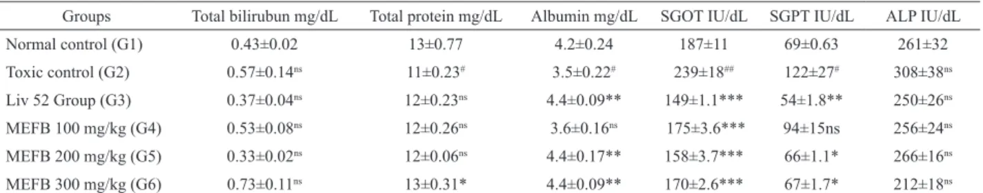
Table 1. Effect of Ficus benghalencis in different biochemical parameters in INH+RIF induced hepatotoxic rats.
Groups Total bilirubun mg/dL Total protein mg/dL Albumin mg/dL SGOT IU/dL SGPT IU/dL ALP IU/dL
Normal control (G1) 0.43±0.02 13±0.77 4.2±0.24 187±11 69±0.63 261±32
Toxic control (G2) 0.57±0.14ns 11±0.23# 3.5±0.22# 239±18## 122±27# 308±38ns
Liv 52 Group (G3) 0.37±0.04ns 12±0.23ns 4.4±0.09** 149±1.1*** 54±1.8** 250±26ns
MEFB 100 mg/kg (G4) 0.53±0.08ns 12±0.26ns 3.6±0.16ns 175±3.6*** 94±15ns 256±24ns
MEFB 200 mg/kg (G5) 0.33±0.02ns 12±0.06ns 4.4±0.17** 158±3.7*** 66±1.1* 266±16ns
MEFB 300 mg/kg (G6) 0.73±0.11ns 13±0.31* 4.4±0.09** 170±2.6*** 67±1.7* 212±18ns
Values are expressed as ean±SEM; Values are ind out by using ONEWAY ANOVA followed by Dunnett’s multiple range tests; #p<0.05 vs Control (G1); ##p<0.01 vs Control (G2); *p<0.05 vs Toxic Control (G2); **p<0.01 vs Toxic Control (G2); ***p<0.001 vs Toxic Control (G2); ns;-non signiicant.
Table 2. The levels of TBARS and GSH after the treatment of rats with isoniazid-rifampicin and MEFB on 21 days treatment.
Group TBARS nmol/g wet wt GSH µg/g wet wt
Normal control (G1) 276.42±0.23 91.13±0.14
Toxic control (G2) 398.75±0.51a 43.6±0.27b
Liv 52 10 mg/kg (G3) 245.21±0.31# 94.52±0.36#
MEFB 100 mg/kg (G4) 231.45±0.2# 87.27±0.12#
MEFB 200 mg/kg (G5) 254.61±0.7# 98.15±0.6#
MEFB 300 mg/kg (G6) 292.91±0.32# 89.6±0.25#
Acute toxicity study
Acute oral toxicity (AOT) of methanolic extract
of F. benghalensis (MEFB) were determined using Swiss
albino mice. The animals were fasted for 3 h prior to the experiment and were administered with single dose of extract dissolved in 5% gum acacia (doses ranges from 500-5000 mg/kg at various dose levels) and observed for mortality up to 48 h (short term toxicity). Based on the short-term toxicity, the dose of next animal was determined as per OECD guideline 425.
Free radical scavenging activity by 1,1-diphenyl-2-picryl hydrazyl (DPPH) method
In vitro antioxidant activity of MEFB was
analyzed by DPPH method (Singh et al., 2002). Different
concentrations (10, 50, 100 and 500 μg) of extract
samples and Butylated hydroxyl anisole (BHA-synthetic antioxidant) were taken in different test tubes. The volume
was adjusted to 500 μL by adding methanol. Five milliliters
of a 0.1 mM methanolic solution of DPPH was added to these tubes and shaken vigorously. A control without the test compound, but with an equivalent amount of methanol was maintained. The tubes were allowed to stand at room temperature for 20 min. The absorbance of the samples was measured at 517 nm. Radical scavenging activity was calculated using the following formula: % radical scavenging activity=[(control Abs-sample Abs)/Control Abs]×100.
Induction of experimental hepatotoxicity
Isoniazid and rifampicin solution were prepared separately in sterile distilled water. Rats were treated with isoniazid (100 mg/kg, i.p.) and co-administered with rifampicin (100 mg/kg, i.p.), for 21 days (Yue et al., 2004; Saleem et al., 2008). In order to study the effect of MEFB in rat, 100, 200 and 300 mg/kg bw, p.o. were used respectively. Liv 52 (10 mg/kg bw, p.o.) was used as a standard drug in this study. Rats were divided into Six different groups (n=6), group 1 was served as a control, group 2 was toxic control receive isoniazid+rifampicin (100 mg/kg bw i.p.), group 3, 4 and 5 were served as extract treatment groups received 100, 200 & 300 mg/kg bw, p.o
MEFB and group 6 served as standard group received Liv 52. Rats were treated as per the treatment protocol. Body weights of these rats were monitored sequentially in control and experimental animals for a period of 21 days.
Biochemical estimation
Rats were sacriiced 1 h after administration on
day 21. The blood was collected by retro-orbital artery bleeding. Blood samples were centrifuged for 10 min at
protein and bilirubin levels were estimated from the serum by using standard kits (Rajesh et al., 2005). Liver was excised immediately, quickly cooled and perfused with cold normal saline. Ten percent homogenate was prepared by homogenizing the liver tissue by using 0.3 m phosphate buffer. TBARS (Okhawa et al., 1979) and GSH (Ellman, 1959) levels were estimated from the liver homogenate by using spectrophotometric determination.
Histopathological studies
The livers were excised quickly and ixed in 10%
formalin and stained with haemotoxylin and eosin and then observed under microscope for degeneration, fatty changes or necrotic changes as evidence of hepatotoxicity.
Statistics
All values were expressed as means±SEM (n=6 in each group). One way ANOVA was applied to test for
signiicance of biochemical data of the different groups. Signiicance is set at p≤0.05.
Results
Phytochemical screening
The freshly prepared extracts were subjected to preliminary phytochemical screening test for various constituents. All the extracts shows the presence of different chemical constituents and the methanolic extract revealed
the presence of lavonoids, alkaloids and terpenoids.
Based on this the methanolic extract was selected for pharmacological evaluation.
Acute toxicity study
Acute oral toxicity studies, the extracts treated animals were observed for mortality up to 48 h. there was no mortality or any signs of behavioral changes observed after oral administration of methanol extract up to 5000 mg/kg body weight.
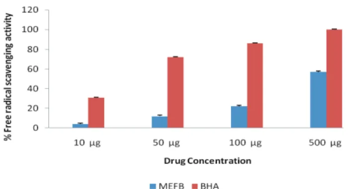
Free radical scavenging activity by DPPH method
The free radical scavenging activity of MEFB
and BHA were like at 10 μg 4.06 and 31.24%, 50 μg 12.41 and 71.68%, 100 μg 22.35, 86.46% and 500 μg 57.43%, respectively. MEFB shows signiicant free radical scavenging activity at higher dose (500 μg) when compared
Figure 1. Free radical scavenging activity of MEFB of Ficus benghalensis.
Mortality data
There was no mortality occurred in control, toxic control and standard group. During the treatment period all the group of extract treatment group shows the death event like 3 in G4, 2 in G5 and 1 in G6, respectively. There
was no signii cant difference in survival proportion. The
body weight and relative liver weights of the experimental animals calculated at the end of the study had no
statistically signii cant difference when compared to the
control animals. The results were present in Figure 2.
Figure 2. Survival proportions of rats of respective group during treatment period.
Biochemical parameters
There was no signii cant difference between
control and other treatment group in total bilirubin level.
There was a signii cant elevation of (p<0.01) AST in
INH+RIF treated rats. It was signii cantly (p<0.001) decreased by the administration of MEFB at a dose of 100,
200 and 300 mg/kg. There was a signii cant elevation of
(p<0.01) ALT in INH+RIF treated rats. It was signii cantly
(p<0.05) decreased by the administration of MEFB at a dose of 200 and 300 mg/kg. In total protein level there
was a signii cant difference (p<0.01) between control
and INH+RIF treated rats. But, there was no signii cant
difference between treatment rats at a dose of 100 and 200 mg/kg and only slightly increased level of (p<0.05) total protein in 300 mg/kg of MEFB treated group. In albumin level also only 200 and 300 mg/kg treated rats shows the
signii cant (p<0.01) rise when compared with toxic control group. All the treatment groups show the data same like standard Liv 52 treated animals. The results were present in Table 1.
Increased liver TBARS level in toxic control group is indication for increased oxidative stress by treatment of INH+RIF. Increased liver TBARS significantly (p<0.001) reduced by co-administration of MEFB in three different doses (100, 200 and 300 mg/kg b.w) and Liv 52 treatment group. The INH+RIF-administered animals exhibited significantly (p<0.01) low levels of hepatic GSH levels significantly increased by co-administration of MEFB and Liv 52 treatment group. The results were shown in Table 2.
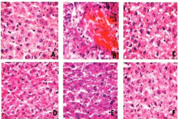
Histopathological studies
The liver of rats from respective groups were subjected to histopathological study. The results were present in Figure 3. Hepatocytes of the normal control group showed a normal lobular architecture of the liver (A). In the INH+RIF treated group the liver showed
hepatocytic necrosis and inl ammation and neutrophil ini ltration also observed in the centrilobular region with
portal triaditis (B). Liv 52 pretreated group showed minimal
inl ammation and hepatic congestion with moderate portal
triditis and their lobular architecture was normal (C). MEFB pretreated group at al dose of 100, 200 and 300 mg/
kg showed minimal inl ammation with moderate portal
triditis and their lobular architecture was normal (D-F).
These above i ndings indicate the hepatoprotective effect
of extracts. However, MEFB showed minimal hepatocytes changes with dose dependent activity.
Discussion
Phytochemical screening of methanolic extract
revealed the presence of l avonoids. We also reported the
same by thin layer chromatography and HPTLC studies. In our previous report, TLC of methanolic extract performed by the mobile phase ethyl acetate: formic acid: glacial acetic acid: water in the ratio of 100:11:11:26 shows the single spot at Rf 0.86 with blue l orescence under UV chamber at 365 nm. The same conformed by HPTLC also (Parameswari et al., 2011). In the present study, hepatotoxicity model in Wistar albino rats was successfully produced by administering INH and RIF (100 mg/kg per day each i.p.). A marked rise above the normal upper limits in the measured serum transaminases in INH+RIF group on day 21 of the experiment was a biochemical indication of liver injury.
Survival proportions
0 5 10 15 20 25
70 80 90 100 110
DAYS
hepatotoxicity (Yue et al., 2004). and also oxidative stress as one of the mechanism for INH+RIF induced hepatic injury (Peretti et al., 1987).
In this study the results suggest that the statistically
signiicant different in biochemical parameters in toxic
control group G2, indicate that hepatic damage has been induced by INH+RIF. Following treatment with Liv 52 and MEFB (100, 200 and 300 mg/kg), all the parameters were reduced and total protein restored to normal value. Thakare et al., (2010) also reported that administration of methanolic extract of F. benghalensis revert the elevated level of serum liver marker enzymes and malondialdehyde (MDA) (an index of lipid peroxidation) formation in edematous tissue. at a dose of 200 and 400 mg/kg.
Histopathology also revealed that the signiicant protection
from the hepatic damage by the treatment of MEFB. Metabolism of chemicals takes place largely in the liver, which accounts for the organ’s susceptibility to metabolism-dependent, drug induced injury. The drug metabolites can be electrophilic chemicals or free radicals that undergo or promote a variety of chemical reactions, such as depletion of reduced glutathione; covalently binding to proteins, lipids, or nucleic acids; or inducing lipid peroxidation (Kaplowitz, 2004). In present study in toxic control group increased level of TBARS (a marker for oxidative stress), reduction in the GSH concentration During the metabolism of INH, hydrazine
is produced directly (from INH) or indirectly (from acetyl hydrazine). From earlier study (Garner et al., 2004) it is evident that hydrazine play a role in INH-induced liver damage in rats, which is consistent with the report by Sarich et al., 1996. The combination of INH and RIF was reported to result in higher rate of inhibition of biliary secretion and an increase in liver cell lipid peroxidation, and cytochrome P450 was thought to be involved the synergistic effect of RIF on INH (Ramaiah et al., 2001). However, its role in INH-induced hepatotoxicity is unclarified, as INH itself is an inducer of CYP2E1 (Skakun & Shmanko, 1985). In previous report also says that there did not seem to be clear evidence that Isoniazid proves much more injuries than Rifampicin and, in this connection, they consider that it is the combination of these two drugs that confer the additive, or even synergistic, potential of liver toxicity than either agent alone, as conjectured (Yasuda et al., 1990; Wu et al., 1990).
INH is metabolized in the liver primarily by acetylation and hydrolysis, and it is these acetylated metabolites that are thought to be hepatotoxins (Steele et al., 1991). Previous report in rats suggest that the hydrazine metabolite of INH and is subsequent effect on CYP2E1 induction is involved in the development of INH-induced
is indication for increased oxidative stress in INH+RIF
treatment group. Elevation of TBARS were signiicantly
reduced by co-administration of MEFB and Liv 52 and elevation of GSH level after MEFB and Liv 52 treatments indicate that the extracts is useful for the treatment of drug injury caused by INH+RIF. Moreover, the MEFB showing
signiicant free radical scavenging activity at a dose of 500
µg also revealed that the extract having the therapeutic value against hepatotoxicity through reduction in oxidative
stress. Recently we reported the same indings by using
Annona squamosa in this INH-RIF induced hepatotoxic
model (Mohamed Saleem et al., 2011).
The reason for hepatoprotective effect of the extracts may be that F. benghalensis contain lavonoids
and terpenoids which might have scavenged the free
radical offering hepato protection. Puriication of extracts and identiication of the active principle may yield a good
hepatoprotective drug.
Conclusion
This study showed that MEFB protective action against the hepatotoxicity induced by the drugs used in the treatment of tuberculosis. The hepatoprotective role of MEFB might be due to its antioxidant potential mechanism suggesting that the extract of plant may be useful to prevent the oxidative stress induced damage. More research is required in this view point to develop a good hepatoprotective drug from aerial root of Ficus
benghalensis. Puriication of extracts and identiication
of the active principle may yield active hepatoprotective ingredients.
References
Achrekar S, Kaklaji GS, Pote MS, Kelkar SM 1991. Hypoglycemic activity of Eugenia Jambolana and Ficus benghalensis: Mechanism of action. In vivo 5: 143-147.
Ellman GL 1959. Tissue sulphydryl groups. Archives. Biochem Biophysics 82: 70-77.
Garg VK, Paliwal SK 2011. Wound-healing activity of ethanolic and aqueous extracts of Ficus benghalensis. J Adv Pharm Tech Res 2: 110-114
Garner P, Holmes A, Ziganahina L 2004. Tuberculosis. Clin Evid 11: 1081-1093.
Gond NY, Khadabadi SS 2008. Hepatoprtotective asctivity of
Ficus carica leaf extract on rifampicin-induced hepatic damage in rats. Indian J Pharm Sci 70: 364-366 Harbone JB, Baxter HH 1993. Phytochemical Dictionary: A hand
Book of Bioactive Compound from plants. Washington: Taylor and Francis; p.237.
Kaplowitz N 2004. Drug induced liver injury. Clin Infect Dis 38(Suppl 2): S44-S48.
Kaplowitz N 2001. Drug-induced liver disorders: implications for drug development and regulation. Drug Safety 24:
483-490.
Kubsad P, Channa B, Shrishailappa B, Suresh B 2008. Hepatoprotective and antioxidant activity of methanol extract of Ficus glomerata. J Nat Med 62: 379-383 Mandal SC, Maity TK, Das J, Pal M, Saha BP 1999.
Hepatoprotective activity of Ficus racemosa leaf extract on liver damage caused by carbon tetrachloride in rats.
Phytother Res 13: 430-432.
Mohamed Saleem TS, Ramkanth S, Alagusundaram S, Gnanaprakash K, Angalaparameswari S, Thiruvengadarajan VS, Gauthaman K 2011. Protective effect of methanolic extract of Annona squamosa Linn. in isoniazid-rifampicin induced hepatotoxicity in rats.
Pak J Pharm Sci 24: 129-134.
Mousa O, Vuorela P, Kiviranta J, Wahab SA, Hiltunen R, Vourela H 1994. Bioactivity of certain Egyptian Ficus species. J Ethnopharmacol 41: 71-76.
Mukherjee PK, Saha K, Murugesan T, Mandal SC, Pal M, Saha BP 1998. Screening of anti diarrhoeal proile of some plant extracts of a speciic region of West Bengal, India. J Ethnopharmacol 60: 85-89.
Okhawa H, Qohishi N, Yagi K 1979. Assay of lipid peroxides in animal tissues by thiobarbituric acid reaction. Anal Biochem 95: 351-358.
Parameswari SA, Chetty CM, Chandra Sekar KB 2011. Phytochemical studies and anti-bacterial activity of Ficus benghalensis.Int J Phytochem Pharmacol 1: 83-87. Peretti E, Karlaganis G, Lauterburg BH 1987. Acetylating of
acetylhydrazine, the toxic metabolite of Isoniazid, in humans: inhibition by concomitant administration of Isoniazid. J Pharmacol Exp Ther 243: 686-689. Rajesh KG, Achyut NK, Geeta W, Murthy PS, Ramesh C,
Vibha T 2005. Nutritional and hypoglycemic effect of fruit pulp of Annona squamosa in normal healthy and alloxan-induced diabetic rabbits. Ann Nutr Metab 49: 407-413.
Ramaiah SK, Apte U, Mehendale HIM 2001. Cytochrome P4502E1 induction increases thioacetamide liver injury in diet-restricted rats. Drug Metab Dispos 29: 1088-1095.
Saleem TSM, Christina AJM, Chidambaranathan N, Ravi V, Gauthaman K 2008. Hepatoprotective activity of Annona squamosa Linn. on experimental animal model. Int J Appl Res Nat Prod 1: 1-7.
Sarich TC, Youssei M, Zhou T, Adams SP, Wall RA, Wright JM 1996. The role of hydrazine in the mechanism of isoniazid hepatotoxicity in rabbits. Arch Toxicol 70: 835-840
Sharma SK 2004. Antituberculosis drugs and hepatotoxicity.
Infect Genet Evol 4: 167-170.
Shukla R, Gupta S, Gambhir JK, Prabhu KM, Murthy PS 2004. Antioxidant effect of methanol extract of the bark of
Ficus benghalensis in hypercholesterolaemic rabbits. J Ethnopharmacol 92: 47-51.
benghalensis aerial roots. Int J Pharma Bio Sci 1: 1-3. Singh RP, Murthy KNC, Jayaprakasha GK 2002. Studies on the
antioxidant activity of Pomegranate (Punica granatum) peel and seed extracts using in vitro models. J Agr Food Chem 50: 81-86.
Skakun NP, Shmanko VV 1985. Synergistic effect of Rifampicin on hepatotoxicity of isoniazid. Antibiot Med Biotek 30: 185-189.
Steele MA, Burk RF, Des Prez RM 1991. Toxic hepatitis with Isoniazid and rifampicin: a meta-analysis. Chest 99: 465-471.
Subramanian PM, Misra GS 1978. Chemical constituents of Ficus benghalensis. Pol J Pharmacol Pharm 30: 559-562.
Tasduq SA, Peerzada K, Koul S, Bhat R, Johri RK 2005. Biochemical manifestation of anti-tuberculosis drugs induced hepatotoxicity and the effect of Silymarin.
Hepatol Res 31: 132-135.
Taur DJ, Nirmal SA, Patil RY, Kharya MD 2007. Antistress and antiallergic effects of Ficus benghalensis bark in asthma.
Nat Prod Res 21: 1266-1270.
Thakare VN, Suralkar AA, Deshpande AD, Naik SR 2010. Stem bark extraction of Ficus benghalensis Linn for anti-inlammatory and analgesic activity in animal models. Ind J Exp Biol 48: 39-45.
The Wealth of India 1999. In: A Dictionary of Indian Raw
Materials and industrial products. Vol. 4. New Delhi: Council of Scientiic and Industrial Research; p. 24-6. Trease GE, Evans MC 1989. Text book of Pharmacognosy.
London: Bailiere Tindall; p. 200-201, 340-348, 419-423, 626-630, 765-775.
Wu J, Leev S, Yeh P 1990. Isoniazid-Rifampicin induced hepatitis in hepatitis B carriers. GSGPTroentrology 98: 502-504.
Yadav S, Mayank K, Mradul G, Chandana VR, Veena S 2011. Elucidation of Analgesic and Antipyretic activities of
Ficus benghalensis Linn. Leaves in rats. J Pharm Sci 1: 38-41.
Yasuda K, Sato A, Chida K 1990. Pulmonary tuberculosis with chemotherapy related liver dysfunction. Kekkadu 65: 407-413.
Yue J, Peng RX, Yang J, Kong R, Liu J 2004. CYP2E1 mediated isoniazid-induced hepatotoxicity in rats. Acta Pharmacol Sin 25: 699-704.
*Correspondence
S. Angalaparameswari
Department of Pharmaceutical Analysis, Annamacharya College of Pharmacy