Epidemiological Risk Factors and Perinatal
Outcomes of Congenital Anomalies
Fatores de risco epidemiológicos e resultados perinatais
das anomalias congênitas
Lissa Fernandes Garcia Almeida
1Edward Araujo Júnior
2Gerson Claudio Crott
1Marcos Masaru Okido
1Aderson Tadeu Berezowski
1Geraldo Duarte
1Alessandra Cristina Marcolin
11Department of Gynecology and Obstetrics, Faculdade de Medicina de Ribeirão Preto, Universidade de São Paulo, Ribeirão Preto, São Paulo, Brazil
2Department of Obstetrics, Universidade Federal de São Paulo, São Paulo, São Paulo, Brazil
Rev Bras Ginecol Obstet 2016;38:348–355.
Address for correspondence Edward Araujo Júnior, PhD, Rua Napoleão de Barros, 875, Vila Clementino, 04024-002, São Paulo, SP, Brazil (e-mail: [email protected]).
Keywords
►
pregnancy
►
congenital anomaly
►
epidemiological risk
factor
►
ultrasound
►
perinatal outcome
Abstract
Objectives
To identify the epidemiological risk factors for congenital anomalies (CAs)
and the impact of these fetal malformations on the perinatal outcomes.
Methods
This prospective cohort study comprised 275 women whose fetuses had
CAs. Maternal variables to establish potential risk factors for each group of CA and
perinatal outcomes were evaluated. The primary outcome was CA. Secondary
out-comes included: fetal growth restriction (FGR); fetal distress (FD); premature rupture of
membranes (PROM); oligohydramnios or polyhydramnios; preterm delivery (PTD);
stillbirth; cesarean section; low birth weight; Apgar score
<
7 at the 1st and 5th
minutes; need for assisted ventilation at birth; neonatal infection; need for surgical
treatment; early neonatal death; and hospitalization time. Chi-square (
2) test and
multilevel regression analysis were applied to compare the groups and determine the
effects of maternal characteristics on the incidence of CAs.
Results
The general prevalence of CAs was of 2.4%. Several maternal characteristics
were associated to CAs, such as: age; skin color; level of education; parity; folic acid
supplementation; tobacco use; and history of previous miscarriage. There were no
signi
fi
cant differences among the CA groups in relation to FGR, FD, PROM, 1-minute
Apgar score
>
7, and need for assisted ventilation at birth. On the other hand, the
prevalence of the other considered outcomes varied signi
fi
cantly among groups.
Preterm delivery was signi
fi
cantly more frequent in gastrointestinal tract/abdominal
wall defects. The stillbirth rate was increased in all CAs, mainly in isolated fetal hydrops
(odds ratio [OR]: 27.13; 95% con
fi
dence interval [95%CI]: 2.90
–
253.47).
Hospitaliza-tion time was higher for the urinary tract and congenital heart disease groups
(
p
<
0.01). Neonatal death was signi
fi
cantly less frequent in the central nervous
system anomalies group.
received
February 25, 2016
accepted
June 10, 2016
published online
July 23, 2016
DOI http://dx.doi.org/ 10.1055/s-0036-1586160.
ISSN 0100-7203.
Copyright © 2016 by Thieme Publicações Ltda, Rio de Janeiro, Brazil
Introduction
Congenital anomalies (CAs), fetal growth restriction and prematurity are the main causes of morbidity and mortality
during childhood.1,2The etiologies of many developmental
disorders are poorly understood; however, some risk factors
have already been identified, such as environmental or
occupational exposures,3medications,4 smoking,5 the use
of illicit drugs6 and alcohol7; maternal diseases, such as
pregestational diabetes mellitus8and thyroid dysfunction;9
and congenital infections.10,11
The European Surveillance of Congenital Anomalies (EURO-CAT) recorded a total prevalence of major CAs of 23.9 per 1,000
births for 2003–2007.12According to this network, 80% of those
were livebirths, 2.0% were stillbirths or fetal deaths from 20 weeks gestation, and 17.6% of all cases were terminations of pregnancy. Among the live births with CAs, 2.5% died during
thefirst week of life. Congenital heart defects were the most
common CA in euploid fetuses, followed by limb defects, urinary tract malformations and central nervous system anomalies. A better understanding of the possible risk factors associated with CAs is crucial for the primary prevention, especially during the
preconceptional period.13Furthermore, prenatal diagnosis of
CAs is important for adequate perinatal management in a tertiary healthcare service with a multidisciplinary team to
decrease morbidity and mortality rates,14–16mainly in
coun-tries where the termination of pregnancy is not allowed.17
The prenatal ultrasound accuracy to detect CAs ranges in
different countries (31–61%), which seems to be related to
the health public policy regarding the prenatal ultrasound
screening programs.18–20Since CAs are highly prevalent and
associated with adverse perinatal outcomes, an adequate prenatal diagnosis is imperative for an appropriate perinatal management, allowing the reduction of perinatal morbidity and mortality rates. Therefore, surveillance networks of CAs able to point out weak points in prenatal screening policies
Conclusion
It was possible to identify several risk factors for CAs. Adverse perinatal
outcomes were presented in all CA groups, and may differ according to the type of CA
considered.
Resumo
Objetivos
Identi
fi
car os fatores epidemiológicos de risco para anomalias congênitas
(ACs) e o impacto destas malformações fetais sobre os resultados perinatais.
Métodos
Este estudo de coorte prospectivo compreendeu 275 mulheres cujos fetos
tinham ACs. Variáveis maternas para estabelecer potenciais fatores de risco para cada
grupo de AC e resultados perinatais foram avaliados. O desfecho primário foi CAs. Os
desfechos secundários incluíram: restrição de crescimento fetal (RCF); sofrimento fetal
(SF); ruptura prematura de membranas (RPM); oligo-hidrâmnio ou polidrâmnio; parto
pré-termo (PPT); morte fetal; parto cesárea; baixo peso ao nascer; índice de Apgar
<
7
no 1° e 5° minutos; necessidade de ventilação assistida no momento do nascimento;
infecção neonatal; necessidade de tratamento cirúrgico; óbito neonatal precoce; e
tempo de internação. Teste de Qui-quadrado (
2) e análise de regressão múltipla foram
aplicados para comparar os resultados entre os grupos e determinar os efeitos das
características maternas sobre a incidência de ACs.
Resultados
A prevalência geral de ACs foi de 2.4%. Várias características maternas
foram associadas às ACs, tais como: idade; cor da pele; escolaridade; paridade;
suplementação com ácido fólico; tabagismo; e histórico de aborto anterior. Não houve
diferenças signi
fi
cativas entre os grupos de ACs com relação à RCF, SF, RPM, índice de
Apgar
<
7 no 1° minuto e necessidade de ventilação assistida no nascimento. Por outro
lado, a prevalência dos demais resultados adversos considerados variou signi
fi
cativa-mente entre os grupos. O parto pré-termo foi signi
fi
cativamente mais frequente nos
casos de defeitos do trato gastrointestinal/parede abdominal. As taxas de óbito fetal
foram elevadas em todos os grupos de ACs, principalmente na hidropsia fetal isolada
(
odds ratio
[OR]: 27.13; intervalo de con
fi
ança de 95% [IC95%]: 2.90
–
253.47). O tempo
de internação foi maior nos casos de anomalias do trato urinário e nas cardiopatias
congênitas (
p
<
0,01). O óbito neonatal foi signi
fi
cativamente menos frequente no
grupo de anomalias do sistema nervoso central.
Conclusão
Foi possível identi
fi
car vários fatores de risco para ACs. Resultados
perinatais adversos foram observados em todos os grupos de ACs, e podem diferir
de acordo com o tipo de AC considerada.
Palavras-chave
►
gestação
►
anomalias congênitas
►
fatores
epidemiológicos de
risco
►
ultrassom
could contribute for implementing the required improve-ments and increase the detection rates of fetal malformations.
Despite the fact that CAs are a highly reported topic in
scientific literature, very little information is available
re-garding the potential risk factors associated with these anomalies and their perinatal outcomes. Thus, the objectives of the present study were to identify the epidemiological risk factors for CAs and evaluate the impact of these fetal defects on the perinatal outcome.
Methods
This prospective cohort study comprised 289 high-risk preg-nant women whose fetuses had CAs. All participants were recruited from the group of women admitted to the university hospital of the Faculdade de Medicina de Ribeirão Preto, São Paulo, Brazil, from September 2011 to July 2013. This 34-bed unit is a Fetal Medicine reference center in Brazil covering an area of 2 million inhabitants in the north of the State of São
Paulo. This tertiary healthcare service serves1,800 high-risk
pregnant women per year within the Brazilian public health system. The aim and methodology of the study was explained to all recruited women. Voluntary participation was requested, and informed consent was obtained. This study was approved by the local Ethics Research Committee (protocol number 6319/ 2011) in agreement with the current procedures and according to the internationally acknowledged Strengthening the Report-ing of Observational Studies in Epidemiology (STROBE) criteria. The inclusion criteria were: 1) pregnant women carrying fetuses with CAs diagnosed at any trimester of pregnancy; and 2) gestational age determined by the last menstrual period
and confirmed by ultrasound exam performed until 13th week.
Following the exclusion of subjects throughout the study, data from 275 pregnant women were used for the current analysis. Fourteen subjects were excluded by the following reasons:
failure to follow-up (n¼9) and inability to obtain all data
from medical records (n¼5).
All recruited pregnant women were referred to our ser-vice from primary or secondary public healthcare serser-vices after an ultrasound level I demonstrating CAs. After admis-sion to the institution, the pregnant women were submitted to an ultrasound level III to properly diagnose the CA. All
scans were transabdominal, using a 4–8 MHz probe (Voluson
730 Expert, GE Medical Systems, Milwaukee, WI, USA) oper-ated by experienced sonographers who had the appropriate
Fetal Medicine Certificate of Competence in the fetal
anom-aly assessment. During the prenatal follow-up, the pregnant women had genetic counselling, psychological support, and additional appointments with a multidisciplinary team (neonatologists, pediatric surgeons, neurosurgeons, cardio-vascular surgeons, and anesthesiologists). Termination of pregnancy was not performed, since it is not allowed by
the country’s laws (except in the case of anencephaly).
Maternal demographic characteristics and ultrasonographic
findings were recorded in a computer database. Details
regarding pregnancy and neonatal outcomes were added to the database as soon as they became available.
De
fi
nitions and Outcomes
For data analysis, CAs were divided into seven groups according to type: 1) central nervous system (CNS); 2) urinary tract (UT); 3) heart and great vessels (HGV); 4) gastrointestinal tract/abdominal wall (GI); 5) musculoskele-tal (ME); (6) isolated femusculoskele-tal hydrops; and 7) others. This latter category comprised fetuses with multiple malformations, congenital diaphragmatic hernia, and tumors.
The primary outcome was CA. The following maternal variables were considered as potential risk factors for each
group of CA: age (<19 years, 20–35 and>35 years); skin color
(white or non-white); level of education (8 years,>8 years);
professional activity (with or without); body mass index (nor-mal, overweight or obese); smoking; use of medications with teratogenic potential; regular folic acid supplementation; parity (primigravida, secundigravida, multigravida); history of previ-ous miscarriage; and chronic diseases.
Secondary outcomes included: fetal growth restriction
(estimated fetal weight< 10th percentile for the gestational
age);21fetal distress followed by cesarean section;
prema-ture rupprema-ture of membranes; oligohydramnios or
polyhy-dramnios (single deepest pocket<5th percentile
or>95th for the gestational age);22preterm delivery (birth
before 37 weeks of gestation); stillbirth (fetal death after 20 weeks of gestation); cesarean section; low birth weight
(below 2,500 g); Apgar score<7 at the 1st and 5th minutes;
need for assisted ventilation at birth; neonatal infection; need for surgical treatment; and early neonatal death.
Statistical Analysis
A sample size of 250 fetuses (subjects) was estimated based on the prevalence of CAs of 1.5% in the general population, and the detection rate of 3.0% in the high-risk population,
considering a significance level of 5% and power of 80%.
However, considering a 10% of failure to follow-up, a sample size of 275 would be enough to perform this study.
Mean, standard deviation (SD), median, minimum and maximum were used to describe the variable hospitalization time. Percentages were used to describe qualitative variables.
The Chi-square (2) test was applied to verify the association
between the categorical variables and CAs. Simple and multi-ple logistic regression analyses were used to determine the effects of maternal characteristics on the incidence of CAs at
birth and the influence of types of CA on the perinatal
out-comes.23 The Kruskal-Wallis or Mann-Whitney tests were
applied to verify the differences in secondary outcomes among CA groups. All analyses were performed using the SAS software
version 9.0 (Cary, North Carolina, USA). Ap<0.05 was
con-sidered statistically significant.
Results
The general prevalence of CAs was of 2.4%. The following
groups of birth defects were identified: CNS (n¼78, 28.4%);
UT (n¼59, 21.5%); HGV (n¼32, 11.6%); GI (n¼38, 13.8%);
(n¼35, 12.7%).►Table 1shows the maternal demographic variables taking into account different CA groups. We ob-served a higher rate of pregnant women with higher levels of
education (>8 years) in the HGV defects and hydrops groups
(p¼0.041). Other maternal variables did not differ signifi
-cantly among the CA groups; however, somefindings should
be highlighted. There were more teenagers in the GI and ME groups. The proportions of women who used medications
were higher in the CNS and UT groups. Furthermore, we noticed higher rates of pregnant women smokers in the GI and ME groups. In addition, folic acid supplementation was less common in the GI group; there was a higher rate of multigravida in the HGV group, and a higher rate of women with previous miscarriages in the hydrops group.
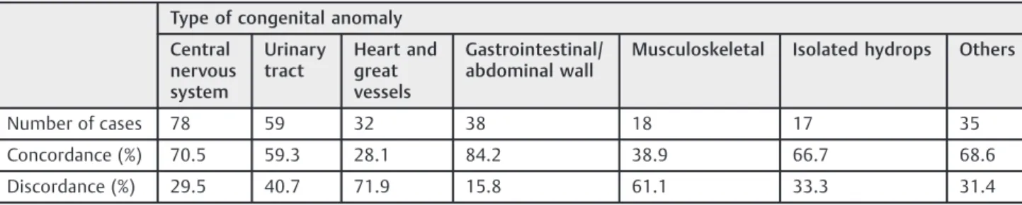
►Table 2shows the concordance between the ultrasound
levels I III performed by a Maternal-Fetal Medicine specialist.
Table 1 Maternal demographic variables taking into account different groups of congenital anomalies
Type of congenital anomaly p
Central nervous system (%)
Urinary tract (%)
Heart and great vessels (%)
Gastrointestinal/ abdominal wall (%)
Musculoskeletal (%)
Isolated hydrops (%)
Others (%)
Maternal age (meanSD)
25.556.0 26.246.6 27.237.0 23.327.8 24.505.8 28.475.1 28.077.4
19 years 14.1 18.6 12.5 28.9 27.8 0.0 8.6 NS
20–35 years 78.2 69.5 68.7 63.2 66.7 93.3 71.4
>35 years 7.7 11.9 18.8 7.9 5.5 6.7 20.0
Skin color
White 88.5 79.7 75.0 76.3 72.2 86.7 77.1 NS
Non-white 11.5 20.3 25.0 23.7 27.8 13.3 22.9
Level of education
8 years 44.9 61.0 34.4 60.5 55.6 26.7 40.0 0.041
>8 years 55.1 39.0 65.6 39.5 44.4 73.3 60.0
Occupation
Without 53.8 67.8 43.7 65.8 72.2 40.0 51.4 NS
With 46.2 32.2 56.3 34.2 27.8 60.0 48.6
Chronic diseases
Yes 15.4 20.3 12.5 13.2 22.2 0.0 25.7 NS
No 84.6 79.7 87.5 86.8 77.8 100.0 74.3
Smoking
Yes 11.5 6.8 9.4 18.4 27.8 20.0 2.8 NS
No 88.5 93.2 90.6 81.6 72.2 80.0 97.2
Body mass index (kg/m2)
Normal 56.4 54.3 62.5 52.6 44.4 40.0 42.9 NS
Overweight 24.3 28.8 28.1 34.2 44.4 40.0 40.0
Obese 19.3 16.9 9.4 13.2 11.2 20.0 17.1
Medication use
Yes 14.1 8.5 9.4 10.5 5.6 6.7 14.3 NS
No 85.9 91.5 90.6 89.5 94.4 93.3 85.7
Folic acid supplementation
Yes 32.1 25.4 37.5 13.2 38.9 20.0 31.4 NS
No 67.9 74.6 62.5 86.8 61.1 80.0 68.6
Parity
Primigravida 46.2 35.6 31.2 42.1 38.9 33.3 37.1 NS
Secundigravida 28.2 28.8 21.9 23.7 22.2 46.7 31.4
Multigravida 25.6 35.6 48.9 34.2 38.9 20.0 31.5
Previous miscarriage
Yes 14.1 20.3 21.9 26.3 22.2 33.3 22.9 NS
No 85.9 79.7 78.1 73.7 77.8 66.7 77.1
The highest concordance occurred in the GI defect group (84.2%), and the lowest concordance was detected in the HGV defect group (28.1%).
Multiple logistic regression analyses were applied to deter-mine the effects of maternal parameters on the prevalence of a
specific type of CA. Non-white skin color decreased the risk of
CNS anomalies by nearly 60% (OR: 0.43; 95%CI: 0.19–0.97;
p¼0.04). High levels of education decreased the risk of UT
defects by almost 50% (OR: 0.52; 95%CI: 0.29–0.94;p¼0.03).
Primigravida showed a reduced risk of having a newborn with
HGV defects (OR: 0.26; 95%CI: 0.08–0.80;p¼0.02). In addition,
maternal age>19 years and regular folic acid supplementation
were associated with decreased risk of GI malformations by
nearly 60% (OR: 0.42; 95%CI: 0.19–0.95; p¼0.04) and 65%
(OR: 0.34; 95%CI: 0.13–0.91;p¼0.03) respectively. Smoking
increased the risk of ME anomalies by 3 times (OR: 3.28; 95%CI:
1.08–9.90;p¼0.04). Furthermore, history of previous
miscar-riage increased the risk of hydrops by almost 8 times (OR: 7.65;
95%CI: 1.40–41.66;p¼0.02).
►Table 3shows the perinatal outcomes considering
differ-ent CA groups. The following perinatal complications were associated with CAs: polyhydramnios; oligohydramnios; still-birth; preterm delivery; cesarean section; low birth weight; need for pressure support; neonatal infection; need for surgi-cal treatment; and early neonatal death. Polyhydramnios was
more common in the ME and hydrops groups specifically,
with a prevalence of 38.9% (OR: 4.46; 95%CI: 1.09–18.29) and
40% (OR: 4.67; 95%CI: 1.07–20.32) respectively. On the other
hand, oligohydramnios was more common in the UT
malfor-mation group (OR: 12.55; 95%CI: 1.58–99.38). The prevalence
of stillbirth was high in all CA groups, mainly in the hydrops
(OR: 27.13; 95%CI: 2.90–253.47). Preterm delivery was very
common in all CA groups (18.7–86.7%), especially in the GI
defects (OR: 5.96; 95%CI: 1.99–17.84) and hydrops groups
(OR: 28.16; 95%CI: 4.98–159.38). The prevalence of low birth
weight was high in all CA groups, mainly in the GI defects
group (OR: 2.08; 95%CI: 1.08–27.83), the ME anomalies group
(OR: 3.34; 95%CI: 1.34–38.52) and in the others group (OR:
Table 2 Concordance between the ultrasound scan performed by non-specialists (level I) and Maternal-Fetal Medicine specialists (level III)
Type of congenital anomaly
Central nervous system
Urinary tract
Heart and great vessels
Gastrointestinal/ abdominal wall
Musculoskeletal Isolated hydrops Others
Number of cases 78 59 32 38 18 17 35
Concordance (%) 70.5 59.3 28.1 84.2 38.9 66.7 68.6
Discordance (%) 29.5 40.7 71.9 15.8 61.1 33.3 31.4
Table 3 Perinatal outcomes considering different groups of congenital anomalies
Type of congenital anomaly p
Central nervous system (%)
Urinary tract (%)
Heart and great vessels (%)
Gastrointestinal/ abdominal wall (%)
Musculoskeletal (%)
Isolated hydrops (%)
Others (%)
Fetal growth restriction 3.8 1.7 6.3 2.6 11.1 6.7 14.3 NS
Fetal distress 2.6 1.7 3.1 5.3 0 0 14.3 NS
Premature rupture of membranes
10.3 11.9 3.1 18.4 22.2 20.0 17.1 NS
Polyhydramnios 10.3 5.1 12.5 13.2 38.9 40.0 20.0 <0.01
Oligohydramnios 3.8 28.8 3.1 13.2 5.6 13.3 11.4 <0.01
Fetal death 9.0 6.8 3.1 7.9 11.1 46.7 8.6 <0.01
Preterm delivery 23.1 33.9 18.7 57.9 33.3 86.7 31.4 <0.01
Cesarean section 75.6 39.0 81.3 84.2 72.2 46.7 60.0 <0.01
Low birth weight 24.4 20.3 21.9 42.1 66.7 40.0 42.9 <0.01
Apgar<7 at the 1st minute 35.2 34.5 29.0 40.0 68.7 62.5 43.7 NS
Apgar<7 at the 5th minute 12.7 27.3 6.5 20.0 37.5 50.0 21.9 0.02
Need for assisted ventilation 43.7 47.3 38.7 71.4 68.7 62.5 56.3 NS
Neonatal infection 19.7 10.9 48.4 48.6 25.0 12.5 21.9 <0.01
Need for surgical treatment 54.9 27.3 61.3 91.4 25.0 37.5 25.0 <0.01
Early neonatal death 12.7 25.5 41.9 37.1 62.5 50.0 28.1 <0.01
1.98; 95%CI: 1.05–7.25). Additionally, Apgar score<7 at the
5th minute was high in all types of CA, especially in the UT
malformations group (OR: 5.44; 95%CI: 1.15–25.64), the ME
anomalies group (OR: 8.70; 95%CI: 1.51–50.28) and the
hydrops group (OR: 14.50; 95%CI: 1.98–106.44). Infections
were less prevalent in the CNS defects group (OR: 0.26; 95%CI:
0.11–0.65), as well as in the UT anomalies group (OR: 0.13;
95%CI: 0.04–0.39) and in the others group (OR: 0.30; 95%CI:
0.10–0.89). Surgeries were less necessary in the UT anomalies
group (OR: 0.24; 95%CI: 0.09–0.60), in the ME group (OR: 0.21;
95%CI: 0.06–0.81), and in the others group (OR: 0.21; 95%CI:
0.07–0.62). In contrast, it was more common in the GI group
(OR: 6.74; 95%CI: 1.68–26.96). Early neonatal death rate was
high in all CA groups; however, it was significantly
less common in the CNS defects group (OR: 0.20; 95%CI:
0.07–0.55).
►Table 4shows the hospitalization time of all CA groups.
In general, the hospitalization time was higher in the
sub-groups of GI anomalies and HGV defects (p<0.01).
Hospi-talization time was also higher in the subgroups in which surgery was required or had neonatal infection. On the other hand, hospitalization time was lower in the subgroups with
Apgar scores<7 at the 5th minute, probably because of
their high mortality rate.
Discussion
In the present study, the prevalence of CAs was of 2.4%,
considering 10 thousand ultrasound scans performed at
our institution between 2011 and 2013. This rate is similar to
the one from Dolk et al,12who described the prevalence of CAs
in Europe. Central nervous system anomalies, including open neural tube defects, were the most common CA detected in
fetuses, afinding similar to those reported on other studies.
The majority of the CAs of the fetal CNS is identified by the
second-trimester ultrasound at 20–24 weeks of gestation,
which makes them the most common.24Furthermore, CNS
defect was a CA group with high concordance (71%) between ultrasound scans performed by non-specialists (level I) and Maternal-Fetal Medicine specialists (level III).
Urinary tract malformation constitutes20% of all CAs,
which is coincident with the data presented here.18
Howev-er, the concordance between the diagnoses made by sonog-raphers with different levels of experience is lower than CNS anomalies. A possible explanation for this result would be that up to 80% of UT malformations can be solved spontane-ously during fetal life, or worsened with advancing
gesta-tional age and impaired fetal renal function.25 Multicystic
dysplasia might not be identified in the second trimester
Table 4 Hospitalization time of live newborns according to the congenital anomaly group and their perinatal outcomes
Hospitalization time (days) p
n Mean Standard deviation Minimum Median Maximum
Congenital anomaly
Central nervous system 71 24.9 31.9 1.0 14.0 201.0
Heart and great vessels 31 28.3 35.5 1.0 12.0 130.0 <0.01§
Gastrointestinal/ abdominal wall 35 34.5 36.2 1.0 26.0 160.0
Urinary tract 55 12.8 27.2 1.0 3.0 150.0
Isolated hydrops 8 9.9 12.6 1.0 5.0 36.0
Musculoskeletal 16 20.9 42.4 1.0 4.0 152.0
Others 32 24.8 51.1 1.0 9.0 274.0
Early neonatal death
No 176 28.0 37.5 1.0 14.0 274.0 <0.01
R
Yes 72 11.6 27.9 1.0 1.0 160.0
Need for surgical treatment
No 128 10.1 26.5 1.0 4.0 274.0 <0.01
R
Yes 120 37.3 38.9 1.0 24.0 201.0
Neonatal infection
No 184 14.3 23.7 1.0 6.0 150.0 <0.01
R
Yes 64 49.0 49.7 1.0 33.5 274.0
Apgar score at the 5th minute
<7 50 19.4 47.6 1.0 1.0 274.0 <0.01
R
7 198 24.2 32.1 1.0 12.0 201.0
Number of live newborns;
§Kruskal-Wallis test; R
scan; on the other hand, renal agenesis and lower urinary
tract obstruction can be identified early, while milder
obstructions are diagnosed later.26
In the present study, the prevalence of congenital heart
disease was of 11.6%, which is in agreement with thefindings
of studies conducted in tertiary reference centers. However, the analysis of this CA group showed the lowest concordance (28.1%) between ultrasound scans level I and III. This can be
explained by the difficulty of non-specialist sonographers in
achieving a proper examination of the fetal heart and great vessels. It is well known that the detection rates of HGV
defects increase with the examiner’s ultrasound experience
and training, and with the adoption of a systematic
ultra-sound examination of the fetal heart.27,28
The prevalence of GI malformations was coincident with
the data presented in other studies (13.8%).29Furthermore, GI
anomaly was the CA group with the highest concordance
(84%) betweenfindings of ultrasound scans level I and III. The
most common GI malformations were abdominal wall defects,
which can be easily identified by ultrasound scan performed
after 13 weeks of gestation. In addition, esophageal atresia and
small bowel obstructions are readily identified in the third
trimester due to the presence of polyhydramnios.
Because of the multifactorial etiology of CAs, we proposed to assess the effect of maternal demographic factors on their occurrence; however few factors showed to have a positive correlation with CAs. This result is probably due to the small sample size. Moreover, the appropriate process of gathering and measuring information on targeted variables, such as skin color, smoking, use of medications and folic acid sup-plementation is not very reliable because of two main reasons: the occurrence of a mixed population in our coun-try, and the low socioeconomic status of our patients.
According to our data, parity was a maternal risk factor for HGV defects. Multigravida have a higher risk of having children affected by this type of CA compared with primigravida or
secundigravida. Thisfinding is similar to the Csermely et al30
study that assessed 21,494 fetuses with different isolated malformations, and compared them to 34,311 normal
con-trols. The authors showed that multiparity was a significant
risk factor for the following five types of HGV anomalies:
ventricular septal defect; ostium secundum atrial septal de-fect; persistence of arteriosus ductus; conotruncal cardiac
defect; and ventricular outflow tract obstructions.
Smoking was a risk factor for ME anomalies. Overall, the prevalence of CAs does not seem to be increased among children of women who smoked during pregnancy. However,
Morales-Suárez-Varela et al31 demonstrated that pregnant
women who did not smoke but used nicotine patches had the
risk of having children with ME anomalies (95%CI: 1.53–4.52)
increased by 2.6 times. The authors suggested that nicotine can interfere with the mechanism of genomic "imprinting"
and lead to this type of CA. Anotherfinding of the present
study was the history of previous miscarriage as a risk factor for hydrops. It is well known that a large proportion of miscarriages is caused by genetic abnormalities, and their recurrence could be one of the causes of hydrops in the
current pregnancy.32
There were two variables that provided protection against
CAs. Maternal age>19 years and folic acid supplementation
were associated with a decreased risk of GI malformations.
Eckmann-Scholz et al33found that teenagers have an increased
risk of having newborns with GI malformations, especially gastroschisis. This can be explained by the fact that pregnancy during adolescence may be associated with several risk con-ditions for CAs, such as use of illicit drugs, alcoholism, and
nutritional deficiencies.33Many studies point out to the
effec-tive prevention of fetal CAs with the regular folic acid
supple-mentation mainly open neural tube defects.12,34
Changes of amniotic volumefluid were more frequent in
fetuses with skeletal dysplasia and isolated hydrops. In skeletal malformations, a small thorax causes increased intrathoracic
pressure and decreased fetal swallowing.32 In hydropic
fetuses, polyhydramnios may be a consequence of increased
urine production.32In contrast, oligohydramnios was more
common in the UT group, in which urine production is impaired by the existence of dysplastic kidneys or distal
obstructions of the UT.26
In the present study, fetal death rates were high for all types of CAs compared with the general population due to the
severity of malformations.35 In addition, preterm delivery
rates were very high mainly for the GI anomalies and hydrops groups because of spontaneous labor caused by polyhydram-nios or suspicion of ischemic bowel and compromised fetal wellbeing respectively. As a consequence, the elective cesarean section rates were also increased for those reasons, and also to obtain a successful perinatal management of neonates through delivery planning with a multi-professional team.
Neonatal adverse outcomes were extremely common in all CA groups. Hospitalization time was increased for all of them as a result of preterm delivery, low birth weight, Apgar score
<7 at the 5th minute, neonatal infection, and need for surgical
treatment. Neonatal death rates were significantly increased
in all CA groups as a consequence of all perinatal complications previously described. The causes behind the low Apgar scores
may be listed as difficult fetal extraction at the cesarean
section or labor dystocia in skeletal dysplasia and hydrops; respiratory distress due to pulmonary hypoplasia, particularly in UT anomalies linked to oligohydramnios; and the presence
of a small thorax or a large pleuralfluid collection possible in
ME CAs and hydrops respectively. Neonatal infections were more common among fetuses with HGS or GI anomalies because they usually require surgical treatments, blood vessel catheterization for parenteral nutrition, blood transfusions,
and medications orfluid administration.
In summary, it was possible to identify several maternal risk factors for CAs. High rates of adverse perinatal outcomes were presented in all CA groups, and may differ according to the type of CA considered.
References
1 Liu X, Roth J. Development and validation of an infant morbidity
index using latent variable models. Stat Med 2008;27(7):971–989
2 Mattison DR. Environmental exposures and development. Curr
3 Brent RL. Environmental causes of human congenital
malforma-tions: the pediatrician’s role in dealing with these complex clinical problems caused by a multiplicity of environmental and genetic factors. Pediatrics 2004;113(4, Suppl)957–968
4 Jentink J, Dolk H, Loane MA, et al; EUROCAT Antiepileptic Study
Working Group. Intrauterine exposure to carbamazepine and specific congenital malformations: systematic review and case-control study. BMJ 2010;341:c6581
5 Little J, Cardy A, Arslan MT, Gilmour M, Mossey PA. Smoking and
orofacial clefts: a United Kingdom-based case-control study. Cleft Palate Craniofac J 2004;41(4):381–386
6 Boix H, Ortega-Aznar A, Vazquez E, Salcedo S, Roig-Quilis M.
Brainstem dysgenesis in an infant prenatally exposed to cocaine. Pediatr Neurol 2010;42(4):295–297
7 Wattendorf DJ, Muenke M. Fetal alcohol spectrum disorders. Am
Fam Physician 2005;72(2):279–282, 285
8 Zhao Z, Reece EA. Experimental mechanisms of diabetic
embry-opathy and strategies for developing therapeutic interventions. J Soc Gynecol Investig 2005;12(8):549–557
9 Gilbert-Barness E. Teratogenic causes of malformations. Ann Clin
Lab Sci 2010;40(2):99–114
10 Marecki MA, Bozzette M. Infections in the perinatal period.
J Perinat Neonatal Nurs 2008;22(3):173–174
11 Oster ME, Riehle-Colarusso T, Correa A. An update on
cardiovas-cular malformations in congenital rubella syndrome. Birth De-fects Res A Clin Mol Teratol 2010;88(1):1–8
12 Dolk H, Loane M, Garne E. The prevalence of congenital anomalies
in Europe. Adv Exp Med Biol 2010;686:349–364
13 Arroll N, Sadler L, Stone P, Masson V, Farquhar C. Can we improve
the prevention and detection of congenital abnormalities? An audit of early pregnancy care in New Zealand. N Z Med J 2013; 126(1380):46–56
14 Ruano R, Ali RA, Patel P, Cass D, Olutoye O, Belfort MA. Fetal
endoscopic tracheal occlusion for congenital diaphragmatic her-nia: indications, outcomes, and future directions. Obstet Gynecol Surv 2014;69(3):147–158
15 Gajewska-Knapik K, Impey L. Congenital lung lesions: Prenatal
diagnosis and intervention. Semin Pediatr Surg 2015;24(4):156–159 16 Sanapo L, Moon-Grady AJ, Donofrio MT. Perinatal and delivery
management of infants with congenital heart disease. Clin Peri-natol 2016;43(1):55–71
17 Al-Matary A, Ali J. Controversies and considerations regarding the
termination of pregnancy for foetal anomalies in Islam. BMC Med Ethics 2014;15:10
18 Grandjean H, Larroque D, Levi S. The performance of routine
ultrasonographic screening of pregnancies in the Eurofetus Study. Am J Obstet Gynecol 1999;181(2):446–454
19 Stoll C, Tenconi R, Clementi M. Detection of congenital anomalies
by fetal ultrasonographic examination across Europe. Community Genet 2001;4(4):225–232
20 European Surveillance of Congenital Anomalies (EUROCAT)
[Inter-net]. Prenatal screening and diagnosis: prenatal detection rates. 2016 [cited 2016 May 23]. Available from:
http://www.eurocat-network.eu/prenatalscreeninganddiagnosis/prenataldetection(pd) rates
21 Hadlock FP, Harrist RB, Martinez-Poyer J. In utero analysis of fetal
growth: a sonographic weight standard. Radiology 1991;181(1): 129–133
22 Magann EF, Sanderson M, Martin JN, Chauhan S. The amniotic
fluid index, single deepest pocket, and two-diameter pocket in normal human pregnancy. Am J Obstet Gynecol 2000;182(6): 1581–1588
23 Hosmer DW, Lemeshow S. Applied logistic regression. 2nd ed.
New York: John Willey & Sons; 2000
24 Population Screening Programmes. NHS Fetal Anomaly Screening
Programme (FASP) [Internet]. 2016 [cited 2016 May 23]. Avail-able from: https://www.gov.uk/topic/population-screening-pro-grammes/fetal-anomaly
25 Sairam S, Al-Habib A, Sasson S, Thilaganathan B. Natural history of
fetal hydronephrosis diagnosed on mid-trimester ultrasound. Ultrasound Obstet Gynecol 2001;17(3):191–196
26 Dias T, Sairam S, Kumarasiri S. Ultrasound diagnosis of fetal renal
abnormalities. Best Pract Res Clin Obstet Gynaecol 2014;28(3): 403–415
27 Tegnander E, Eik-Nes SH. The examiner’s ultrasound experience
has a significant impact on the detection rate of congenital heart defects at the second-trimester fetal examination. Ultrasound Obstet Gynecol 2006;28(1):8–14
28 Pinto NM, Nelson R, Puchalski M, Metz TD, Smith KJ.
Cost-effectiveness of prenatal screening strategies for congenital heart disease. Ultrasound Obstet Gynecol 2014;44(1):50–57 29 Barisic I, Clementi M, Häusler M, Gjergja R, Kern J, Stoll C;
Euroscan Study Group. Evaluation of prenatal ultrasound diagno-sis of fetal abdominal wall defects by 19 European registries. Ultrasound Obstet Gynecol 2001;18(4):309–316
30 Csermely G, Susánszky É, Czeizel AE, Veszprémi B. Possible
association of first and high birth order of pregnant women with the risk of isolated congenital abnormalities in Hungary -a popul-ation-b-ased c-ase-m-atched control study. Eur J Obstet Gynecol Reprod Biol 2014;179:181–186
31 Morales-Suárez-Varela MM, Bille C, Christensen K, Olsen J.
Smok-ing habits, nicotine use, and congenital malformations. Obstet Gynecol 2006;107(1):51–57
32 Bellini C, Hennekam RC. Non-immune hydrops fetalis: a short
review of etiology and pathophysiology. Am J Med Genet A 2012; 158A(3):597–605
33 Eckmann-Scholz C, von Kaisenberg CS, Alkatout I, Jonat W,
Rajabi-Wieckhorst A. Pathologic ultrasoundfindings and risk for con-genital anomalies in teenage pregnancies. J Matern Fetal Neonatal Med 2012;25(10):1950–1952
34 Youngblood ME, Williamson R, Bell KN, Johnson Q, Kancherla V,
Oakley GP Jr. 2012 Update on global prevention of folic acid-preventable spina bifida and anencephaly. Birth Defects Res A Clin Mol Teratol 2013;97(10):658–663
35 Goldenberg RL, Kirby R, Culhane JF. Stillbirth: a review. J Matern