Immunophenotype of lung granulomas in HIV and
non-HIV associated tuberculosis
Andre´ia A. Silva,IThais Mauad,IIPaulo H.N. Saldiva,IIRuy C. Pires-Neto,IIRicardo D. Coletta,IEdgard Graner,I Pablo A. VargasI
IOral Pathology, Oral Diagnosis Department, Piracicaba Dental School, University of Campinas, Piracicaba-SP, BrazilIILaboratory of Pollution (LIM05),
Department of Pathology, School of Medicine of Sa˜o Paulo-University of Sa˜o Paulo, Sa˜o Paulo-SP, Brazil
OBJECTIVE: To describe the immunophenotype of pulmonary TB granulomas from autopsied patients with tuberculosis (TB group) and from HIV patients with tuberculosis (TB/HIV group), and to identify the
Mycobacteriumspecies using polymerase chain reaction (PCR) technique.
METHODS:Lung samples of 15 TB group patients and 23 TB/HIV group patients were selected. Histopathologic analyses and immunohistochemistry tests were performed to describe the granulomas and to detect the infectious agent (anti-BCG). CD4, CD8, CD20 and CD68 were evaluated to characterize the immnophenotype of the granulomas. Polymerase Chain Reaction was performed to identify the mycobacterium species.
RESULTS:CD4þT lymphocytes were the cells with highest density in the TB group, whereas CD68 cells exhibited the highest density in the TB/HIV group. Comparison between groups showed that the CD4þT density was significantly higher in the TB patients; whereas, CD68 density was significantly higher in the TB/HIV patients.
M. tuberculosiswas identified in 8 cases of each group;M. aviumwas only found in one case of the TB/HIV group.
CONCLUSION:With the advent of AIDS, the immunological profile of TB has changed. This may be associated with the depletion of CD4þT lymphocytes in lung granulomas.M. tuberculosiswas the major etiological agent of TB in both groups.
KEYWORDS: tuberculosis; HIV; AIDS; autopsy.
Silva AA, Mauad T, Saldiva PHN, Pires-Neto RC, Coletta RD, Graner E, Vargas PA. Immunophenotype of lung granulomas in HIV and non-HIV associated tuberculosis. MEDICALEXPRESS. 2014 Aug;1(4):174-179.
Received for publication onApril 28 2014;First review completed onMay 5 2014;Accepted for publication onMay 14 2014
Email: [email protected]
B INTRODUCTION
Tuberculosis (TB) is responsible for 2 million deaths globally each year. Overall, the World Health Organization estimates that one-third of the world’s population is currently infected with the Mycobacterium tuberculosis bacillus without demonstrable symptoms.1Despite
success-ful efforts in treatment, TB remains a pandemic disease, linked to poverty, dynamic population migration, and, especially, to the HIV co-pandemic.2
Before the HIV pandemic, 80 to 85% of new reported cases of TB referred to the isolated pulmonary disease.3,4Today, millions of people are living with AIDS and TB in developing and developed countries.5HIV has been responsible for the
resurgence of TB over the past decades in several countries.6 Compared to non-HIV infected persons, the probability of developing active TB is 100 times higher in individuals with HIV co-infection. In addition, TB co-infection accounted for almost 25% of global AIDS-related mortality in 2007.7The
first AIDS case registered in Brazil was in 1981.8Co-infection
rates of HIV/TB have been reported as being as high as 51%
in mortality surveillance systems in Rio de Janeiro, Brazil.9 More recently, a study in Rio de Janeiro showed that 18.5% of the AIDS patients were co-infected with TB during the period 1995–2004.10
Complex host-pathogen interactions occur during TB infection and the development of a latent/active disease, which seem to be tied to the condition of the host immune system. It is well established that HIV impairs the ability to control TB infection; HIV infection impairs the formation of granulomas and therefore the capability to restrain disease. The mechanisms by which HIV impairs the immune response to TB are likely to be several: 1) increase in viral load within the tissue; 2) decrease in the total number of CD4þT lymphocites; 3) impairment in macrophage func-tion; 4) changes to the T-cell responses (e.g. cytokine production) to the mycobacterium.4,5,11 – 13
Most of our knowledge on lung immune responses in patients with TB and AIDS are derived from studies which come from blood, bronchoalveolar lavage cells and animal models.13Few studies compared the immunophenotype of lung granulomas of patients with and without AIDS. The available studies describe a different cytokine pattern in the granulomas of patients with AIDS,14 – 16but few studies described the profile of immune cells within these lesions. DOI:10.5935/MedicalExpress.2014.04.03
Understanding lung tissue-based reactions is important because less invasive approaches may, or may not, mirror the local pulmonary immune reactions.15
Therefore, the aim of this work was to describe the immunophenotype of the granulomas of autopsied patients with confirmed TB with and without HIV co-infection. The diagnosis and identification ofmycobacteriumspecies of TB was confirmed by immunohistochemistry and polymerase chain reaction (PCR).
B MATERIAL AND METHODS
The Ethics Committees of the Sa˜o Paulo University Medical School and of the Dental School of Campinas University approved the use of the autopsy samples for the present study (protocol n. 440/05).
Patient population
We selected 38 cases with the autopsy diagnosis of tuberculosis in the lungs affecting non-HIV and HIVþpatients from University Hospital of the Sa˜o Paulo Medical School between 1974 and 2004. Cases were divided in two groups: patients with TB affecting the lungs before the HIV period (1974 to 1980) (TB group) and patients showing concomitant TB/HIVþ(1981 to 2004) with lung involvement (TB/HIV group).
The clinical and autopsy records were revised in order to obtain information regarding age, gender, basis disease and cause of death. In the TB/HIV group, we obtained results of laboratory tests including CD4þT and CD8þ T cells blood counts and viral titer, when available.
Histopathological and immunohistochemical analysis
Archival paraffin blocks where retrieved and new sections were stained for Hematoxylin and Eosin (H&E) and Ziehl-Neelsen (ZN). Immunohistochemistry was performed to detect the Mycobacterium genus with the BCG anti-body (1/100.000, Dako Dako Corporation, Glostrup, A/S Denmark), and to characterize the immunophenotype of the inflammatory cells around the granulomas. The following antibodies were used: CD4 (1/200, Dako Corporation, Glostrup, A/S Denmark), CD8 (1/200, Dako Corporation, Glostrup, A/S Denmark) CD20 (1/10.000, Dako Corpor-ation, Glostrup, A/S Denmark) and CD68 (1/400, Dako Corporation, Glostrup, A/S Denmark).
Briefly, sections were deparaffinized, rehydrated, and treated with 3% hydrogen peroxide to inhibit endogenous peroxidase. For antigen retrieval, the specimens were incubated with citrate buffer, pH 6, in a microwave, followed by incubation with the primary antibodies for 16 h, and then the secondary antibodies conjugated with streptavidin-biotin-peroxidase (Strept ABComplex/HRP Duet, Mouse/
Rabbit, Dako A/S,Glostrup, Denmark). Reactions were developed with diaminobenzidine chromogen and counter-stained with Carazzi’s hematoxylin. Positive (lymph node with bacilli) and negative (pulmonary carcinoma) controls were performed.
Positive immunomarked cells were counted in 10 fields of 0.21 mm2each in the peripheral areas of the granulomas. The area of the field was determined with the aid of the software System KS-400, 2.1 Carl Zeiss (Carl Zeiss, Mu¨nchen, Germany). We tried to analyze the same granuloma for all antibodies. Data were presented as the median (ranges) of cells/field. BCG staining was scored as positive or negative.
Identification and characterization of the Mycobacterium species
The DNA extractions and Nested-PCR with specific primers for M. tuberculosis and M. avium diagnosis were performed according the methods described by Rangel et al, as shown in Table 1.16Ten cases from group TB and ten
from group TB/HIV were selected based on an intense positivity in the ZN stain for nested-PCR.
Statistical analysis
Data distributions were verified with Kolmogorov-Smirnov test. Student’s t-test or Mann-Whitney test was performed according to data distribution and expressed as mean^SD or median/ranges, respectively. The differences were considered significant with p,0.05. Correlations were performed using the Spearman test. The software SPSS 15.0 (Chicago, USA) was used for the analyses.
B RESULTS Study population
Fifteen patients (10 males) were studied in the TB group. The median age was 33.5 years, ranging from 1 to 62 years. The basicdiseases were tuberculosis (n¼7), alcoholism (n¼2), Systemic lupus erythematosus with corticoid therapy (n¼2), epilepsy (n¼1), adenocarcinoma of the colon (n¼1), hydrocephaly (n¼1), and malnutrition (n¼1). The causes mortis were milliary TB (n¼9), pulmonary TB (n¼4), and pulmonary TBþ neural TB (n¼2) as shown in Table 2.
Twenty-three patients (18 males) comprised the TB/HIV group. The median age was 36.0 years, ranging from 17 to 62 years. CD4 levels were obtained in 17 patients, with a mean of 25 cells/ml^SD of 81^129 cells/ml (ranging from 4 to 514 cells/ml). Eighty percent of the patients had less than 200 cells/ml. The CD4/CD8 ratio was 0.53^0.25 (ranging from 0.1 to 0.98 cells/ml). The causes of death were milliary TB (n¼21), pulmonary TB (n¼1), and disseminated lymphoma þpulmonary TB (n¼1), as shown in Table 2. There were no
Table 1 -Primer sequences and product sizes used in this study
Primer Forward (50– 30) Reverse (50– 30) Product size (bp)
PCR1
Outer primer CGGGACCACCCGCGGCAAAGCCCGCAGGAC CATCGTGGAAGCGACCCGCCAGCCCAGGAT 220
Inner primer CCTGCGAGCGTAGGCGTCGG CTCGTCCAGCGCCGCTTCGG 123
PCR 2 TGGACCAGTCTGCCTT CGTCGATCAAGGCTTGGTAG 106
statistical differences in age and gender between groups. Demographic and clinical information of both study groups are presented in Table 2.
Histopathological findings
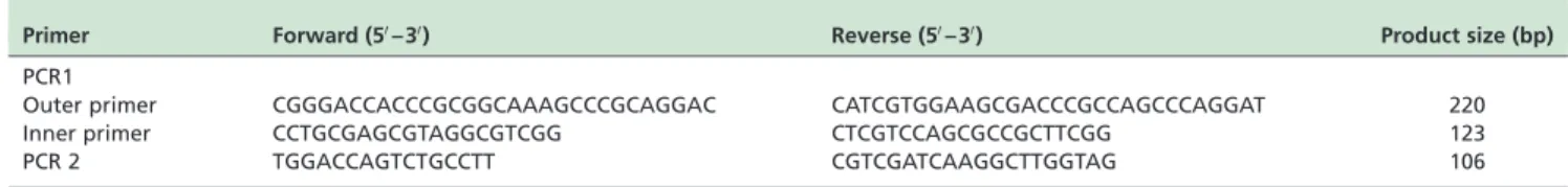
Hematoxilin & eosin analysis showed that the pulmonary parenchyma was affected in all cases by granulomas in different stages of composition with extensive areas of caseous necrosis. In all cases, Ziehl-Neelsen (ZN) and/or Bacillus Calmette-Gue´rin (BCG) staining revealed bacilli within the granulomas (Fig. 1).
TB group
Fourteen out of the 15 cases presented classic, well formed granulomas with giant cells, comprising a nodular structure with a central area of necrosis surrounded by lymphocytes and Langhans type giant cells. A single case of a patient diagnosed with Systemic lupus erythematosus receiving corticosteroids presented a poorly formed granuloma characterized by a large area of caseous necrosis and a few inflammatory cells. Thirteen of the fifteen cases showed variable ZN positivity for acid fast bacilli, and all cases were positive for the BCG antibody by immunohistochemistry.
TB/HIV group
Twenty cases showed poorly-formed granulomas with large areas of necrosis and cell debris, very few giant cells and lymphocytes. Only three cases had better formed granulomas. In general, most cases had larger number of bacilli (Fig. 1). Four cases were negative for ZN staining but positive for the BCG antibody. One case showed co-infection by TB and cytomegalovirus.
Immunohistochemical findings
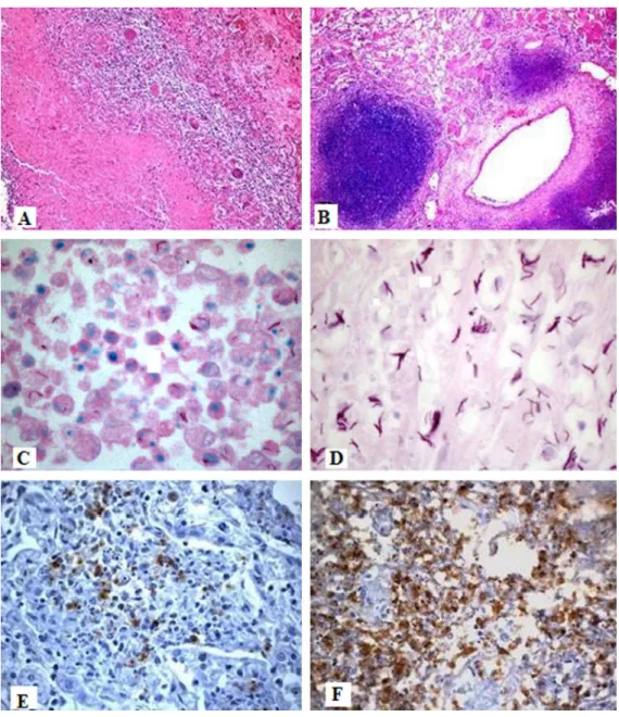
The cells with the highest density in the TB group were the CD4þT lymphocytes, followed by CD8þT lymphocytes, CD68þ macrophages, and CD20þB lymphocytes (Fig. 2). In the AIDS/HIV group the cells with the highest density were the macrophages, followed by CD4þT lymphocytes, CD8þT lymphocytes, and CD 20þB lymphocytes.
When both groups were compared, there was a higher density of CD4þT cells in the group TB than in the TB/HIV group (p¼0.001). On the other hand, macrophages density was significantly higher in patients with TB/HIV (p¼0.002) (Fig. 2). There were no significant differences for CD8þT cells (TB¼12.0þ10.47; TB/HIV ¼17.3þ11.64; p¼0.408) and CD20þB cells (TB¼4.74þ6.0; TB/HIV¼2.25þ4.67; p¼0.064).
There were no correlations between CD4 cell counts or CD4/CD8 ratio in the blood and tissue: CD4þT cell (p¼0.086/r¼0.443; p¼0.233/r¼0.356 respectively) and CD68þ macrophages (p¼0.510, r¼0.178; p¼0.593, r¼0.164 respectively) in the TB/HIV group.
Polymerase chain reaction (PCR)
From both groups, eight out of ten cases gave a positive reaction for M. tuberculosis DNA, as depicted in Fig. 3. M. aviumwas detected in a single case of the group TB/HIV (Fig. 4), which was also positivite for M. tuberculosis DNA (sample 7, Figures 3b and 4). In two cases from TB group and two cases from TB/HIV, no specific band could be detected.
B DISCUSSION
In this study we have shown that the immunopheno-type of the pulmonary lesions in patients with tuberculosis and patients with tuberculosis/HIVþ is different. The poorly formed granulomas in patients with TB/HIV have less CD4þT cells and more macrophages than the granulomas of patients with tuberculosis not associated to HIV.
Our results show that the co-infection with TB/HIV is related to major changes in the histological aspect of the granulomas with a larger number of bacilli being identified in the TB/HIV cases. These findings are in line with the idea that the cellular architecture of the granuloma is important to control bacillus replication at the onset of pulmonary tuberculosis.17 The present study showed that that HIV-1 Table 2 -Demographic and clinical data of TB and AIDS/TB groups
TB group (n¼15) AIDS/TB group (n¼23)
Gender (M/F) 10/5 18/5
Age* (years) 33.5(1 – 62) 36.04 (17 – 62)
CD4þT cells counts#(n¼17) 25.5 (4 – 514) cells/ml
CD4/CD8 counts#(n¼17) 0.53 (0.1 – 0.98) cells/ml
Viral title#(n¼4) 201.500 (920 – 1. 200.000) copies/ml
Basis disease
Tuberculosis 7
Alcoholism 2
SLE with corticoid therapy 2
Epilepsy 1
Adenocarcinoma of the colon 1
Hydrocephaly 1
Malnutrition 1
AIDS 23
Causes Mortis
Miliary TB 9 21
Pulmonary TB 4
Pulmonary TBþ neural TB 2
Disseminated lymphomaþ Pulmonary TB 1
infection alters composition of the pulmonary TB granulomas and is associated with a decreased tissue density of the CD4þT cells and an increase in CD68þ macrophages.17 – 20 The formation and maintenance of granulomas is a complex and dynamic process that requires the interplay of macrophages, CD4þT cells, and an array of cytokines/ chemokines, among which Tumor Necrosis Factor-alpha (TNF-a) seems to play a major role.13 The activation of
a/b T-cell receptor (TcR) expressing CD4þT cells is essential for the formation of granulomas during mycobac-terial infections.13
HIV infection is largely associated with the failure of Tcells to control infection. There are several lines of evidence linking depletion of both mucosal and peripheral CD4þT
cells caused by HIV infection to susceptibility to tuberculosis.13 – 15
Few studies have shown depletion of CD4þT cells at the level of the granuloma. It has been previously shown that there are less CD4þT cells within granulomas in the lymph nodes of patients with AIDS/TB, and in the lungs of monkeys infected with HIV/TB when compared to the granulomas caused by TB alone.11,21To our knowledge, the
current study is the first to show that, at the lung level, human HIV-associated lung granulomas are depleted of CD4þT cells. In addition, bronchoalveolar lavage of HIV-infected adults showed that CD4þT lymphocytes have impaired cytokine responses to virus and (myco) bacterial antigens.21
HIVþ patients in this study presented low CD4þT cell counts in blood, with 80% of them showing less than 200 cell/ml. The HIV population is more susceptible to TB than the general population, regardless of their CD4 level.21 However, HIVþ patients with lower CD4þT cell counts (,200 cell/ml) are in fact more susceptible to TB than HIVþ patients with CD4.500 cell/ml.15We could not detect any correlation between peripheral CD4 and granuloma CD4þT cells in our cases. The lack of correlation is probably related to the fact that most of the patients had CD4þT cell counts within a very low range.
We found a higher density of macrophages in the lung granulomas of the TB/HIV group. Macrophages are pivotal cells in modulating the response against mycobacterium, since alveolar macrophages are likely to be the first cells to be infected by HIV contact in the lungs.15 Macrophages are
infected and can be reservoirs of both HIV and M. tuberculosis. HIV may alter two important components of the responses to tuberculosis: apoptosis and secretion of TNF-a. In alveolar macrophages, HIV seems to decrease apoptosis, a last resort of infected TB macrophages.15 The decrease in HIV-induced apoptosis could explain the increase in macrophages observed in this study. However, these infected macrophages seem to be ineffective in the production of TNF-a, which contributes to failure to contain the infection in HIV patients.
Our study has limitations. We used retrospective autopsy material, with limited information concerning clinical and treatment aspects, especially in the older archival retrieved cases. Therefore, we are not aware of the immunological status of the patients in the TB group, such as the CD4 cells counts in blood. Our PCR results confirmed that M. Figure 3 -Nested polymerase chain reaction. Polyacrylamide gel at 8% stained with ethidium bromide of 10 cases of the TB (a) and
TB/HIV (b) group for amplification of IS6110 gene present in the genome ofMycobacterum tuberculosis(123-bp). M molecular weight
marker (100-bp), P control positive, samples from 1 to 10, N negative control.
Figure 4 -Polymerase chain reaction. Polyacrylamide gel at 8% stained with ethidium bromide for visualization of the nested PCR
reaction of 10 cases of mycobacteriosis group TB/HIV for amplification of IS1245 gene (106 pb) present in genome ofMycobacterium
avium. M molecular weight marker (100-bp), P control positive – sample standard IWGMT 49, PP – paraffin block, samples from 1 to 10
and N negative control.
Figure 2 -CD4þT and macrophage cell densities in the TB and TB/HIV groups. The density of CD4þT cells in the granuloma is higher in
the TB group (A and C) than in TB/HIV group (B). (D) The density of CD68þ macrophages in the granulomas is higher in the TB/HIV
tuberculosis is the major etiological agent of the TB/HIV co-infection in Brazil, as in other developing countries.22 – 25 Lack of PCR positivity in two samples that were intensively positive using other immunohistochemical methods shows that although it is possible to extract DNA from very old archival material, there are important limitations when using autopsy tissue to assess genetic material.26
B CONCLUSION
In summary, the present study showed that HIV infection changed the immunoprofile of lung TB granulomas. This probably contributes to pulmonary disease dissemination in these patients. AIDS and TB are the two most deadly infectious diseases worldwide. Together, these infections kill almost 4 million people every year, mostly in developing nations. Despite this, there is relatively little research on this deadly interaction,6,27 especially research describing the events occurring at tissue level.16Autopsy material may be an extremely useful tool to address this topic.28 Better
understanding of TB/HIV immunopathology may contri-bute to the development of novel disease biomarkers and preventive/therapeutic approaches.
B ACKNOWLEDGEMENTS
Supported by: This work was supported by the Coordination for the Improvement of Higher Educational (CAPES) and the National Council for Scientific and Technological Development (CNPq). There are no conflicts of interest.
B RESUMO
OBJETIVO:Descrever a imunofenotipagem de granulomas TB pulmonar de pacientes autopsiados com tuberculose (grupo TB) e de pacientes infectados pelo HIV com tuberculose (grupo TB/HIV) e identificar as espe´cies de Mycobacteriumem cadeia da polimerase utilizando reaca˜o PCR.
ME´TODOS:Foram selecionadas amostras pulmonares de 15 pacientes do grupo TB e 23 pacientes do grupo TB/HIV. Realizamos Histopatolo´gia e imuno-histoquı´mica para descrever os granulomas e para detectar o agente infeccioso (anti-BCG). Avaliamos CD4, CD8, CD20 e CD68 para caracterizar a imunofenotipagem dos granulomas e realizamos a reaca˜o PCR para identificar as espe´cies de micobacte´ria.
RESULTADOS:linfo´citos T CD4þ foram as ce´lulas com densidade mais
elevada no grupo de TB, ao passo que as ce´lulas CD68 exibiram maior densidade no grupo TB/HIV. A comparaca˜o entre os grupos mostrou que a densidade de linfo´citos T CD4þ foi significativamente maior nos pacientes com TB, ao passo que a densidade CD68 foi significativamente maior nos pacientes com TB/HIV. M. tuberculosis foi identificado em 8 casos de cada grupo; M. avium so´ foi encontrado em um caso do grupo TB/HIV. CONCLUSA˜ O:Com o advento da AIDS, o perfil imunolo´gico da tuberculose se alterou. Isto pode estar associado com a depleca˜o de linfo´citos T CD4þem
granulomas pulmonares. M. tuberculosis foi o principal agente etiolo´gico da tuberculose em ambos os grupos.
B REFERENCES
1. World Health Organization. Global tuberculosis control: WHO report 2010. Geneva: World Health Organization; 2010; p.7.
2. Schwander S, Dheda K. Human lung immunity against Mycobacterium tuberculosis: Insights into Pathogenesis and Protection. Am J Respir Crit Care Med. 2011;183(6):696-707.
3. Mello FA. Changes in the tuberculosis profile in Brazil: a new reality? J Bras Penumol. 2010;36(4):397-8.
4. Uemura M, Yahagi A, Hamada S, Begem D, Watanabe K, Kawakami K, et al., IL-17 mediated regulation of innate and acquired immune response against pulmonary mycobacterium bovis Bacilli Calmette-Gue´rin infec-tion. J Immunol. 2007;178(6):3786-96.
5. Johnson MD, Decker CF. Tuberculosis and HIV infection. Dis Mon. 2006;52(11 – 12):420-7.
6. Brust JCM, O´Donnell MR, Metcalfe JZ. TB/HIV: An orphan disease? Am J Respir Care Med. 2011;183(11):1441-2.
7. Bekker LG, Wood R. The changing natural history of tuberculosis and HIV co-infection in an urban area of hyperendemecity. Clin Infec Dis. 2010;50(Suppl 3):S208-14.
8. Goncalves AP, de Sa CA, Rubini N. HIV/AIDS infection. The Brazilian view. AIDS in Brazil. An R Acad Nac Med (Madr). 1996; (Spec No:145-56). 9. Miranda AE, Golub JE, Lucena FF, Maciel EM, Gurgel MF, Dietze R. Tuberculosis and AIDS co-morbity in Brazil linkage of the tuberculosis and AIDS database. Braz J Infect Dis. 2009;13(2):137-41.
10. Oliveira HB, Marı´n-Leo´n L, Cardoso JC. Differences in mortality profile of tuberculosis patients related to tuberculosis-AIDS co-morbidity. Rev Saude Publica. 2004;38(4):503-10.
11. Diedrich CR, Mattela JT, Klein E, Janssen CJ, Phuch J, Sturgeon TJ, et al., Reaction of latent tuberculosis in Cynomologus macaque infected with SIV is associated with early peripheral T cell depletion and not virus load. PlosOne. 2010;5(3):e9611.
12. Meya DB, McAdam KP. The TB pandemic: an old problem seeking solution. J Intern Med. 2007;261(4):309-29.
13. Sauders MS, Britton W. Life and death in the granuloma: immunopatho-logy of tuberculosis. Immunol Cel Biol. 2007;85(2):103-11.
14. Bai X, Wilson SE, Chmura K, Feldman NE, Chan ED. Morphometric analysis of Th1 and Th2 cytokine expression in human pulmonary tuberculosis. Tuberculosis. 2004;84(6):375-85.
15. Diedrich CR, Flynn JL. HIV-1/Mycobacterium tuberculosis coinfection immunology: How does HIV-1 exacerbate tuberculosis? Infect Immun. 2011;79(4):1407-17.
16. Rangel ALCA, Coletta RD, Almeida OP, Graner E, Lucena A, Saldiva PH, et al., Parotid mycobacteriosis is frequently caused by Mycobacterium tuberculosis in advanced AIDS. J Oral Pathol Med. 2005;34(7):407-12. 17. de Noronha ALL, Ba´fica A, Nogueira L, Barral A, Barral-Netto M. Lung
granulomas from Mycobacterium tuberculosis/HIV-1 co-infected patients display decreased in situ TNF production. Pathol Res Pract. 2008;204(3):155-61.
18. de Castro Cunha RM, Kallas EG, Rodrigues DS, Nascimento B, Saloma˜o R. Interferon-gamma and tumor necrosis factor-alpha production by CD4þT and CD8þT lymphocytes in AIDS patients with tuberculosis. Clin Exp Immunol. 2005;140(3):491-7.
19. Patzina RA, de Andrade HF Jr, de Brito T, Filho HC, Kauffman MR, Pagliari C, et al., Molecular and standard approaches to the diagnosis of mycobacterial granulomatous lymphadenitis in paraffin-embedded tissue. Lab Invest. 2002;82(8):1095-7.
20. Shen JY, Barnes PF, Rea TH, Meyer PP. Immunohistology of tuberculosis adenitis in symptomatic HIV infection. Clin Exp Immunol. 1988;72(2):186-9. 21. Jambo KC, Sepako E, Fullerton DG, Mzinza D, Glennie S, Wright AK, et al., Bronchoalveolar CD4þT cell responses to respiratory antigens are impaired in HIV-infected adults. Thorax. 2011;66(5):375-82.
22. Marchetti G, Gori A, Catozzi L, Vago L, Nebuloni M, Rossi MC, et al., Evalution of PCR in detection of Mycobacterium tuberculosis from formalin-fixed, paraffin-embedded tissue: comparison of four amplifica-tion assay. J Clin Microbiol. 1998;36(6):1512-7.
23. Nagesh BS, Sehgal S, Jindal SK, Arora SK. Evaluation of polymerase chain reaction for detection of Mycobacterium tuberculosis in pleural fluid. Chest. 2001;119(6):1737-41.
24. Ohtomo K, Wang S, Masunaga A, Aikichi, Iwamoto, Sugawara I. Secondary infections of AIDS autopsy cases in Japan with special emphasis on Mycobacterium avium intracellulare complex infection. Tohoku J Exp Med. 2000;192(2):99-109.
25. Salian NV, Rish JA, Eisenach KD, Cave MD, Bates JH. Polymerase chain reaction to detect Mycobacterium tuberculosis in histologic specimens. Am J Respir Crit Care Med. 1998;158(4):1150-5.
26. Azov AG, Koch J, Hamilton-Dutoit SJ. Improved diagnosis of mycobacterial infections in formalin-fixed and paraffin-embedded sections with nested polymerase chain reaction. APMIS. 2005;113 (9):589-93.
27. Mayer KH, Dukes Hamilton C. Synergistic pandemics: confronting the global HIV and tuberculosis epidemics. Clin Infect Dis. 2010;50(Suppl 3): S67-70.