2016
UNIVERSIDADE DE LISBOA
FACULDADE DE CIÊNCIAS
DEPARTAMENTO DE QUÍMICA E BIOQUÍMICA
Revisiting Vitis vinifera subtilase gene family: links to
grapevine resistance against Plasmopara viticola
Mestrado em Bioquímica
Especialização em Bioquímica
Joana Frazão Figueiredo
Dissertação orientada por:
Doutora Marta Sousa Silva
Doutora Andreia Figueiredo
Declaração
De acordo com o disposto no artigo n.º 19 do Regulamento de Estudos de Pós–Graduação da Universidade de Lisboa, Despacho n.º 2950/2015, publicado no Diário da República, 2.ª série — N.º 57 — 23 de março de 2015, esclarece-se ser da minha responsabilidade a execução das experiências que estiverem na base dos resultados apresentados (exceto quando referido em contrário), assim como a interpretação e discussão dos mesmos.
Lisboa, 20 de junho de 2016 Joana Frazão Figueiredo
III
Acknowledgements / Agradecimentos
Este ano a desenvolver trabalho experimental na área científica foi fundamental para o meu crescimento enquanto pessoa e investigadora. Várias foram as pessoas que contribuíram, direta ou indiretamente, para o sucesso deste projeto e que tiveram um papel essencial na minha vida durante este período.
Como tal, quero agradecer em primeiro lugar às minhas orientadoras Andreia Figueiredo e Marta Sousa Silva por me terem aceite neste projeto e na sua equipa. Um muito obrigado por todo o apoio, todos os ensinamentos, por me terem proporcionado o melhor ambiente de trabalho, por terem sido as melhores orientadoras e que sem elas este projeto não teria sido tão bem-sucedido. Obrigada por todos os conselhos, todas as respostas certas nas alturas certas e por sempre acreditarem em mim e no meu trabalho. Ficarei para sempre grata a estas duas excelentes profissionais e amigas.
Em segundo lugar, quero agradecer à extrovertida da minha colega de laboratório e grande amiga, Marisa Maia, que me acompanhou nesta jornada desde o primeiro dia e que partilhou comigo todos os bons, os maus, os mais felizes e os mais stressantes momentos. Obrigada pelo apoio incondicional, por toda a motivação, pela constante boa disposição, por todos os conselhos nas alturas certas, por me acalmar nos momentos mais difíceis e por toda a comida carinhosamente doada, que fizerem este meu ano muito melhor.
Em terceiro, agradeço do fundo do coração aos meus pais, Anabela e Luís, e à minha irmã, Filipa, que foram essenciais nesta fase da minha vida. Obrigada por todo o amor e carinho, pelo apoio incondicional, pela coragem e motivação, por sempre acreditarem no meu sucesso pessoal e profissional, por me ouvirem e aconselharem todos os dias. Sem eles teria sido muito mais difícil a realização deste projeto pessoal.
A todos os meus amigos, mas em especial ao Marcelo Coelho, um grande obrigado pelo apoio, pela paciência e por todo o carinho que me deu forças para continuar sempre a acreditar e a lutar pelos meus objetivos.
Quero agradecer também ao meu colega Gonçalo Costa, aos investigadores Mónica Sebastiana e Fernando Vaz Dias e aos professores Carlos Cordeiro e António Ferreira, pela ajuda que prestaram, por todos os conselhos e ensinamentos que foram fundamentais para a realização deste projeto.
IV
Por último, um agradecimento à Fundação para a Ciência e a Tecnologia (Projetos EXPL/BBB-BIO/0439/2013, UID/MULTI/00612/2013, OE/QUI/UI0612/2013 e PEst-OE/BIA/UI4046/2014), Rede Nacional de Espectrometria de Massa (REDE/1501/REM/2005) e à comissão europeia (Projeto Europeu FP7 PERSSILAA, grant agreement 610359) pelo apoio financeiro. Agradeço também ao Plant Functional Genomics Group – Biosystems and Integrative
Sciences Institute (BioISI) e Centro de Química e Bioquímica da Universidade de Lisboa por me
V
Table of contents
Conteúdo
Acknowledgements / Agradecimentos ... III Table of contents ... V Resumo ... VII Summary ... XI Abbreviations ... XV
1. Introduction... 1
1.1 Subtilisin-like proteases: classification and basic features ... 3
1.2 Structure and biochemistry of plant subtilases ... 4
1.3 Subtilase participation in plant-specific developmental processes ... 6
1.4 Subtilases in environment stimulus and pathogen attack responses ... 7
1.5 Study model: the interaction between Vitis vinifera and Plasmopara viticola... 9
1.6 Grapevine resistance to Plasmopara viticola ... 10
1.7 Participation of subtilases in grapevine resistance to Plasmopara viticola ... 10
1.8 Aims ... 12
2. Materials and Methods ... 13
2.1 Updating grapevine subtilase gene family ... 15
2.1.1 Identification and retrieval of Grapevine Subtilase gene sequences ... 15
2.1.2 Chromosomal location ... 15
2.1.3 Protein Sequence Alignment and Phylogenetic Analysis ... 15
2.1.4 Sequence properties ... 16
2.1.5 Selection of subtilase sequences involved in grapevine immunity ... 16
2.1.6 Biochemical predictions of grapevine subtilases possibly involved in immunity ... 17
2.1.7 Prediction of protein–protein interaction network for the subtilases putatively involved in grapevine immunity ... 17
2.2 Expression analysis by qPCR ... 17
2.2.1 Plant Material for inoculation experiments ... 17
2.2.2 Plant material for species comparison ... 18
2.2.3 RNA extraction and cDNA synthesis ... 19
2.2.4 Primer Design ... 19
2.2.5 Quantitative Real time PCR ... 20
VI
2.3.1 Primer Design ... 22
2.3.2 Gene amplification ... 22
2.3.3 Purification of the Bacterial Expression Vector ... 23
2.3.4 Plasmid and cDNA digestion with restriction enzymes and ligation ... 24
2.3.5 Preparation of chemically competent E. coli cells ... 24
2.3.6 E. coli One Shot TOP10 transformation... 25
2.3.7 Colony PCR and plasmid purification ... 25
3. Results and Discussion ... 27
3.1 Characterization of the subtilase gene family in V. vinifera ... 29
3.1.1 Identification of grapevine subtilase genes ... 29
3.1.2 Phylogenetic analysis of grape subtilases ... 30
3.1.3 Grapevine subtilase proteins: properties prediction ... 32
3.1.4 Putative subtilases involved in grapevine immunity ... 35
3.2 Analysis of subtilases putatively involved in V. vinifera immunity ... 40
3.2.1 Biochemical characterization of grapevine subtilases possibly involved in immunity ... 40
3.2.2 Subtilase expression profiles in V. vinifera-P. viticola pathosystem ... 49
3.2.3 Subtilase expression profiles in Vitis species ... 53
3.3 Cloning of subtilases putatively involved in V. vinifera immunity ... 54
4. Conclusion ... 59 4.1 Future perspectives ... 62 5. References ... 63 6. Appendix ... 81 6.1 Appendix 1... 83 6.2 Appendix 2... 85 6.3 Appendix 3... 91 6.4 Appendix 4... 93 6.5 Appendix 5... 99 6.6 Appendix 6... 101
VII
Resumo
Proteases do tipo subtilisina, também conhecidos por subtilases, são uma família de proteínas muito diversa e a segunda maior dentro das peptidases de serina, e que existe nos mais variados organismos. Nas plantas, as subtilases são especialmente abundantes, como por exemplo, em Oryza sativa (arroz) são conhecidos 63 genes, em Arabidopsis thaliana são conhecidos 56 genes e em Lycopersicum esculentum (tomate) estão presentes pelo menos 15 genes. Estes proteases funcionam como enzimas secretores e são direcionados para a retículo endoplasmático migrando depois para a membrana plasmática da célula. A maioria dos subtilases são sintetizados como pré-pro-proteínas precursoras inativas, apresentando um péptido sinal, um pró-domínio (domínio inibidor I9), um domínio subtilase (domínio peptidase S8) e um domínio associado a proteases (PA, protease-associated) localizado no interior do domínio S8. Curiosamente, alguns subtilases podem ter apenas um ou dois destes domínios ou até domínios adicionais. Os subtilases apresentam uma tríade catalítica extremamente conservada localizada dentro do domínio S8 peptidase, constituída por um resíduo de aminoácido de aspartato (Asp), um de histidina (His) e um de serina (Ser), podendo alguns subtilases apresentar também um outro resíduo catalítico conservado de asparagina (Asn) dentro do mesmo domínio. Normalmente, os subtilases de plantas apresentam uma estrutura monomérica, apesar de vários estudos sugerirem que muitos deles possam sofrer uma homo-dimerização mediada pelo domínio associado a proteases (PA) de modo a serem ativados. Uma outra característica destes proteases é a aparente independência de cálcio (Ca2+),
contrariamente ao que é conhecido para outros proteases. Em plantas, os subtilases estão envolvidos nas mais diversas funções biológicas, desde a mobilização de proteínas de armazenamento durante a germinação de sementes, até à iniciação de programas de senescência e morte celular. Evidências recentes realçaram também a participação de subtilases na resposta a estímulos ambientais bióticos e abióticos, bem como nas interações simbióticas das plantas com outros organismos. Em 1987 foi demonstrado pela primeira vez o envolvimento de subtilasesna resposta de defesa em plantas através da identificação da acumulação de um subtilase, o P69, nas folhas de tomate após infeção com o viroide citrus exocortis. Mais recentemente, estudos em Arabidopsis thaliana identificaram o gene SBT3.3 cuja expressão aumenta muito rapidamente durante a ativação da resposta imune inata, seguida da ativação de genes de resposta ao ácido salicílico (SA). Outros estudos demonstraram também que os subtilases são glicosilados e secretados para a matriz extracelular da planta. Uma vez que é nesta matriz que acontece a primeira interação hospedeiro-patogénio, o respetivo reconhecimento e consequente sinalização, a acumulação de subtilases neste espaço extracelular pode ter um
VIII
papel importante durante a patogenicidade. Recentemente foi relatada a expressão constitutiva de um subtilase numa videira resistente e um aumento da expressão desta proteína após inoculação com o oomycete Plasmopara viticola (Berk. et Curt.) Berl. et de Toni, o agente causador do míldeo em videira (Vitis vinifera). Este subtilase apresenta elevada homologia com o P69 de tomate. Apesar de recentemente várias hipóteses serem colocadas relativamente à presença e importância das subtilases em Vitis vinifera, estas proteínas ainda não foram associadas com a resposta imunitária da videira, particularmente contra o Plasmopara viticola. Uma vez que as videiras cultivadas (Vitis vinifera L.) são atualmente a mais importante planta de fruto cultivada em todo o mundo e altamente suscetível a várias doenças incluindo o míldio causado pelo P. viticola, a importância das subtilases na resistência deve ser explorada.
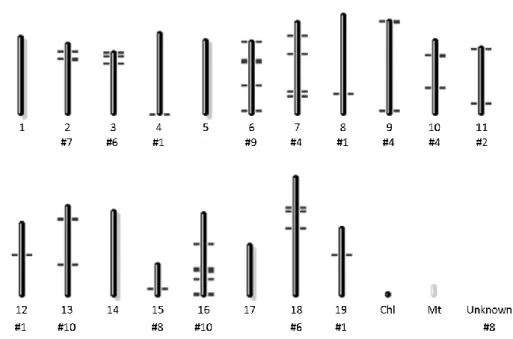
Em 2014, a família de subtilases em Vitis vinifera foi preliminarmente caracterizada. Simultaneamente, uma nova anotação do genoma de videira foi publicada, havendo genes que deixaram de ter anotação e outros que mudaram de nome. Assim, o principal objetivo deste projeto foi realizar uma re-caracterização genómica da família de subtilases de videira, tomando como referência a re-anotação do genoma de videira que ocorreu em 2014, e associar alguns desses genes com a resposta de defesa da Vitis vinifera contra o Plasmopara viticola. Para cumprir este objetivo, foram realizadas pesquisas em bases de dados considerando os três domínios conservados característicos de subtilases (domínios peptidase S8, PA e inibidor I9), que conduziram à identificação de 85 genes de subtilases em videira, desigualmente distribuídos ao longo de 15 dos 19 cromossomas da videira. Verificou-se que estes genes codificam 97 possíveis proteínas (resultantes de eventos de splicing alternativo). Estes subtilases foram organizadas em 6 grupos de acordo com a semelhança entre a sequência das proteínas, resultante de uma análise filogenética. Uma análise a nível da localização subcelular demonstrou que a maioria dos subtilases de videira estão localizados no apoplasto, na parede celular ou na região extracelular. A comparação dos subtilases de videira com subtilases de Arabidopsis
thaliana e Solanum lycopersicum demonstrou que, uma elevada percentagem dos mesmos,
partilham grande semelhança de sequência com os subtilases SBT3.3 e P69. Estes proteases já tinham sido anteriormente relacionados com a resposta de defesa da Arabidopsis e do tomate, respetivamente, contra estímulos ambientais bióticos. Para além disso, foi demonstrado neste estudo que alguns genes de subtilases em videira estão localizados perto de genes associados à resistência da Vitis vinifera contra o Plasmopara viticola.
O segundo objetivo deste projeto foi realizar estudos de expressão de genes de forma a elucidar a participação de subtilases de videira anteriormente selecionadas na resistência da
IX
vinifera (Regent e Trincadeira) após inoculação com o oomycete Plasmopara viticola. Para além
disso, o padrão de expressão constitutivo destes subtilases foi analisado em várias espécies selvagens de Vitis e cultivares de Vitis vinifera que apresentam vários graus de resistência quando infetadas com o P. viticola. Os resultados sugeriram que, sob ataque do patogénio, as cultivares resistentes têm uma rápida resposta através do aumento da expressão de alguns subtilases. Este rápido aumento pode estar relacionado com o estabelecimento imediato de uma estratégia defensiva contra o patogénio invasor. Por outro lado, a cultivar suscetível demonstrou um atraso no aumento da expressão de subtilases, que pode estar relacionado com a sua tentativa de iniciar uma estratégia defensiva, mas que não é rápida nem robusta o suficiente para prevenir a invasão e o crescimento do patogénio. Ao nível constitutivo, em várias espécies e cultivares de videira não foi observado um padrão de expressão de subtilases. Ambos os resultados sugerem que existe uma expressão diferencial de certos subtilases nas espécies resistentes, mas esta só acontece após estimulação da planta com o ataque do patogénio, o que suporta a hipótese de que alguns subtilases de videira podem ter um papel importante na resposta de defesa contra o Plasmopara viticola.
Em último lugar, dois subtilases de videira foram selecionados com base nas suas características e nos seus perfis de expressão e foram clonados com o intuito de expressar a respetiva proteína recombinante.
Do que se sabe até agora, este estudo é o primeiro a realizar uma caracterização em larga escala de subtilases de videira associados à resposta de imunidade contra o patogénio responsável pelo míldeo. Estudos futuros serão realizados com o intuito de caracterizar as proteínas recombinantes, elucidar respetivas estruturas e identificar possíveis substratos, a fim de estabelecer estes subtilases como candidatos para a introgressão de genes em programas de melhoramento.
XI
Summary
Subtilisin-like proteases, also known as subtilases, are a very diverse family of serine peptidases present in many organisms. In plants, subtilases are especially abundant with, e.g., 63 known genes in Oryza sativa, 56 genes in Arabidopsis thaliana and 15 in Lycopersicon
esculentum. These proteins act as secretory enzymes and are targeted to the endoplasmic
reticulum migrating to the cell plasma membrane. The majority of plant subtilases are synthesized as an inactive pre-pro-protein precursor formed by a signal peptide, a pro-domain (I9 inhibitor domain), a subtilase (S8 peptidase) domain, and a protease-associated (PA) domain located within the subtilase domain. Moreover, some of them may have only one or two of these domains or even additional domains. Subtilases present a highly conserved catalytic triad within the S8 peptidase domain, constituted by aspartate (Asp), histidine (His) and serine (Ser) amino acid residues, and some subtilases may have also a conserved catalytic asparagine (Asn) residue. Concerning protein structure, plant subtilases are generally monomeric, although several studies have indicated that many subtilases suffer a homo-dimerization mediated by PA domain in order to become activated. In plants, subtilases are involved in many biological functions from the mobilization of storage proteins during seed germination to the initiation of cell death and senescence programs. Recent evidences have also highlighted the participation of subtilases in response to biotic and abiotic environment stimulus and in symbiotic interactions of plants with other organisms. In 1987 was demonstrated for the first time the involvement of subtilisin-like proteases in plant defence response with the identification of subtilase P69 accumulation in tomato leaves after the infection with citrus exocortis viroid. More recently, studies in A.
thaliana identified the SBT3.3 gene which expression rapidly increases during the activation of
innate immunity preceding the activation of salicylic acid (SA) responsive genes. Studies have also shown that subtilases are glycosylated and secreted to the plant extracellular matrix (ECM). Since ECM is where the first host-pathogen interactions, recognition and signalling events take place, the accumulation of subtilases in this cellular location may account for an important role during pathogenesis.
Recently, was reported a constitutively expression of a subtilisin-like protein, sharing high sequence similarity with the tomato subtilases P69C, in a resistant grapevine and an increase of protein expression after inoculation with the oomycete Plasmopara viticola. Besides these clues of the presence and importance of the subtilases in V. vinifera, these proteins were not yet associated with grapevine immune responses particularly concerning P. viticola resistance. As cultivated grapevine (Vitis vinifera L.) is currently the most important fruit plant cultivated
XII
worldwide and highly susceptible to several disease including downy mildew, caused by
Plasmopara viticola (Berk. et Curt.) Berl. et de Toni, the importance of subtilases in resistance
should be further investigated.
In 2014, the subtilase family in Vitis vinifera was preliminarily characterized but at the same time a new annotation of the grapevine genome was published. Thus, the main purpose of this project was perform a genome-wide update of the grapevine subtilase gene family and associate some subtilase genes with the defence response of the V. vinifera against the P.
viticola, taking as reference the grapevine genome reannotation that occurred in 2014. For that,
several database searches were performed considering the three domains (S8, PA and I9) leading to the identification of 85 grapevine subtilase genes, unevenly distributed among 15 of the 19 grapevine chromosomes. From these genes, it was predicted to obtain 97 subtilase proteins, which were organized into 6 groups accordingly with their similarity. An analysis at subcellular location level, showed that the majority of the grapevine subtilases were located in apoplast, cell wall or extracellular region. Comparison of the grapevine subtilases with
Arabidopsis thaliana and Solanum lycopersicum subtilases showed that a high percentage of
them shared high sequence similarity with SBT3.3 and P69C subtilases. These proteases have been already related to the defence response of Arabidopsis and tomato, respectively, against biotic environment stimulus. Moreover, it was demonstrated in this study that some grapevine subtilases genes were located near of locus associated to Vitis vinifera resistance against
Plasmopara viticola.
The second goal of this project was performed gene expression studies elucidating the role of selected grapevine subtilases in the grapevine resistance against P. viticola. Subtilase gene expression was studied in two Vitis vinifera cultivars (Regent and Trincadeira) after P.
viticola inoculation and the constitutive expression pattern of the subtilases was studied in
several grapevine species/ cultivars showing varying degrees of resistance towards P. viticola. The results suggested that under pathogen attack, resistant grapevine cultivar have an early response increasing the expression of some subtilases. This early increase of subtilase expression may be related to the establishment of a defence strategy against the invading pathogen. On the other hand, the susceptible grapevine cultivar showed a delay of the subtilase expression increase, which may be related to an attempt by the susceptible cultivar to initiate a defence strategy that was not fast or robust enough to prevent pathogen growth. At a constitutive level, in several grapevine species/ cultivars, it was not observed a pattern of subtilases expression. Both results suggest that there was a differential expression of certain
XIII subtilases in resistant species but only after stimulation with the pathogen attack, which supports the hypothesis that some grapevine subtilases may have a role in defence response against P. viticola.
Finally, two subtilases were selected, based on their characteristics and on expression profiles, and cloned for recombinant protein expression.
Up to our knowledge, this study is the first to preformed a large scale characterization of grapevine subtilases associated to immune responses against the downy mildew pathogen. Further studies in order to characterize the recombinant proteins, to access their structure and substrates should be done in order to establish these subtilases as candidates for introgression in breeding programs.
XV
Abbreviations
AICc Akaike Information Criteria
Asp Aspartate
BI Bayesian Inference
bp Base pair
CaCl2 (2H2O) Calcium chloride dihydrate
cDNA Complementary DNA
Cq Quantification cycle DNA Deoxyribonucleic acid dNTP Deoxynucleotide ECM Extracellula matrix
EDTA Ethylenediaminetetraacetic acid EF1α Elongation factor 1-alpha
ER Endoplasmic reticulum
GAPDH Glyceraldehyde-3-phosphate dehydrogenase
gDNA Genomic DNA
His Histidine
hpi Hours post inoculation HR Hypersensitive response
KAC Potassium acetate
LB Luria-Bertani
LRR-RLK Leucine-rich repeat receptor-like kinase MgCl2 (4H2O) Magnesium chloride tetrahydrate
ML Maximum likelihood
MOPS 3-(N-Morpholino)propanesulfonic acid
mRNA Messenger RNA
Mw Molecular weight
N2 Nitrogen
PA Protease-associated
PAMP Pathogen-associated molecular pattern
PCD Programmed cell death
PCR Polymerase chain reaction PCs Proprotein convertases
XVI
pI Isoelectric point
PR Pathogenesis-related
PRR Pattern recognition receptors qPCR Quantitative real time PCR QTL Quantitative trait locus RbCl2 Rubidium chloride
RNA Ribonucleic acid
RPV ‘Resistance Plasmopara viticola’
SA Salicylic acid
SAND SAND family protein
SDD1 Stomatal density and distribution 1
Ser Serine
SOB Super optimal broth
TBE Tris-borate-EDTA
UBQ Ubiquitin-conjugating enzyme
Units
C Celsius degrees
ɡ Earth's gravitational acceleration
kDa kilodalton Mg Milligram mL Millilitre mM Millimolar Ng Nanogram nm Nanometre Ta Annealing temperature Tm Melting temperature μL Microlitre μM Micromolar
1. Introduction
1 | INTRODUCTION
1 | INTRODUCTION
3
1.1 Subtilisin-like proteases: classification and basic features
Subtilisin-like proteases (also known as subtilases) are a very diverse and the second largest family of serine peptidases present in archaea, bacteria, eukarya, fungi and yeast (Siezen et al., 1991). They belong to the S8 family within the SB clan of serine proteases, according to the classification of the peptidase database MEROPS (Rawlings et al., 2014; http://merops.sanger.ac.uk).
In mammals, the subtilase homologs are the proprotein convertases (PCs), responsible for the formation of peptide hormones, growth factors, neuropeptides, and receptor proteins from inactive pro-proteins by limited proteolysis at highly specific sites (Barr, 1991; Schaller and Ryan, 1994; Seidah et al., 1999, 1994). Beyond the highly specific maturation of peptide hormones and processing of protein precursors, by the kexin in Saccharomyces cerevisiae and AtSBT1.1 in
Arabidopsis thaliana (Srivastava et al., 2008), subtilases can have also a non-selective
degradation function in general protein turnover, like the subtilisin Carlsberg from Bacillus
licheniformis (Rose et al., 2010). There are several examples of degradative subtilases from the
cucumisin from Cucumis melo that cleaves a broad variety of peptide and protein substrates (Kaneda and Tominaga, 1975; Uchikoba et al., 1995; Yamagata et al., 1994), to macluralisin from
Maclura pomifera for which similar characteristics have been reported (Rudenskaya et al.,
1995).
Unlike mammals on which only nine subtilases have been identify, subtilases from plants are especially abundant, with 63 known genes in Oryza sativa (Tripathi and Sowdhamini, 2006), 56 genes in Arabidopsis thaliana (Rautengarten et al., 2005), 15 genes in Lycopersicon
esculentum genome (Meichtry et al., 1999), 23 genes in the moss Physcomitrella patens, 90
genes in Populus trichocarpa (Schaller et al., 2012) and 80 genes in Vitis vinifera genome (Cao et al., 2014). In plants, subtilases act as secretory enzymes and are targeted to the endoplasmic reticulum (ER) (Siezen and Leunissen, 1997) migrating to the cell plasma membrane, e.g, subtilisin-like serine protease SDD1 from Arabidopsis thaliana that is located at the plasma membrane, mediate cell-to-cell signalling and controls stomatal distribution and density during leaf development (von Groll, 2002).
4
1.2 Structure and biochemistry of plant subtilases
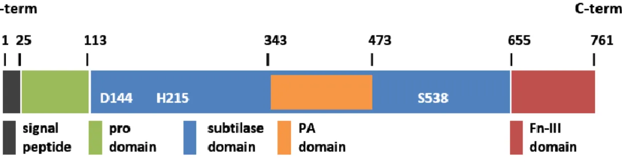
The majority of the subtilases from plants are synthesized as an inactive pre-pro-protein precursor. Their structure usually presents a signal peptide, a pro-domain (also known as I9 inhibitor domain), a subtilase domain (also known as S8 peptidase domain) and a protease-associated (PA) domain located within the subtilase domain (Figure 1), although some of them may have only one or even additional domains (Antão and Malcata, 2005; Dodson and Wlodawer, 1998; Siezen et al., 2007; Siezen and Leunissen, 1997; Vartapetian et al., 2011). The presence of a highly conserved catalytic triad within the S8 peptidase domain, composed by aspartate (Asp), histidine (His) and serine (Ser) amino acid residues (Dodson and Wlodawer, 1998), is characteristic of the subtilase family (Figure 1Figure 1). Additionally, certain subtilases may also have a conserved catalytic asparagine (Asn) residue in the same S8 peptidase domain (Dodson and Wlodawer, 1998; Jordá et al., 1999; Siezen and Leunissen, 1997). Contrary to other organisms, PA domain in plants is found within the S8 peptidase domain. The PA domains is an insertion of 120–160 amino acids between the His and Ser active site residues that cause a displacement of the reactive Ser from the catalytic triad to the C-terminal (Siezen and Leunissen, 1997), (Figure 1Figure 1). Most plant subtilases also contain a fibronectin (Fn) III-like domain, required for the activity of some of these enzymes, but dispensable in others (Rawlings and Salvesen, 2013).
Figure 1 – Example of a subtilase domain architecture showing the four characteristics domains (adapted from Rose et al 2010). This in particular is the architectural domain of SlSBt3, a subtilase from tomato (Solanum lycopersicum). The main domains are represented, as well as the 3 amino acid residues [aspartate (D), histidine (H) and serine (S)] in the subtilase domain.
As consequence from the pre-pro-protein structure, the active enzyme maturation from its inactive precursor requires the removal of the signal peptide, that is responsible for targeting the protease for secretion (Cedzich et al., 2009; Feliciangeli et al., 2006; Nour et al., 2003), and
5 of the pro-domain. In plants, this process occurs late in the ER or in the early Golgi, either as an intramolecular autocatalytic reaction, or as result of the interaction with a secondary peptidase (Bergeron et al., 2000; Cedzich et al., 2009; Chichkova et al., 2010). The I9 inhibitor domain (also known as pro-domain) works as an auto-inhibitory domain maintaining the inactive state of the zymogen and preventing the access of the substrate to the active site. It also works as an intramolecular chaperone that is required transiently to assist in folding of the catalytic domain (Baker et al., 1993; Bryan, 2002; Huang et al., 1997; Li and Inouye, 1994; Zhu et al., 1989). After cleavage, the pro-domain remains non-covalently bound, acting as a specific inhibitor of proteolytic activity (Anderson et al., 2002; Nour et al., 2003; Steiner, 1998), and thus protecting proteins involved in the secretory pathway from nonspecific proteolytic degradation (Yamagata et al., 1994). Hence, the cleavage of the N-terminal of the subtilase is a prerequisite for the protease to acquire its functional conformation and perform its function at the action site. Once the N-terminal inhibitory domain is removed, the activity of the subtilase is therefore stimulated (Bergeron et al., 2000).
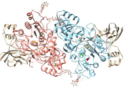
Concerning protein structure, plant subtilases generally present a monomeric structure (Figure 2), although several studies have indicated that many subtilases suffer a homo-dimerization mediated by PA domain in order to activate it (Rose et al., 2010; Siezen and Leunissen, 1997). Without this activation process subtilases cannot perform their function (Ottmann et al., 2009).
Figure 2 – Dimeric structure of the subtilase SISBt3 (PDB ID 3I6S) from S. lycopersicum, highlighting the two S8 peptidase domains (coral and blue) and the three catalytic residues (aspartate [D144] in red, histidine [H215] in green and serine [S538] in yellow). This image was prepared with the UCSF Chimera package [Chimera is developed by the Resource for Biocomputing, Visualization, and Informatics at the University of California, San Francisco (supported by NIGMS P41-GM103311), (Pettersen et al., 2004)].
6
The PA domain also interacts with the I9 inhibitor domain, leading to the cleavage of the N-terminal and allowing the access of the substrates to the catalytic site, stimulating the subtilase activity (Bergeron et al., 2000). The PA domain has been also implicated in protein-protein interactions and substrate recognition (Rautengarten et al., 2005; Schaller et al., 2012). Studies in soybean have shown that PA domain plays an important role in substrate selection, namely in substrate length determination (Rautengarten et al., 2005; Tan-Wilson and Wilson, 2012).
Another feature of subtilases is the apparent Ca2+-independence of most of them,
contrarily to what was expected by modelling studies of the first subtilases described such as the bacterial protease subtilisin BPN’ (Alexander et al., 2001; Siezen and Leunissen, 1997). In 2009, Ottmann and co-workers demonstrated that the thermostability and activity of the subtilase SISBT3 from Solanum lycopersicum were not influenced by the addition of Ca2+ or
chelating agents. Instead, in SISBT3, another subtilase from tomato, a positively charged site chain of Lys498 mimics the calcium ion bound function. This region, including the stabilizing lysine residue, is highly conserved in all the plant subtilases studied so far (Rose et al., 2010).
1.3 Subtilase participation in plant-specific developmental processes
In plants, subtilases are involved in many biological functions from the mobilization of storage proteins during seed germination to the initiation of cell death and senescence programs (Schaller et al., 2012). There are several examples of the involvement of subtilases in plant development, as the Arabidopsis thaliana AtSBT1.7 that participates in seed germination (Rautengarten et al., 2008), LIM9 from Lilium longiflorum that is involved in microspore development (Riggs and Horsch, 1995; Taylor et al., 1997), the papaya CpSUB1 that is involved in fruit ripening (Othman and Nuraziyan, 2010), the ARA12 in Arabidopsis and SCS1 in soybean that participate in seed coat development (Batchelor et al., 2000). Moreover, previous studies in Arabidopsis thaliana have shown the participation of the subtilases XSP1 and AIR1 in xylem differentiation (Zhao et al., 2000) and lateral root formation (Neuteboom et al., 1999). In 2001, Tanaka and co-workers have suggested that ALE1 protease is necessary for cuticle formation and epidermal differentiation during embryo development in A. thaliana. Also in A. thaliana, von Groll and co-workers have elucidated the role of the SDD1 (stomatal density and distribution 1) protease, a apoplast-secreted protein that acts as a processing protease in the generation of a signal responsible for regulation of stomata distribution and density during leaf development
7 (von Groll, 2002). Apparently, the SDD1 is involved in cell-to-cell communication through the processing of the ligand for the TMM (Too Many Mouths), a member of the large leucine-rich repeat receptor-like kinase (LRR-RLK).
Besides the cellular functions already mentioned, plant subtilases may also contribute to the cell wall dynamics, either directly by cleavage of structural protein, or indirectly by the regulation of cell wall remodelling enzymes (Schaller et al., 2012).
1.4 Subtilases in environment stimulus and pathogen attack responses
It is evident the importance and the participation of subtilases in a lot of plant development functions. Recent evidences have also highlighted the participation of subtilases in response to biotic and abiotic environment stimulus (Chichkova et al., 2004; Liu et al., 2007; Tian, 2005; Tian and Kamoun, 2005). One example is the participation in the response to abiotic environment of the subtilase AtSBT6.1 from A. thaliana, the ortholog of mammalian site-1-protease (S1P). This site-1-protease is involved in the unfolded protein response through the cleavage of an ER-resident type II membrane protein (bZIP28). The bZIP28 protein moves to Golgi where unfolded proteins accumulate, cleavage releases the bZIP domain that translocates to the nucleus in order to activate gene expression related to the stress response (Che et al., 2010; Liu et al., 2007; Liu and Howell, 2010a, 2010b). Additional substrates of AtSBT6.1 include the precursor proteins of a peptide growth factor (Rapid Alkalinization Factor 13) and pectin methylesterases (Srivastava et al., 2009; Wolf et al., 2009). In recent years, subtilases were shown to be involved in symbiotic interactions of plants with other organisms, such as the mutualistic interactions of plant roots with fungi resulting in as arbuscular mycorrhiza or with nitrogen-fixing Rhizobia as nodule symbiosis (Takeda et al., 2007).
The first evidence of the involvement of plant subtilisin-like proteases in plant-pathogen interactions was reported by Granell and co-workers (1987) in tomato plants. They identified the accumulation of the subtilase P69 in tomato leaves after infection with citrus exocortis viroid (CEV) (Granell et al., 1987). Two years later, Christ and Mösinger (1989) and Fischer and co-workers (1989) associated the same subtilase with the response of tomato leaves to
Phytophothora infestans (Christ and Mösinger, 1989; Fischer et al., 1989). The P69 subtilase was
characterized as an alkaline proteinase located in the vacuole and intercellular spaces of leaf parenchyma cells (Vera et al., 1989; Vera and Conejero, 1988). Tornero and co-workers (1996)
8
cloned for the first time a P69 subtilase from tomato and revealed the presence of at least six closely related genes in tomato (Jordá et al., 2000, 1999; Meichtry et al., 1999; Tornero et al., 1997). Two of them, P69A and P69D, are constitutively expressed (Jordá et al., 1999), P69E and P69F have a specific developmental expression pattern (Jordá et al., 2000). P69B and P69C were shown to behave as pathogenesis-related (PR) genes being induced by pathogen infection and salicylic acid (Jordá et al., 1999; Tornero et al., 1997). P69 was also the first plant subtilase for which protein substrates were identified: the systemin, a travelling peptide hormone mediating signalling processes during wound response in plants (Schaller and Ryan, 1994), and the leucine-rich repeat (LRP) protein (Tornero et al., 1996a), an extracellular matrix associated leucine-leucine-rich repeat (LRR) protein that mediates molecular recognition and/or protein interaction processes (Kobe and Deisenhofer, 1995). However, the consequences of these substrates processing events for plant pathogen interaction, remains unknown.
More recently, studies in A. thaliana identified the SBT3.3 gene as encoding a serine protease homologue to the P69C subtilase from tomato (Ramírez et al., 2013). Like P69C in tomato, SBT3.3 can specifically process an extracellular LRP containing-protein, suggesting the involvement of the SBT3.3 on the LRR-containing proteins’ cleavage, including pattern recognition receptors (PRR) as PRR-type receptors and activation of plasma membrane receptors and downstream signalling processes (Ramírez et al., 2013). Thus, like the tomato P69C, A. thaliana SBT3.3 may be linked to pathogen recognition. Ramirez and co-workers (2013) have shown that the expression of SBT3.3 rapidly increases during the activation of innate immunity preceding the activation of salicylic acid (SA) responsive genes, responding very rapidly to H2O2, a common ROS species generated very early during pathogen-associated
molecular pattern (PAMP) recognition by PRR leading to activation of innate immune responses. Despite the SBT3.3 substrate is not yet identified, this subtilase may have activity on the extracellular domain (ectodomain) of a larger protein that works like a receptor located in the plasma membrane. Thus, as consequence of the proteolytic shedding of the ectodomain, the receptor could become activated and initiate a downstream immune signalling process. After that, a positive feedback loop circuit would maintain the SBT3.3 expression in a level sufficient to keep cells in a sustained sensitized mode (Ramírez et al., 2013). This expression pattern would consequently be the basis to explain the memory-based characteristics of priming and induced resistance (Ramírez et al., 2013).
Studies have also shown that subtilases are glycosylated and secreted to the plant extracellular matrix (ECM) where they accumulate and presumably exert their biochemical
9 functions by recognition and processing pericellular substrates (Siezen and Leunissen, 1997; Taylor et al., 1997; Tornero et al., 1997, 1996a, 1996b; Yamagata et al., 1994). Considering that ECM is where the first host-pathogen interaction, recognition and signalling events take place (Dixon and Lamb, 1990), the accumulation of subtilases in plant ECM may account for an important role during pathogenesis. This seems to be the case for grapevine infection with the mildew causing agent, Plasmopara viticola, in which some signalling events might occur in ECM. The first clues were given by Figueiredo and co-workers (2008) when comparing resistant and susceptible genotypes prior and post-inoculation with P. viticola. The results showed constitutively expression of a subtilisin-like protein, sharing sequence similarity with the tomato P69C, in the resistant genotype and an increase of protein expression after inoculation with P.
viticola (Figueiredo et al., 2012, 2008; Monteiro et al., 2013).
1.5 Study model: the interaction between Vitis vinifera and Plasmopara viticola
Grapevine (Vitis vinifera L.) is currently the most important fruit plant cultivated worldwide due to its economic importance in the wine industry. In Portugal this industry accounts over 890 million euro per year of exports (Bettini, 2014). Cultivated grapevine is highly susceptible to downy mildew disease which is caused by the obligate oomycete Plasmoparaviticola (Berk. et Curt.) Berl. et de Toni. P. viticola requires the genus Vitis to complete its life
cycle once it cannot survive outside its host except as oospores (Yu et al., 2012). This oomycete, under optimal conditions, such as high humidity and warm temperatures, can spread rapidly over large areas within a very short period of time (Müller and Sleumer, 1934). For infection, upon contact with water, P. viticola release several flagellate zoospores that swarm within the water film on the lower surface of the leaf. On susceptible hosts, the zoospores are targeted to the stomata, where they shed their flagella, attach and encyst (Kiefer et al., 2002). Then, a germ tube is formed that reaches into the substomatal cavity, where it dilates into a substomatal vesicle. From this vesicle a primary hypha emerges developing a mycelium that spreads within the leaf tissue, extending mainly into the intercellular spaces of the spongy parenchyma and forming haustoria that penetrate into the cell wall of the host (Unger et al., 2007). The P. viticola infection interfere with the normal regulation of the stomata guard cells resulting in water loss (Allègre et al., 2007), cause tissue damage and reduces the functional green area of the leaf as well as assimilation rates by the leaf remainder (Moriondo et al., 2005). In resistant species, the infection progress is slowed down, inhibited, or completely stopped (Yu et al., 2012).
10
The attack by this pathogen leads to heavy crop losses which represent a cost of several million euro each year. The current strategy for downy mildew disease control is the massive use of pesticides in each growing season. However, as for the crops, excessive use of pesticides is highly prejudicial to human health. So, the search for alternative methods to control grapevine downy mildew is crucial (reviewed in Gessler et al., 2011).
1.6 Grapevine resistance to Plasmopara viticola
Contrary to V. vinifera cultivars that have no known natural resistance to downy mildew infection (Cadle-Davidson, 2008; Staudt and Kassemeyer, 2015), sources of resistance against the mildews were identified in a range of American wild species, like V. labrusca, V. riparia and
V. rupestris (Alleweldt and Possingham, 1988; Eibach et al., 2010; Wan et al., 2015). With this in
mind, grape breeders tried to combine the resistance traits from the American wild species with the quality of V. vinifera cultivars, resulting in new cultivars with high-quality features and considerable mildew resistance characteristics (Bundessortenamt, 2008). Resistance mechanisms elucidated so far vary from physical barriers such as hairs and stomatal closure, accumulation of phenolic antimicrobial compounds, increase of peroxidase activity, accumulation of pathogenesis-related proteins (PRs) and hypersensitive response (HR) (Allègre et al., 2007; Greenberg and Yao, 2004; Kortekamp, 2006; Kortekamp et al., 1998; Kortekamp and Zyprian, 2003).
Also, until now, quantitative trait locus (QTL) for resistance to downy mildew have been reported, e.g., Rpv3 (Bellin et al., 2009; Welter et al., 2007), Rpv8 (Blasi et al., 2011) and Rpv11 (Bellin et al., 2009; Fischer et al., 2004; Schwander et al., 2011) (Supplementary Table 1). With the grapevine genome sequencing and physical mapping (Jaillon et al., 2007; Moroldo et al., 2008; Velasco et al., 2007), the distribution pattern of candidate genes for disease resistance across the whole genome of V. vinifera has been recently elucidated.
1.7 Participation of subtilases in grapevine resistance to Plasmopara viticola
The first clues regarding subtilase participation in grapevine - P. viticola interaction were reported by Figueiredo and co-workers (2008), when comparing resistant and susceptible genotypes prior and post-inoculation with this pathogen. A subtilisin-like protein, sharing
11 sequence similarity with the tomato P69C, was constitutively expressed in resistant genotype and increased its expression after P. viticola inoculation (Figueiredo et al., 2012, 2008; Monteiro et al., 2013). Studies on the grapevine- P. viticola interaction with serine protease inhibitors, shown that after the treatment with the inhibitor, an immune cultivar becomes resistant, a resistant cultivar reaches the level of a susceptible one, and a susceptible cultivar becomes even more sensitive (Gindro et al., 2012). Therefore, after treatment with a serine protease inhibitor, the infection rate could rise due to the inhibition of the proteases involved either in the regulation of stomatal density (Berger and Altmann, 2000) or in plant defence against pathogens (Oh et al., 2008; van der Hoorn and Jones, 2004; van der Hoorn, 2008). These results point to a possible involvement of subtilases given what is known about their cellular functions.
In grapevine - P. viticola interaction, inhibition of phytaspases, a subgroup of plant subtilases, could partially inhibit the overall activation of programmed cell death (PCD) and thereby change the level of susceptibility of resistant cultivars to the pathogen (Gindro et al., 2012). The secretome of P. viticola is specific, but it could also be tailored to the host plant to a certain extent. Therefore, under natural conditions, the secretome of P. viticola could inhibit the endogenous subtilases of susceptible varieties, thereby inhibiting the plant’s normal defence reaction. By contrast, it is possible to hypothesize that resistant or immune varieties possess endogenous subtilases that are not recognized by the secretome of P. viticola due to slight structural modifications of the protein patterns of these cultivars. In this case, plant defence mechanisms would continue to operate, with fatal consequences for the pathogen and restricting its development.
In 2014, the subtilase family was preliminarily characterized by Cao and co-workers (2014). These authors identified 80 subtilase genes in Vitis vinifera that were divided into 8 groups based on their phylogenetic relationships. Besides these clues of the presence and importance of the subtilases in V. vinifera, these proteins were not yet associated with grapevine immune responses particularly concerning P. viticola resistance.
12
1.8 Aims
The main purpose of this project is perform a genome-wide update of the grapevine subtilase gene family and associate some subtilase genes with the defence response of the V.
vinifera against the P. viticola, taking as reference the grapevine genome reannotation that
occurred in 2014.
The second goal of this project was to perform gene expression studies elucidating the role of selected grapevine subtilases in the grapevine resistance against P. viticola. Subtilase gene expression will be studied in two Vitis vinifera cultivars (Regent and Trincadeira) after P.
viticola inoculation and the constitutive expression pattern of the subtilases will be studied in
several vine species and grapevine cultivars showing different degrees of resistance towards P.
viticola.
Finally, based on the most promising results of qPCR, selected subtilases were cloned for further recombinant protein expression.
2. Materials and Methods
1 | INTRODUCTION
1 | INTRODUCTION
15
2.1 Updating grapevine subtilase gene family
2.1.1 Identification and retrieval of Grapevine Subtilase gene sequences
Grapevine subtilase gene family has been previously characterized (Cao et al., 2014). As the grapevine genome annotation has been recently updated (2014), new database searches were performed to identify and select the members of the subtilase gene family in Vitis vinifera. The conserved domains of subtilases, i.e., PA (PF02225), S8 Peptidase (PF00082) and I9 Inhibitor (PF05922) were used as query for blast searches at NCBI (http://www.ncbi.nlm.nih.gov/) and MEROPS (Rawlings et al., 2014, https://merops.sanger.ac.uk/) databases. MEROPS database was also used to identify S8 peptidase domain containing-proteins in Vitis vinifera. The retrieved sequences were compared with the sequences obtained from the NCBI database. The results of both databases were manually curated. A new search using the NCBI BLAST tool (Manual and Altschul, 1990, https://blast.ncbi.nlm.nih.gov/Blast.cgi) was performed using the selected subtilase sequences as query in order to retrieve both mRNA and protein sequence identifiers.
2.1.2 Chromosomal location
The subtilase genes were mapped in V. vinifera chromosomes with the Map Viewer tool from NCBI (http://www.ncbi.nlm.nih.gov/mapview/).
2.1.3 Protein Sequence Alignment and Phylogenetic Analysis
Protein sequences were obtained from the NCBI Database using the protein sequence identifiers (prefix XP), for each of the subtilase genes (Supplementary Table 2). The protein sequences were initially aligned using the MAFFT software with the L-INS-i option (version 7, Katoh and Standley, 2013, http://mafft.cbrc.jp/alignment/software/) and gaps were manually checked and edited in BioEdit v. 7.2.5 (Hall, 1999).
A preliminary maximum likelihood (ML) phylogenetic analysis was performed with RAxML-HPC v.8, on CIPRES Science Gateway (Miller et al., 2010, https://www.phylo.org), with the following parameters: protein substitution model PROTCAT; protein substitution model + BLOSUM62; bootstrap 1000 iterations with rapid bootstrap analysis (-f a). No outgroups were selected.
16
The best amino acid substitution model (WAG +G) was selected with ProtTest3.4 (Darriba et al., 2011) under the corrected Akaike Information Criteria (AICc), removing gamma-invariable mixture (+I+G) models from the analysis.
Dataset matrixes consisting of 97 aligned protein sequences were converted from Fasta to Nexus format with Concatenator 1.1.0 (Pina-Martins and Paulo, 2008).
A Bayesian Inference (BI) phylogenetic tree was generated by MrBayes 3.2.1 (Ronquist et al., 2012), outgroups were set according to the basal proteins obtained from RAxML and amino acid substitution model, WAG+G, with four rate categories for the gamma distribution. The Metropolis-coupled Markov chain Monte Carlo analysis was carried out with four chains. The posterior probabilities for each node were generated from 108 generations, sampling at every
1000th iteration. The burn-in was set to the first 10% trees, and the remaining trees were used
to generate a consensus tree by the 50% majority rule.
ML and BI trees were viewed on FIGTree (http://tree.bio.ed.ac.uk/software/figtree/) and edited on Inkscape (Harrington, B. et al., 2004; http://www.inkscape.org/).
2.1.4 Sequence properties
Molecular weight (Mw) and theoretical isoelectric point (pI) were predicted using the Protparam tool from ExPASy (Gasteiger et al., 2005; http://web.expasy.org/protparam/). Signal peptide prediction of subtilase proteins was performed using the MemPype software (Pierleoni et al., 2011; http://mu2py.biocomp.unibo.it/mempype). Subcellular location of proteins was predicted using Blast2GO (version 3.3, Conesa et al., 2005; https://www.blast2go.com/), TargetP (Emanuelsson et al., 2000; http://www.cbs.dtu.dk/services/TargetP/), PredoTar (Small et al., 2004; https://urgi.versailles.inra.fr/predotar/predotar.html) and LocTree3 (Goldberg et al., 2014, https://rostlab.org/services/loctree3/). Putative function was predicted using the Blast2GO tool. The presence of the PA, S8 peptidase and I9 inhibitor domains was confirmed using the Pfam tool (Finn et al., 2016; http://pfam.xfam.org/). All the molecular predictions were manually curated and compiled.
2.1.5 Selection of subtilase sequences involved in grapevine immunity
Previous studies in plants associated some subtilases with the defence response to pathogen attack, like the subtilase SBT3.3 in A. thaliana (Ramírez et al., 2013), the P69 in S.
17
lycopersicum (Jordá et al., 1999; Tornero et al., 1996a) and cucumisin in grapevine (Figueiredo
et al., 2012, 2008). The subtilase genes from V. vinifera were blasted against the A. thaliana genome (using the TAIR database, https://www.arabidopsis.org/) and the tomato genome (using the SolGenomics database, https://solgenomics.net/) to retrieve the grapevine sequences presenting higher homology to A. thaliana SBT3.3 and tomato P69 genes. The Solgenomics results were corroborated in NCBI BLAST tool, restricting to Solanum lycopersicum organism, and was assumed the NCBI accession for further studies. Moreover, subtilase sequences with a chromosomal location near ‘Resistance to Plasmopara viticola’ (RPV) locus (Bellin et al., 2009; Blasi et al., 2011; Fischer et al., 2004; Schwander et al., 2011; Welter et al., 2007) on grapevine genome were also selected for further studies. Grapevine subtilase genes were selected for analysis by qPCR (Real-time polymerase chain reaction).
2.1.6 Biochemical predictions of grapevine subtilases possibly involved in immunity
Multiple alignment of the grapevine subtilases putatively involved in immunity was performed in DNASTAR software (version 13, Burland, 1999, http://www.dnastar.com/) and prediction of protein glycosylation was done using the NetNGlyc online server (version 1.0, Ramneek and Brunak, 2001). The presence of signal peptides was searched using SignalP server (version 4.1, Petersen et al., 2011), automatically run on all sequences analysed with NetNGlyc.
2.1.7 Prediction of protein–protein interaction network for the subtilases putatively
involved in grapevine immunity
The protein interaction network of the selected subtilases was obtained (STRING, version 10.0, http://string-db.org/). The gene accessions for all the interacting proteins were queried at the NCBI database. The GO terms for all the interacting proteins were also obtained.
2.2 Expression analysis by qPCR
2.2.1 Plant Material for inoculation experiments
Two Vitis vinifera cultivars were selected to access subtilase expression during interaction with P. viticola. The cultivar Regent, bread by multiple introgressions from resistant wild genotypes (Welter et al., 2007), presenting a high degree of resistance to downy and powdery
18
mildews (Anonymous, 2000), and Trincadeira, a Portuguese traditional grapevine cultivar widely used for quality wine production and highly sensitive to Plasmopara viticola (Figueiredo et al., 2008). Plant material was already available, grown and inoculated as described in Figueiredo et al. (2012). The third to fifth fully expanded leaves beneath the shoot apex were harvested at 0, 6, 12 and 24 hpi (hours post inoculation), immediately frozen in liquid nitrogen and stored at −80 °C. For each genotype and condition (inoculated and mock inoculated), three independent biological replicates were collected, being each biological replicate a pool of three leaves from three different plants.
2.2.2 Plant material for species comparison
In order to access if subtilases are constitutively expressed, young leaves from several
Vitis species and Vitis vinifera cultivars (Table 1), showing different degrees of resistance
towards P. viticola, were harvested from five different plants, at the Portuguese Grapevine Germplasm Bank at INIA - Estação Vitivinícola Nacional (Dois Portos), immediately frozen in liquid nitrogen and stored at -80 C.
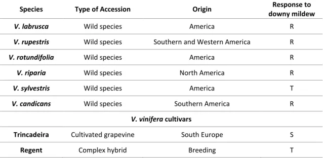
19 Table 1 - Vitis species and Vitis vinifera cultivars used in qPCR analysis of subtilase expression; R (resistant), T (tolerant), S (susceptible)
Species Type of Accession Origin Response to
downy mildew
V. labrusca Wild species America R
V. rupestris Wild species Southern and Western America R
V. rotundifolia Wild species America R
V. riparia Wild species North America R
V. sylvestris Wild species America T
V. candicans Wild species Southern America R
V. vinifera cultivars
Trincadeira Cultivated grapevine South Europe S
Regent Complex hybrid Breeding T
2.2.3 RNA extraction and cDNA synthesis
Total RNA was isolated from frozen leaves with the Spectrum™ Plant Total RNA Kit (Sigma-Aldrich, USA), according to manufacturer's instructions. Residual genomic DNA was digested with DNase I (On-Column DNase I Digestion Set, Sigma-Aldrich, USA). RNA purity and concentration were measured at 260/280 nm using a spectrophotometer (NanoDrop-1000, Thermo Scientific) while RNA integrity was verified by agarose gel electrophoresis (1.2% agarose in TBE buffer). Genomic DNA (gDNA) contamination was checked by qPCR analysis of a target on the crude RNA (Vandesompele et al. 2002). Complementary DNA (cDNA) was synthesized from 2.5 µg of total RNA using RevertAid®H Minus Reverse Transcriptase (Fermentas, Ontario,
Canada) anchored with Oligo(dT)23 primer (Fermentas, Ontario, Canada), according to
manufacturer's instructions.
2.2.4 Primer Design
Grapevine subtilases specific primers were designed with Primer Express software version 3.0 (Applied Biosystems, Sourceforge, USA) using the following parameters: amplicon length between 75 and 200 bp; size: 20 ± 2 bp; melting temperature (Tm): 58 ± 2 C; GC content ± 50 % (Table 2).
20
The reference genes used for the normalization of the target genes results were the previously described in Monteiro et al. (2013).
2.2.5 Quantitative Real time PCR
Quantitative RT-PCR (qPCR) experiments were carried out using Maxima™ SYBR Green qPCR Master Mix (2×) kit (Fermentas, Ontario, Canada) in a StepOne™ Real-Time PCR system (Applied Biosystems, Sourceforge, USA). A final concentration of 2.5 mM MgCl2 and 0.2 μM of
each primer were used in 25 μL volume reactions, together with 4 μL of cDNA as template. The amplification efficiency of each candidate/target gene was determined using a pool representing all cDNA samples. The pool was used to generate a five-point standard curve based on a ten-fold dilution series. Each standard curve was amplified in two independent qPCR runs and each dilution was run in duplicate. Amplification efficiency (E) was calculated from the slope of the standard curve (E = 10(-1/a)), where a is the slope of the linear regression model (y=a log(x)+
b) fitted over log-transformed data of the input cDNA concentration (y) plotted against quantification cycle (Cq) values (x).
Thermal cycling for all genes started with a denaturation step at 95 °C for 10 minutes followed by 40 cycles of denaturation at 95 °C for 15 seconds and annealing for 30 seconds (annealing temperatures are indicated in (Table 2). Each set of reactions included a control without cDNA template. Dissociation curves were used to analyse non-specific PCR products. Three biological replicates and two technical replicates were used for each sample. Gene expression (fold change) was calculated by the Hellemans et al. method* (2007) for both compatible (‘Trincadeira’ versus mock inoculated control samples) and incompatible interactions (‘Regent’ versus mock inoculated control samples). Statistical significance (p < 0.05) of gene expression between the two genotypes was determined by the Mann–Whitney U test using IBM® SPSS® Statistics version 23.0 software (SPSS Inc., USA).
*
The Hellemans et al. method was also applied by me in the qPCR analysis of the gene expression results of ‘Guerreiro, A., Figueiredo, J., Silva, M. S., & Figueiredo, A. (2016). Linking Jasmonic Acid to Grapevine Resistance against the Biotrophic Oomycete Plasmopara viticola. Frontiers in Plant Science, 7: 565.’
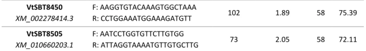
21 Table 2 - Reference genes and target gene primer sequences, amplicon length, amplification efficiency, annealing and melting temperature are represented.
Adopted Identifier/
Accession Number Primer sequence
Amplicon length (bp) Amplification efficiency (E) Ta (C) Tm (C) Reference genes (Monteiro et al. 2013)
EF1α (elongation factor 1-alpha) XM_002284888.2 F: GAACTGGGTGCTTGATAGGC R: ACCAAAATATCCGGAGTAAAAGA 164 1.89 60 79.16 GAPDH (glyceraldehyde-3-phosphate dehydrogenase) XM_002263109.3 F: TCAAGGTCAAGGACTCTAACACC R: CCAACAACGAACATAGGAGCA 226 1.99 60 80.95 UBQ (ubiquitin-conjugating enzyme) XM_002284161.3 F: GAGGGTCGTCAGGATTTGGA R: GCCCTGCACTTACCATCTTTAAG 75 1.95 60 78.86 SAND
(SAND family protein)
XM_002285134.2 F: CAACATCCTTTACCCATTGACAGA R: GCATTTGATCCACTTGCAGATAAG 76 1.93 60 79.16 Target genes VtSBT1922 XM_010663620.1 F: CCATTATACACGACTCCCTT R: TAACCGTCGCCACCAAACA 88 1.96 58 77.48 VtSBT3195 XM_002273159.3 F: CAAGCCCCATTAGCACAC R: TTAGAATCAAGATCAAAGAAG 87 1.92 56 72.95 VtSBT4101 XM_002284065.3 F: CCAGTCCCTACAGTTTATC R: ACACGCCGGAGTAGTTCTT 120 1.96 58 83.45 VtSBT4152 XM_003634104.2 F: GGCGTTCCCATTGCTTGAT R: TTCCCTCGTCTTTGATTATTC 111 1.96 58 76.59 VtSBT4153 XM_003634105.2 F: CCTCCCAATGGAAAAATCTG R: GGCTCATGCTATACAACAAG 170 2.01 58 77.93 VtSBT5381 XM_002275345.2 F: GCCGGAGGGTGGAGTTTTT R: CATGCGTTCTTGCTGTTTTGA 100 1.95 58 80.34 VtSBT5410 XM_002275374.2 F: GGACGGCCTGCAACAACAA R: ATGGCCCTCTTCATCAATAG 86 1.90 58 79.28 VtSBT5429 XM_002275393.2 F: TTGCATAAGGGGTCAGGGTT R: CATTTCGCAGGTGGAGGTG 134 - 60 - VtSBT5471 XM_002275435.2 F: TGACGGAGGAAGAAGTGAGA R: GGGTGAATGCGTTGTTAGTA 95 2.04 58 76.13 VtSBT7502 XM_010659200.1 F: CAGCGAGTTTTAGTGATGAAG R: GGGGTATGGAAGGAAGAGT 172 1.96 58 79.58 VtSBT7672 XM_010649370.1 F: GGGATATGGCCTGAGTCTGA R: CAACGCGCACCGATTATTTT 134 2.03 60 79.44 VtSBT7899 XM_002277863.3 F: GTCCAACCTCACACTACC R: GTTTTCCCATACCCTCGTC 160 - 58 -
22 VtSBT8450 XM_002278414.3 F: AAGGTGTACAAAGTGGCTAAA R: CCTGGAAATGGAAAGATGTT 102 1.89 58 75.39 VtSBT8505 XM_010660203.1 F: AATCCTGGTGTTCTTGTGG R: ATTAGGTAAAATGTTGTGCTTG 73 2.05 58 72.11
2.3 Cloning of the immunity-related grapevine subtilases
2.3.1 Primer Design
Based on the qPCR results, candidate genes were selected for cloning. Grapevine specific primers were designed with Primer Express software version 3.0 and restriction sites for EcoRI and XhoI were added (Table 3).
Table 3 – Primer sequences, amplicon length and annealing temperature for VtSBT7502 and VtSBT8505 subtilases. The forward and reverse primers contained EcoRI and XhoI restriction sites (underlined), respectively.
Adopted identifier Primer sequence Amplicon length (bp) Ta (C)
VtSBT7502 F: CCGGAATTCGAAGTAGCTGCATGACAAC
R: CCGCTCGAGTGAGAACTAATAAGGCAAGAA 1972 56
VtSBT8505 F: CCGGAATTCATGTGCATAGCTTACCTTCTA
R: CACCGCTCGAGGTGCTTGCCGCATCATTTA 2307 56
2.3.2 Gene amplification
VtSBT7502 and VtSBT8505 cDNA’s were amplified from a cDNA sample of the Vitis vinifera cv Regent by polymerase chain reaction (PCR) using the forward and reverse gene-specific primers and 0.02 U/L of Phusion® High-Fidelity DNA Polymerase (Thermo Scientific, Waltham,
Massachusetts, EUA). The 50 L PCR reactions contained 0.5 M of each forward and reverse primers, 2 mM dNTPs and 1 μL of cDNA sample. The PCR were performed on a thermal cycler (Thermal Cycler 2720, Applied Biosystem), using the following conditions: initial denaturation at 98 C for 30 seconds; 35 cycles of 98 C for 10 seconds, annealing for 30 seconds (see Table 3 for annealing temperature (Ta) of each gene), and 72 C for 2.5 minutes; final extension at 72 C for 10 minutes.
23 The PCR products were separated by agarose gel electrophoresis (1.2% agarose in TBE) to confirm the amplified gene sizes. The corresponding band for each gene was excised from the gel and purified using the QIAquick Gel Extraction Kit (Qiagen, Hilden, Germany), according to manufacturer's instructions. Each purified gene was quantified at 260/280 nm using a spectrophotometer.
2.3.3 Purification of the Bacterial Expression Vector
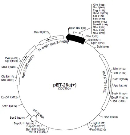
The prokaryotic expression vector pET28a (+) (Novagen) (Figure 3) was used for cloning the selected subtilases. The vector was extracted from Escherichia coli DH5α cells (Novagen), stored in glycerol stock. Briefly, cells were plated in LB (Luria-Bertani) medium supplemented with 50 μg/mL of kanamycin and incubated at 37 C overnight. Five bacterial colonies were inoculated into liquid LB medium supplemented with 50 μg/mL of kanamycin and incubated overnight at 37 C, 200 rpm. The vector was extracted from bacteria with the NZYMiniprep Kit (NZYTech, Lisbon, Portugal), according to manufacturer’s instructions. The purified vector concentration was measured at 260/280 nm using a spectrophotometer.
24
2.3.4 Plasmid and cDNA digestion with restriction enzymes and ligation
Subtilase genes and pET28a (+) expression vector were digested with EcoRI and XhoI restriction enzymes (Thermo Scientific, Waltham, Massachusetts, EUA), according to manufacturer’s instructions, at 37 C for 3 hours. The enzymes were inactivated at 80 C for 20 minutes. The digestion products were purified with the QIAquick® PCR Purification Kit (Qiagen,
Hilden, Germany), according to manufacturer's instructions, and the DNA concentration was measured at 260/280 nm using a spectrophotometer. The efficiency of the digestion for pET28a (+) expression vector was confirmed by agarose gel electrophoresis (1.2% agarose in TBE).
For the construction of the plasmids containing each amplified gene (see Equation 1), subtilase genes were cloned into the pET28a (+) vector using the T4 DNA Ligase enzyme (Thermo Scientific, Waltham, Massachusetts, EUA), according to manufacturer’s instructions. The ligation was performed at 22 C during 4 hours. The obtained constructs were named after the plasmid and the respective subtilase: pET28a – VtSBT7502 and pET28a – VtSBT8505.
𝑛𝑔 𝑖𝑛𝑠𝑒𝑟𝑡 = 𝑏𝑝 𝑖𝑛𝑠𝑒𝑟𝑡 𝑏𝑝 𝑣𝑒𝑐𝑡𝑜𝑟 ×
2
1 × 𝑛𝑔 𝑣𝑒𝑐𝑡𝑜𝑟 Equation 1 - Calculation formula for vector-insert proportion.
2.3.5 Preparation of chemically competent E. coli cells
E. coli One Shot TOP10 (Novagen) was submitted to a protocol for competence induction
and used as host for amplification of recombinant plasmids. Cells were plated in SOB (Super Optimal Broth) medium at 37 C overnight. One colony was inoculated in 225 mL of SOB medium and grown for 2 hours at 37 C, 250 rpm until the OD600nm reached 0.7. Cells were kept on ice for
10 minutes and centrifuged at 1400 g for 5 minutes. The supernatant was discarded and the pellet re-suspended inRF1 buffer (100 mM RbCl2, 50 mM MgCl2 (4H2O), 30 mM KAC, 10 mM
CaCl2 (2H2O), 15 % glycerol). Cells were kept on ice for 15 minutes and centrifuged at 1400 g for
5 minutes. The supernatant was discarded and the pellet re-suspended in RF2 buffer (100 mM MOPS, 10 mM RbCl2, 75 mM CaCl2-2H2O, 15 % glycerol). Cells were divided in 200 L aliquots