MESTRADO EM ONCOLOGIA
ESPECIALIZAÇÃO EM ONCOLOGIA LABORATORIAL
Cooperation between IL-7R signaling
and HOXA9 in leukemogenesis
Beatriz Raposo
M
2019B
e
a
tri
z Ra
p
o
so
A
S
B
e
a
tri
z Ca
rr
inh
o
Ra
p
o
so
IN S TI TU T O D E C IÊ N C IA S B IO MÉ DI CA S A BE L S A LA Z A RBeatriz Carrinho Raposo
Cooperation between IL-7R signaling and HOXA9 in
leukemogenesis
Dissertação de Candidatura ao grau de Mestre em
Oncologia
– Especialização em Oncologia Laboratorial
submetida ao Instituto de Ciências Biomédicas de Abel
Salazar da Universidade do Porto
Orientador: Professor Doutor João Pedro Taborda
Barata
Categoria: Investigador principal/Director de
Laboratório
Afiliação: Instituto de Medicina Molecular João Lobo
Antunes
Categoria: Professor Associado Convidado
Afiliação: Faculdade de Medicina, Universidade de
Lisboa
Coorientador: Doutor Afonso Rocha Martins de
Almeida
Categoria: Investigador Pós-doutorado Sénior
Afiliação: Instituto de Medicina Molecular João Lobo
1
Acknowledgments
First of all, I would like to thank my supervisor, Prof. Dr. João Barata, for accepting me as master’s student in his laboratory, for believing in me and in my capabilities for the development of this challenging work. Thank you for the guidance, the constant support and for being a role model and inspiration.
My personal and professional development during this year and a half was marked by the people in the laboratory who also had a huge importance for me. I would like to thank to my dream mice team: Afonso Almeida and Ana Cachucho, which have been my co-workers and mentors. I am grateful for the knowledge that I gained from you. A special thanks to Afonso, for everything you have taught me, for the patience, for the discussions and the ideas throughout the experiments, for being a positive person in the rare moments I wasn’t. Marta Fernandes, Mafalda Duque and Sofia Ramalho, my gym’s partners, thank you for the snack breaks and the coffees after lunch, for the long talks, for the encouragements, for helping me in my experiments and also thank you for the chocolates and the ice cream moments. Last, but not least, thank you Bruno Cardoso, Rita Fragoso and Daniel Ribeiro for helping me with your advices, Mariana Oliveira, Eunice Paisana, Carlos Custódia, Rita Cascão, Cláudia Faria, Luís Monteiro, Diana Pereira, João Neto, Mafalda Matos and Cláudia Prego thank you all for being such sweet and peculiar persons at the same time and thank you for being a great part of this journey, I couldn’t ask for a better group. I’m lucky to say that I was able to spend a big part of my scientific journey with all of you. I also want to mention the IMM facilities, especially to the people working in the animal facility and flow cytometry, thank you for helping with my work, I also learned so much from you.
I also would like to thank my parents, my grandparents and my sister for providing me the necessary and more that I needed to pursue my dreams, thank you for being always there for me. Cláudia thank you for the love and for put up with my singing moments sister! Thank you to my boyfriend, Renato, for believing in me when I had doubts, for the long brain-storming calls, for listening to my extensive description of my experiments and advising even though you are more of neuroscience person and for calming me down during my stress moments that unfortunately were not few. Lastly, to my friends thank you for being there, for the dinners, the birthdays, the talks and especially for being always who you are, for still being part of my life and for always making me laugh.
2
Resumo
Leucemia linfoblástica aguda (LLA) é o cancro mais frequente em pacientes pediátricos sendo caracterizados por um crescimento anormal de linfócitos imaturos que se inicia na medula óssea e pode evoluir para outros órgãos. O subtipo LLA de linfócitos B é o mais comum, representando 85% dos casos em crianças e 75% em adultos. Têm sido feitos grandes desenvolvimentos no tratamento, no entanto, 20% dos pacientes pediátricos ainda têm recidivas e o tratamento ainda resulta em efeitos secundários importantes, existindo a necessidade de desenvolver novas abordagens terapêuticas.
A Interleucina-7 (IL-7) é uma citocina importante no desenvolvimento de células B e T. Quando a IL-7 se liga ao seu receptor (IL-7R), que é composto pela subunidade alfa e a cadeia gama comum, ativa vias de sinalização incluindo JAK/STAT, MEK/ERK e PI3K/Akt e que desempenham um papel fundamental na manutenção da viabilidade e progressão do ciclo celular. A via de sinalização IL-7/IL-7R tem sido descrita como sendo crucial na LLA, papel particularmente caracterizado em LLA subtipo de linfócitos T. O nosso laboratório descobriu uma mutação de ganho de função no exão 6 do gene IL7R, que codifica para a subunidade alfa do IL-7R, em pacientes com LLA do subtipo T. Tendo todas estas descobertas interessantes em mente, era relevante investigar a sinalização anormal da via IL-7/IL-7R em contexto de leucemia. Para isso, o nosso laboratório desenvolveu um modelo de ratinho com um knock-in condicional onde a mutação de ganho de função do IL-7R está no seu contexto genómico correto, e onde a expressão é mediada pela recombinação de Cre, que por sua vez é controlada pelo promotor CD2. Posto isto, as células hematopoiéticas irão expressar a forma mutada do receptor desde a fase de célula progenitora da linhagem linfóide até à sua maturação.
O primeiro objectivo desta tese que iremos abordar é a caracterização do nosso modelo de ratinho em que obtivemos grandes resultados pois notámos um impacto da mutação IL-7Rα nas células B, levando então ao desenvolvimento de leucemia linfoblástica aguda de células B. Após análises destes resultados, fomos mais além e através das análises do transcriptoma das células B leucémicas descobrimos o gene
Hoxa9 com baixa expressão, que nos levou ao segundo objectivo – explorar o papel do
gene HOXA9. HOXA9 é amplamente caracterizado como sendo essencial na hematopoiese e apesar de ser geralmente encontrado como sobre-expresso em leucemia, alguns artigos mostram o oposto. Através da sobre-expressão deste gene nas células B leucémicas dos nossos ratinhos nós obtivemos resultados promissores
3 mostrando efeitos anti-proliferativos e apoptóticos revelando uma nova função de supressor de tumor.
Palavras-chave: leukemia linfoblástica aguda de células B (LLA-B); interleucina-7 (IL-7);
4
Abstract
Acute lymphoblastic leukemia (ALL) is the most frequent cancer in pediatric patients being characterized by an abnormal proliferation of immature lymphoid cells which initiates in the bone marrow and can evolve to extramedullary sites. The subtype B-ALL is the most common, consisting in 85% of the children cases and 75% of the adults. There have been made great developments in the treatment field, however, 20% of pediatric patients still relapse and treatment still presents relevant secondary effects, therefore, there is an urge to develop novel therapeutic approaches.
Interleukin-7 (IL-7) is a cytokine with vital importance in B- and T-cell development. Upon IL-7 binding to its receptor (IL-7R), which itself is composed by alpha and common cytokine gamma chain subunit, it activates a signaling landscape comprising the JAK/STAT, MEK/ERK and PI3K/Akt pathways that play a key role in maintaining cell viability and cell cycle progression. IL-7/IL-7R signaling has been described as being crucial in ALL, particularly characterized in T-ALL. Our laboratory discovered a gain-of-function mutation in the exon 6 of the gene IL7R, which encodes for the alpha subunit of IL-7R, in T-ALL patients. Having all these interesting findings in mind, it was relevant to investigate the aberrant IL-7/IL-7R signaling in leukemia context. For that reason, our laboratory developed a conditional knock-in mouse model where IL7Rα gain-of-function mutation is in its correct genomic context, where expression is mediated upon Cre recombination, which in its turn is controlled by the promoter CD2. That being said, IL-7R expressing hematopoietic cells will express the mutated form of the receptor from the common lymphoid stage until maturation.
The first aim of this thesis is the characterization of our mouse model in which we achieved great results since we noticed an impact of IL-7Rα mutation on B cells eventually leading to the development of B cell acute lymphoblastic leukemia. Upon these findings we went further and through the transcriptome analysis of leukemic B cells we discovered Hoxa9 downregulated, which led us to the second goal – explore the role of
HOXA9 gene. HOXA9 is widely characterized as being essential in hematopoiesis and
although is generally found overexpressed in leukemia, a few reports indicate the opposite. Through overexpression of this gene in our mice B-leukemic cells we obtained promising results showing anti-proliferative and apoptotic effects revealing a novel tumor suppressor function.
Keywords: B cell acute lymphoblastic leukemia (B-ALL); interleukin-7 (IL-7); IL-7
5
Contents
Acknowledgments ... 1 Resumo ... 2 Abstract ... 4 Contents ... 5 Index of Figures ... 7 Index of Tables ... 8 Abbreviations ... 9 Introduction ...14 B cell development ...14Acute lymphoblastic leukemia ...16
B cell acute lymphoblastic leukemia (B-ALL) ...17
Genetic features in B-ALL ...17
Symptoms and Prognosis ...18
Treatment in B-ALL ...18
IL-7/IL-7R- mediated signaling in normal hematopoiesis and leukemogenesis ....19
IL-7/IL-7R signaling in hematopoiesis ...19
IL-7R-mediated signaling pathways in leukemogenesis ...23
Gain-of-function IL7R mutation ...24
Conditional knock-in mouse model ...24
Homeobox genes and HOXA9 ...27
HOXA9 in hematopoiesis ...28
HOXA9 cofactors ...29
HOXA9 in leukemia ...30
Objectives ...32
Material and Methods ...34
Experimental animals used ...34
Mouse genotyping ...34
Mice leukemia characterization ...35
Blood samples analysis ...35
Leukemia characterization ...36
Cell viability and proliferation assays ...37
6
Cell culture...39
Transfection– Retroviral production ...40
Transduction of the primary cells ...40
Sorting of transduced cells ...41
Treatment with epigenetic inhibitors ...41
Quantitative Real-Time PCR analysis ...42
cDNA synthesis and RT-PCR ...42
Statistical analysis ...42
Results...44
Mutant IL-7R expression from the CLP stage ...44
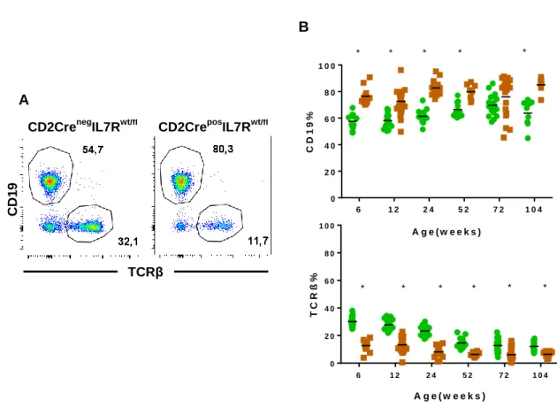
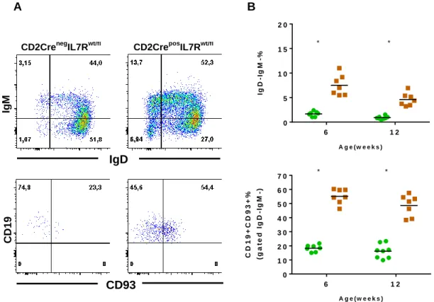
CD2CreposIL7Rwt/fl animals display increased frequency of B-lineage cells ...45
Increase in B lymphoid cells is due to the expansion of immature/precursor B-lineage cells ...47
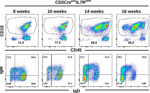
CD2CreposIL7Rwt/fl animals develop B-ALL...49
HOXA9 in CD2CreposIL7Rwt/fl mice leukemia ...53
Overexpressing HOXA9 in CD2CreposIL7Rwt/fl mouse B-ALL ...55
Proliferation differences in leukemic cells overexpressing HOXA9 ...56
In vivo impact of HOXA9 overexpression in leukemic cells ...59
Epigenetic regulation of Hoxa9 ...62
Discussion and future directions ...64
Conclusion ...71
7
Index of Figures
Figure 1. Schematic representation of B cell developmental stages. ...16
Figure 2. IL-7R expression in B and T cell development. ...21
Figure 3. Schematic summarized representation of the IL-7/IL-7R signaling. ...23
Figure 4. Cre-LoxP conditional knock-in system. ...26
Figure 5. Genomic organization of the HOX genes. ...27
Figure 6. Modulation of HOXA9 expression in hematopoiesis. ...29
Figure 7. MSCV‐IRES‐GFP and HOXA9-MSCV-short plasmid structure. ...39
Figure 8. FACS analysis of blood samples from CD2CreposIL7Rwt/fl and CD2CrenegIL7Rwt/fl mice. ...46
Figure 9. B-lineage cells analysis from blood samples. ...48
Figure 10. FACS analysis of CD2CreposIL7Rwt/fl leukemic mice. ...50
Figure 11. Leukemia Free Survival. ...50
Figure 12. FACS analysis of leukemic phenotype of CD2CreposIL7Rwt/fl mice.. ...51
Figure 13. Proliferation and cell viability assay in leukemic mice.. ...52
Figure 14. Next-Generation Sequencing (RNA-sequencing) data of huCD2Cre.IL7Rfxcys mice………. ...53
Figure 15. FACS analysis of the CD2posIL7Rwt/fl leukemic transduced cells in vitro and Hoxa9 expression quantification. ...58
Figure 16. Analysis of the mice injected with transduced cells.. ...61
8
Index of Tables
Table 1. Overall characterization of leukemias developed by CD2posIL7Rwt/fl mice by
FACS. ...52
Table 2. Hoxa mRNA data from Next-Generation Sequencing (RNA-sequencing) of
9
Abbreviations
γc Common gamma chain
7-AAD 7-Aminoactinomycin
Akt/PKB v-akt murine thymoma viral oncogene homolog/Protein kinase B
ALL Acute lymphoblastic leukemia
Allo Allogeneic
AML Acute Myeloid Leukemia
APC Allophycocyanin
APC-CY7 Allophycocyanin Cy-7 Tandem
BAD BCL2-associated agonist of cell death
B-ALL B cell Acute lymphoblastic leukemia
Bcl-2 B cell CLL/Lymphoma 2
BCR B cell receptor
BCR- ABL1 Breakpoint cluster region - Abelson murine leukemia viral oncogene
homolog 1
BM Bone marrow
BTG B cell translocational gene
BV Brilliant violet
CD Cluster of differentiation
CDKNB2A/2B
Cyclin-dependent kinase inhibitor 2A/2B
cDNA coding Deoxyribonucleic acid
CEBPA CCAAT Enhancer Binding Protein Alpha
CLP Common lymphoid progenitor
CMP Common myeloid progenitor
c-Myc Myelocytomatosis viral oncogene
10
Col1a1 Collagen Type I Alpha 1 Chain
CREB cAMP response element binding
DMSO Dimethyl sulfoxide
DNA Deoxyribonucleic acid
E2A Immunoglobulin enhancer-binding factor
EBF Early B cell factor
EDTA Ethylenediaminetetraacetic acid
ERK Extracellular-signal-regulated kinase
ETV6 ETS variant 6
FACS Fluorescence activated cell sorting
FBS Fetal bovine serum
FITC Fluorescein Isothiocyanate
FLT3 Fms-like tyrosine kinase 3
FOXO Forkhead box class O
FSC-A Forward scatter area
GFP Green fluorescent protein
GSK3 Glycogen synthase-3
HEK 293T Human embryonic kidney 293T cell line
HSC Hematopoietic stem cells
iAMP21 Intrachromosomal amplification of chromosome 21
Ig Immunoglobulin
IKZF1 Ikaros zinc finger family protein 1
IL Interleukin
IL-7R Interleukin-7 receptor
IL-7Rα Alpha subunit of interleukin-7 receptor
ILF2 Interleukin Enhancer Binding Factor 2
11
ITGAM Integrin Subunit Alpha M
JAK Janus kinase
KLF4 Kruppel-like factor 4
KMT2A Histone-lysine N-methyltransferase 2A
MAPK Mitogen activated protein kinase
MEF2C/2D Myocyte-specific enhancer factor2c/2d
MEIS1 Myeloid Ecotropic Viral Integration Site 1 Homolog
MLL Mixed lineage leukemia
MRD Minimal residual disease
mTOR Mammalian target of rapamycin
NK Natural killer cells
PAX5 Paired box protein 5
PBS Phosphate buffered saline
PBX Pre-B-Cell Leukemia Homeobox
PCR Polymerase chain reaction
PDK1 Phosphoinositide-dependent kinase-1
PE Phycoerythrin
PE-CY7 Phycoerythrin Cy-7 Tandem
PerCP Peridinin-chlorophyll-protein
Ph Philadelphia chromosome
PI3K Phosphatidylinositol 3-kinase
PIP2 Phosphatidylinositol (4,5)-bisphosphate
PIP3 Phosphatidylinositol (3,4,5)-trisphosphate
Pre-B Precursor B cells
Pro-B Progenitor B cells
RAG Recombination-activating gene
12
RNA Ribonucleic acid
RSK p90 ribosomal kinase
RUNX1 Runt-related transcription factor 1
SCT Stem cell transplantation
SDS Sodium dodecyl sulfate
SSC-A Side scatter area
STAT Signal transducer and activator of transcription
TAE Tris-acetate-EDTA
T-ALL T cell acute lymphoblastic leukemia
TCRβ T cell antigen receptor beta chain
14
Introduction
B cell development
Hematopoiesis is a highly regulated process by which blood cells are formed, developed and differentiated from pluripotent hematopoietic stem cells (pHSCs)1. During embryonic development, hematopoiesis occurs in the fetal liver, while after birth the major hematopoietic site is the bone marrow (BM)2, with B cells able to complete their development within the BM, in contrast to T cells, that require the thymus in order to develop. Functional maturation for both subsets occurs mainly in secondary lymphoid tissues such as the spleen and lymph nodes3.
While pHSCs cells have the ability to self-renew, thus providing lifelong hematopoietic and regenerative potential, it is their differentiation that will provide the immediate precursors of lymphoid and myeloid lineages, namely the common myeloid progenitor (CMP) and common lymphoid progenitor (CLP). The latter is thus the cell stage where B-lineage development starts4.
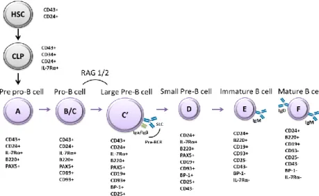
B cell development is a process in which a wide repertoire of cell-intrinsic and extrinsic signaling components such as cytokines, receptors and transcription factors collaborate in the maturation and functional diversity of B lymphocytes5. This implies an important degree of regulation in both the expression of molecules and receptors for environmental clues and in internal machinery dynamics, such as transcription factor expression and chromatin accessibility6. B cell lymphopoiesis can be characterized by the stage and maturation of the B cell and can be identified by fractions (from A to F) based on Richard Hardy’s classification on the early 1990s, from pre-pro to mature B cells7(Figure 1).
Early stages of B cell development are mainly regulated by signaling pathways of three important receptors: c-kit and fms-like tyrosine kinase-3 (FLT3) receptors and the interleukin-7 receptor8, which are controlled by the transcription factors Ikaros and PU.14. The first phases of B-lineage commitment are characterized by the critical process of rearrangement of the immunoglobulin (Ig, encoded by IG genes) heavy and light chain gene segments (variable V, diversity D, joining J)9, although in the very early pre-pro B cell (Fraction A) this rearrangement is practically null2. The rearrangement starts with the pro-B cell stage (Fraction B/C) where cells start to express the CD19 marker4, while rearranging the D and J segments of the heavy chain gene. Afterwards, a V region connects with the DJ segment9. In addition, in this stage, transcriptions factors paired box
15 protein 5 (PAX5) and early B cell factor 1 (EBF1) are expressed and have an important role in B-lineage specification10. Importantly, IG gene rearrangements are mediated by recombinase-activating genes RAG1 and RAG2, known to be regulated by the immunoglobulin enhancer-binding (E2A) transcription factor2,11.
Following the rearrangement of the μ-heavy chain genes, the large pre-B cell stage (Fraction C’) arises with the association of the heavy chain to the surrogate light chain, composed by λ5 and VpreB, that allows the formation of pre-B cell receptor complex (pre-BCR). Assembling with accessory proteins such as Igα and Igβ is necessary for pre-BCR-mediated signaling contributing to further development of B cells2,12. That being said, if the heavy chains produced are effectively associated with the surrogate light chain, the pre-BCR signaling induces a proliferative burst of pre-B cells2, the components of the pre-BCR complex are downregulated and immunoglobulin light chain genes are recombined12.
Afterwards, the cells enter a non-proliferative stage and become small pre-B cells (Fraction D) where light chains are rearranged with the μ-heavy chains selected culminating in the first mature BCR13, characterized by the expression of IgM immunoglobulin on the cell surface9 of immature cells (Fraction E). These cells are now prepared to leave the bone marrow and enter the spleen, where they will further differentiate into mature functional B cells expressing a variety of receptors and different functions (Fraction F)14.
IL-7 plays an important role in B cell development along with E2A, EBF, PAX5 transcription factors in the commitment of cells to B-lineage development and during IG rearrangement through modulation of RAG activity15. The IL-7/IL-7R axis in B lymphopoiesis is thus vital in proliferation, survival and differentiation16, as further highlighted in the rest of the Introduction.
16
Acute lymphoblastic leukemia
Acute lymphoblastic leukemia (ALL) is a hematological malignancy characterized by malignant transformation and abnormal proliferation of B or T cell precursors that invade the bone marrow and extramedullary sites. ALL is the most common cancer among pediatric patients and is the second most prevalent acute leukemia in adults. In the latter, the prognosis is considerably poorer. ALL has a peak of incidence around 2 to 5 years of age17 and also in adulthood around the age of 5018. When the malignant transformation occurs, during B cell development, it gives rise to a specific type of ALL: B cell acute lymphoblastic leukemia (B-ALL)18. In the adult population, B-ALL comprises the majority of the cases, about 75% of ALL cases, while T-ALL comprises the remaining 25%18. In pediatric patients, B-ALL accounts for 85% of cases, whereas T-ALL comprises 15% of the cases19. Relatively to treatment, in pediatric patients, has made great progress, with 5-year overall survival rate from 85% to 90%, however, 20% of the patients still experience relapse17, while in adults the long-term overall survival rate is about 30% to 40%20.
Figure 1 - Schematic representation of B cell developmental stages. B cell development
initiates from a hematopoietic stem cell (HSC). B cell lineage starts with the expression of PAX5 in pre-pro-B cell, which then differentiates and expresses CD19 to become a pro-B cell. At this stage RAGs are expressed and promote IG gene rearrangements. Large pre-B cells express pre-BCR, and, after several rounds of proliferation, differentiate into small pre-B cells and subsequently into immature B cells that express an IgM molecule on the cell surface. SLC, surrogate light chain.
17 B cell acute lymphoblastic leukemia (B-ALL)
B-ALL can be classified into early-pre-B, common B and pre-B-ALL, according to B cell differentiation markers21. Nevertheless, as more patients are studied and transcriptomics and genomics analyses increasingly performed in a routine fashion, other classifications, based on the expression of particular genetic aberrations and/or transcriptional profiles rather than immunophenotyping, gained increasing relevance22. An important and recently identified subtype is Philadelphia (Ph)-like ALL, lacking the hallmark BCR-ABL1 oncoprotein but with similar molecular features to Ph+ B-ALL23–25. Ph-like ALL is more prevalent in the adolescent and adult population21. Patients with Ph-like ALL exhibit poor prognosis and harbor a diverse range of genetic abnormalities, mainly divided in ABL-class and janus kinase-signal transducer and activator of transcription (JAK-STAT) alterations, but with a large fraction including genetic alterations in IKAROS zinc finger family protein 1 (IKZF1)26. Interestingly, IL7R insertions or deletions are also present27. As such, we have a particular interest in this subtype.
Genetic features in B-ALL
Several chromosomal aberrations and genetic alterations have been identified as being involved in the malignant leukemic transformation of B cells including numerical abnormalities, hiperdiploidy and hypodiploidy, being the latter less frequent (3%) but known to be a negative prognostic factor28. Chromosomal translocations such as those leading to the fusions breakpoint cluster region - abelson murine leukemia viral oncogene homolog 1 (BCR-ABL1) and ETS variant 6-Runt-related transcription factor 1 (ETV6-RUNX1) (which define important B-ALL subgroups), as well as deregulated tumor suppressor genes including cyclin-dependent kinase inhibitor 2A/2B (CDKN2A/B) and
TP53, along with mutations affecting IL7R, JAK2 and others have also been identified19.
Importantly, lesions affecting transcriptions factors such as IKZF1, EBF1 and PAX5, leading to loss of function in B-ALL29, constitute a hallmark of this malignancy, being present in more than 60% of the cases30. This includes a recently described mutation in PAX5 (P80R)31. Also, histone-lysine N-methyltransferase 2A (KMT2A) rearrangements are recurrent in children, being linked with poor outcome28.
18 Symptoms and Prognosis
Patients often present symptoms non-related to the disease such as anemia, thrombocytopenia, and leukopenia and others present more B-leukemia-specific for instance fever, weight loss, fatigue and infection. When the cells invade other organs such as the spleen, lymph nodes and liver there is generally an increase in size, due to the accumulation of malignant cells, leading to splenomegaly, lymphadenopathy and hepatomegaly, respectively18.
Patients can be classified into groups based on a range of biological, clinical and genetic features. For example, infants (less than 1 year) have more probability to develop aggressive disease, contrary to children from age 1 to 1032, while older patients over the age of 60 present poor prognosis (10-15% long-term survival as compared to 30-40% for patients less than 60 years old20). White blood cell counts, immunophenotype as well as central nervous system (CNS) disease involvement are also high risk features, being the latter associated with the increased difficulty of chemotherapy penetrance. Additionally, presence of cytogenetic abnormalities can define favorable and unfavorable outcomes. For instance, high hyperdiploidy and ETV6/RUNX1 translocation are associated with the favorable outcome, while hypodiploidy, BCR-ABL fusion, MLL rearrangements and intrachromosomal amplification of chromosome 21 (iAMP21) can be associated with poor outcome32. Relatively to BCR-ABL fusions, Philadelphia chromosome prevalence in adults can range from 15-30%33. The prognostic factors that are more related with adverse prognosis in Ph+ ALL are the deletion of chromosome 9, the presence of +der(22) and trisomy 834. Historically, Ph+ ALL patients are associated with very poor prognosis. However, introduction of tyrosine kinase inhibitors targeted therapeutics has improved treatment outcome18. Related to treatment, the response to initial therapy is also prognosis predictor, being complete remission linked with good outcome while induction failure and detection of minimum residual disease (MRD) at the end of first treatment phase induction are associated with low percentage of overall survival32.
Treatment in B-ALL
Current therapies have a success rate of long-term survival of 80%-90% in children, consequently, being part of the category that have better prognosis. The treatment in adults is more challenging due to the low tolerance to standard chemotherapy, medical comorbidities and the higher probability to develop cytogenetic
19 unfavorable profile, for example, carrying the Ph chromosome21. As for the population in between these two main groups (adolescents and young adults) studies have revealed favorable outcomes in administering controlled pediatric-inspired chemotherapy protocols35. Overall, B-ALL chemotherapy treatment comprises 3 phases: induction, consolidation and long-term maintenance18. Firstly, induction therapy normally includes corticosteroids, vincristine and an anthracycline, afterwards, are given three series of methotrexate with leucovorin rescue and then L-asparaginase. After induction phase, high-risk disease eligible patients are selected to allogeneic stem cell transplantation (Allo-SCT), whilst others may go to consolidation and maintenance. Consolidation step recurs to similar agents as in the induction and in patients with involvement of CNS includes intrathecal therapy and/or radiation. Maintenance phase consists of using mercaptopurine and weekly oral methotrexate and it can last for 2 to 3 years18,36.
IL-7/IL-7R- mediated signaling in normal hematopoiesis and leukemogenesis
IL-7/IL-7R signaling in hematopoiesis
Interleukin-7 (IL-7) is a cytokine, initially discovered in 1988 as growth factor of murine B cell precursors37, essential for the development of T and B cells11. IL-7 is a member of the hematopoietin family, together with other interleukins such as 2, 3, IL-5, IL-9, IL-1IL-5, or stem cell factor11. It is produced by non-lymphoid cells localized in the thymic and liver stroma, bone marrow, spleen and kidney, fetal intestine, embryonic brain, keratinocytes along with other types of cells such as endothelial, fibroblasts and peripheral dendritic cells38.
IL-7-mediated signaling requires a receptor complex, formed with the IL-7Rα chain, and the common cytokine gamma chain (γc-chain), shared with the receptors of several other cytokines (IL-2, IL-4, IL-9, IL-15, IL-21)11. The binding of IL-7 and heterodimerization of the IL-7 receptor (IL-7R) subunits activates a plethora of signaling pathways responsible for proliferation and survival39, as described in further detail below.
The requirement of IL-7 in lymphopoiesis in vivo was first noticed when the use of IL-7 blocking antibodies in mice led to inhibitory effects on the development of B and T cells40. Later studies showed that the elimination of IL-7 or of its receptor in mice led to the loss of both T and B cells29. The absence of IL-7/IL-7R signaling resulted in arrest of B cell
20 development from the pro-B cell stage41 and studies with Il7 knock-out (Il7-/-) mice displayed an interruption from the pro-B cell to the pre-B cell transition42, while mice with knockdown of IL-7Rα expression the interruption occurs at earlier stage, at the pre-pro-B cell stage43. Nonetheless, the common lymphoid progenitors already show dysfunctional development in γc-/- and Il7rα-/- mouse models44, suggesting an even earlier role. Interestingly, it is known that IL-7 requirements differ between humans and mice, B cell development seems little affected in human subjects with non-functional IL-7Rα or common γc chain45. However, in vitro experiments with human B cells, from adult human
bone marrow suggest that IL-7 can still play a role in human B cell differentiation46,47. Thus, there are still unsolved issues concerning the exact physiological role of IL-7 in human B-lymphopoiesis.
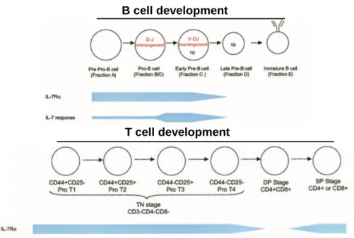
Although the role of the expression of IL-7Rα in very early B-lineage lymphocytes may have not been completely elucidated, it is quite clear that upon differentiation the IL-7R is not expressed in mature B cells48. Of relevance for this thesis, the cooperation between IL-7R and pre-B cell receptor is crucial at critical stages during B-lymphopoiesis49 highlighting the importance of a high degree of regulation in control of IL-7Rα expression, as it occurs in T cell development, where IL-7Rα expression is also under tight regulation (Figure 2). In early mouse B cell development, the transcription factor PU.1 is required to initiate the transcription of Il7r locus and in both human and mouse developing B cells the receptor is expressed until the cells differentiate to mature cells29.
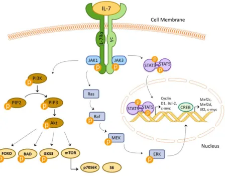
21 IL-7/IL-7R signaling initiates with the binding of IL-7 causing the heterodimerization of the two IL-7R subunits (i.e. IL-7Rα and γc), that results in a conformational change bringing JAK enzymes into close proximity and thereby phosphorylating the cytoplasmic tail of IL-7Rα and allowing a series of downstream intracellular phosphorylation events to occur through different signaling cascades: JAK/STAT, mitogen-activated protein kinase kinase/extracellular-signal-regulated kinase MEK/ERK, phosphatidylinositol 3-kinase/v-akt murine thymoma viral oncogene homolog (PI3K/Akt) and Src family tyrosine kinases - key pathways to promote gene transcription and cellular responses29,39 (Figure 3). IL-7R and pre-BCR collaborate in the development of B cells and each receptor is responsible to activate specific signaling pathways, however, both receptors activate MEK/ERK and PI3K pathways10.
PI3K signals have been stated to be crucial in B cell development50, acting by phosphorylating phosphatidylinositol (4,5)-bisphosphate (PIP2) and leading to the generation of the second messenger phosphatidylinositol (3,4,5)-trisphosphate (PIP3), that in its turn recruits v-akt murine thymoma viral oncogene homolog/protein kinase B
Figure 2 – IL-7R expression in B and T cell development. IL-7/IL-7R signaling is
crucial to lymphopoiesis and acts differently in each lineage. In B cell development, IL-7R is expressed from the pre-pro-B cell stage and continues until maturation. In T cell development the receptor is expressed from the initial Pro T1 stage onwards, except at the double positive (DP) stage, where its transient downregulation is essential for proper T cell selection. Adapted from Fry et al 200211.
B cell development
22 (Akt/PKB) to the plasma membrane, allowing the phosphorylation of Akt by phosphoinositide-dependent kinase-1 (PDK1) in Thr308 and by mammalian target of rapamycin mTOR complex-2 (mTORC2) in Ser47310,51. Activated Akt/PKB then negatively regulates apoptosis by targeting BAD protein, a member of Bcl-2 family, promotes cell survival and proliferation by phosphorylating of forkhead box class O (FOXO) family proteins, and regulates cell metabolism, and, again, viability, through phosphorylation of glycogen synthase-3 (GSK3)52, amongst other targets.
MEK/ERK signals are relevant already in pro-B-cells where the stimulus of anti-Igβ antibodies and exposure to IL-7 were shown to activate this pathway53. ERK phosphorylates several targets in the cytoplasm and nucleus, including the p90 ribosomal kinase (RSK) that in turn phosphorylates cAMP response element binding (CREB). This transcription factor upregulates other transcription factors associated with pre-B cell proliferation, such as c-Myc, myocyte-specific enhancer factor-2c (MEF2C), MEF2D and interleukin enhancer binding factor-2 (ILF2). MEF2C is a target of the p38 mitogen activated protein kinase (MAPK) pathway, being required for mature B cell proliferation. ERK proteins can be involved in proliferation, inhibiting the activity of anti-proliferation agents such as Tob1 and B cell translocation gene (BTG), as well as survival, specifically in pre-B cells10.
As referred above, the other most relevant IL-7R-dependent signaling pathway in B cell development is the JAK/STAT pathway, especially involving the activation of STAT5. Upon IL-7 binding and receptor dimerization, the tyrosine kinases JAK1 and 354 phosphorylate each other and become fully activated, leading to the transmission of intracellular signals by subsequently phosphorylating and thus activating the STAT family of transcription factors55. Activation and dimerization of STAT proteins are conducted by phosphorylation of a tyrosine residue localized in their C-terminal, transactivation domain54, allowing for subsequent translocation to the nucleus, where STATs act as transcription factors controlling proliferation, differentiation and survival55. Particularly, it has been shown that STAT5 restores B cell development in Il7r-/- mice56. As noted already, JAK kinases are responsible for activating also other key signaling pathways, such as MEK/ERK or PI3K/Akt57.
23 IL-7R-mediated signaling pathways in leukemogenesis
The signaling pathways associated with IL-7 binding to IL-7R that were described above are known to be aberrantly active in leukemia. Given their importance in promoting proliferation, cell survival and metabolism, it is not surprising that abnormalities leading to the hyperactivation of these pathways should contribute to leukemogenesis. Indeed, the PI3K/Akt pathway58, as well as its downstream target mTOR59, have been found to be involved in T-ALL, and importantly PI3K/Akt is the dominant signaling pathway associated with IL-7 in T-ALL60. Moreover, STAT5 and STAT3 are considered important in the pathogenesis of lymphoid malignancies, including T-ALL61,62. Other studies showed that IL-7 induced upregulation of Bcl-2 that promoted leukemia progression in human T-ALL cells63 and MEK/ERK cascade was found activated at relapse in pediatric ALL64. While IL-7/IL-7R signaling is widely characterized in T-ALL65,66, PI3K/Akt pathway is also known to be aberrantly activated in B-ALL67.
Figure 3 – Schematic summarized representation of the IL-7/IL-7R signaling.
Upon binding of IL-7, the subunits IL-7Rα and γc dimerize and JAK1 and JAK3 proteins are phosphorylated and activate several pathways. The main pathways here represented are PI3K/Akt/mTOR, JAK/STAT and MEK/ERK that are essential in cell cycle progression, proliferation and cell viability.
24 Overall, these findings suggest that the IL-7/IL-7R axis contributes to the progression and expansion of leukemia, although, the exact mechanisms by which IL-7/IL-7R contribute to leukemogenesis are not clear. For that reason, more studies are necessary to understand the processes underlying malignant transformation downstream from IL-7R signaling, particularly in the context of B cell development.
Gain-of-function IL7R mutation
In accordance with a role for these signaling axis in leukemogenesis, somatic gain-of-function mutations in IL7R (encoding IL-7Rα subunit) were found in pediatric B68 and T
acute lymphoblastic leukemia39,66. Our laboratory demonstrated exon 6 oncogenic gain-of-function IL7R mutations in around 10% of patients with T-ALL39. Other studies found that around 1% of patients with B-ALL also display mutations in exon 6, as well as in exon 5 (S185C), of IL7R68. Exon 6 gain-of-function mutations take place at the extracellular juxtamembrane-transmembrane interface and the majority of them result in an unpaired cysteine residue that is necessary for IL-7Rα homodimerization, and downstream constitutive signaling, independently of IL-7 or the γc subunit39,68. Cysteine-dependent
signaling hyperactivation was further shown to be required for cell transformation39,68.
Conditional knock-in mouse model
In order to investigate the putative role of IL7R gain-of-function mutations in leukemogenesis, several different experimental strategies have been attempted69–71. In
vitro, we have shown that these mutations can be transforming39, while in vivo
experiments using transduced BM or thymic precursors provided only mixed evidence on the role of the mutation in ALL leukemogenesis. Although B and T cell leukemias developed in some instances69,70 strategies used display caveats that can lead to misinterpretations. Indeed, these models include several artificial features, with important consequences on their capacity to mimic true leukemogenesis settings in patients. First, they rely on transplantation of transduced cells to recipient mice, which requires conditioning of the hosts and may interfere in leukemogenesis. Second, retroviral transduction does not allow for control of copy number or integration site. Last but not least, having the IL7R cDNA under the control of a strong promoter rather than of
25 endogenous regulatory elements will result in overexpression/ectopic expression of the receptor, which may have significant consequences in terms of IL-7R-signaling strength and downstream consequences. A good example of this is the fact that Yokoyama et al reported the development of mature “B-ALL”/lymphoma in their in vivo model of IL7R mutational activation70, instead of bona fide B-ALL affecting B-cell precursors, as well as the development of myeloproliferative neoplasms – none of which have been reported in humans in association with IL7R mutation.
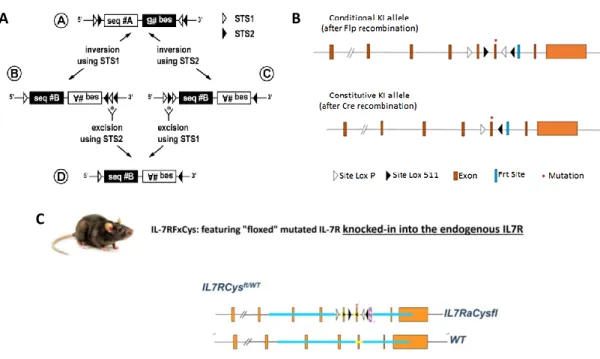
The Cre-LoxP system is a powerful tool that has been used to study molecular genetics through generation of tissue or cell-specific and development-regulated conditional mutants, where genes of interest can be modulated in a defined spatial and temporal context, avoiding resultant indirect effects72. Given the power of this method, our laboratory developed a conditional knock-in mouse mutated for IL-7Rα, in which Cre recombinase is under control of the promoter huCD2 and, most importantly, expression of the mutant IL-7Rα will still be under control of endogenous Il7r regulation (Figure 4). The huCD2 promoter is known to stimulate expression from the stage of common lymphoid progenitors, therefore we are able to target the consequences of such expression to the lymphocyte (CLP-onwards) lineage, thus to both B and T lymphocytes72,73. In this way, we ensure that the mutated receptor is expressed only in lymphoid cells and at physiological levels.
In detail, the mutation selected to characterize IL-7R impact on lymphopoiesis and leukemogenesis was previously identified in our laboratory in T-ALL patients, namely, c.731_732insTTGTCCCAC mutation that led to p.Thr244_Ile245insCysProThr alteration resulting in an insertion of a cysteine39. The mutation was further integrated into the genome of a C57BL/6J mouse strain, connoted IL-7RfxCys mouse. The exon 6 mutated sequence is inverted and integrated in the endogenous Il7r locus of the mouse where it is flanked by two lox sites, LoxP and Lox511, to avoid uncontrolled expression of the mutant
IL7Rα. Since the mutation is flanked by lox sequences, Cre recombinase needs to be
present in order to activate gene expression. Taking this into account, IL-7RfxCys mice with the flanked mutation were crossed with Cre-expressing mice, where Cre is under the control of the promoter CD2 (CD2CRE). Therefore, when the CD2 promoter is active it allows expression of the Cre recombinase, which in turn will drive the expression of the
IL7Rα mutation. This knock-in is generated through a FLEX switch, Cre-mediated,
recombination process74, where the mutated exon 6 is reversed to its original orientation in order to be properly transcribed, while the wild type exon 6 is deleted, culminating in the
26
Figure 4 – Cre-LoxP conditional knock-in system A) Schematic of the FLEX switch system.
Composition of Flex Switch mechanism by image order (A) a first specific target site 1(STS1) and a first specific target site 2 (STS2) and a A DNA sequence all in a given orientation, followed by a B DNA sequence and two others STS1 and STS2 with an inversed orientation when compared to the first sequences referred. Importantly, both STS1 and STS2 are heterotypes therefore there is no recombination between them. Then, B and C are Cre-mediated intermediate sequences, B is formed by an inversion mediated by recombinase STS2 and C is formed after inversion mediated by recombinase STS2. (D) Final product after Cre-mediated excision between homotypic STS2 and homotypic STS1. This reaction is irreversible. Adapted from Schnütgen et al., 200774. B) Organization of the IL-7R knock-in allele before and after CRE recombination. C) Genotype of the heterozygous mice carrying the mutated allele. These mice were then crossed with huCD2CRE mice.
knock-in of the mutated sequence into the allele. The first cells expressing CD2 during hematopoietic development are the common lymphoid progenitors (CLP). As such these will be the first to recombine the Il7r locus leading to the expression of the mutated form of the receptor in all of their progeny, while normal IL-7R function will occur in all other cells that may naturally express the receptor.
This strategy is crucial to mimic a mutated IL-7R in normal lymphocyte maturation and in leukemia development, allowing a correct characterization of how much impact this mutation actually has in the absence of artefactual overexpression of the receptor. The knowledge generated should therefore be closer to the real pathophysiology of the disease downstream from IL7R mutational activation and, as such, it should constitute also an excellent tool to discover novel therapies.
A B
27
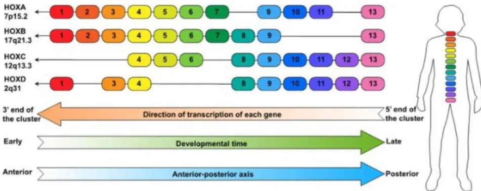
Homeobox genes and HOXA9
The HOX genes encode a family of homeodomain-containing transcription factors that are evolutionarily conserved between species75 and were originally found in
Drosophila melanogaster as regulators of trunk and tail development during
embryogenesis. In mammals, these genes are divided in 4 clusters (A, B, C and D), located in 4 chromosomes76 (Figure 5). HOX genes are very important in shaping animal morphology, more precisely, in specifying cell identity and positioning throughout embryonic development77. HOX genes are relevant in normal and aberrant hematopoiesis78 being differently expressed in hematopoietic cells. For instance, HOXB8 and HOXA10 are expressed in myeloid cells, whereas HOXB3, HOXB4 and HOXA9 have high levels of expression in non-differentiated cells76. HOXA9 has been implicated in ALL and I will go into further detail below.
Although, regulation of expression of HOX genes is not well established, there is some evidence regarding post-transcriptional regulation by micro RNAs, miR-19675 and miR-1080 as well as epigenetic regulators. Polycomb group and trithorax group proteins are crucial to regulate gene expression, have a role in silencing and activating by binding to DNA and induce post-translational modification of histones, respectively81. Studies
Figure 5 – Genomic organization of the HOX genes. The genes are divided
in four clusters HOXA, HOXB, HOXC, and HOXD in which in total contain 39 genes and are expressed in certain body segments during different development phases. It is represented the direction of the transcription of each gene as well as the direction of the spatial and temporal line in which the HOX genes are expressed. From Luo, Z. et al 201979.
28 showed that mutations in the genes belonging to these groups presented similar phenotypes to HOX mutants, suggesting that these genes may be involved in the regulation of Hox genes82. Moreover, it was found myogenic hypermethylation in regions of HOX genes83 and also methylation of HOXA gene was revealed in normal and malignant tissues84, while others studies suggest that histone (de)acetylation can be involved in the modulation of HOX gene transcriptional activity85.
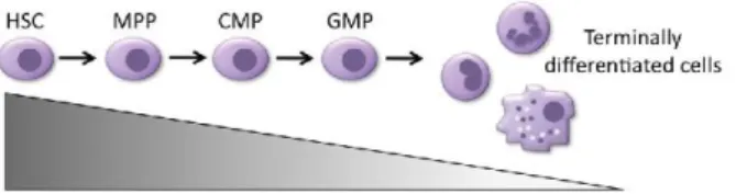
HOXA9 in hematopoiesis
HOXA9 is known to play a role in hematopoiesis (Figure 6), displaying higher expression in HSCs and being the HOX gene most expressed in CD34+ HSCs and early hematopoietic progenitors76, being downregulated upon differentiation. Studies conducted with Hoxa9 knock-out mice revealed that Hoxa9 is necessary for lymphoid development since CLP or earlier phases78, while in others demonstrated impaired hematopoiesis leading to reduced spleen and thymus size as well as decreased numbers of lymphocytes, granulocytes and committed progenitors86. Interestingly, HOXA9 knockdown abolishes the differentiation of HSCs while preserving a pluripotency profile of the cells87. In addition to these findings, Hoxa9-/- HSCs present deficiencies in repopulating bone marrow in irradiated mice88.
As HOXA9 is most highly expressed in the earlier precursors, it is not surprising that in a study, where a Cre-Lox model was developed in which Hoxa was conditionally deleted in a homozygous manner, the most notable finding was a decrease of the repopulation potential of Hoxa-deficient HSCs, a defect restored upon HOXA9 overexpression. At a molecular level, several genes were shown to be upregulated upon HOXA9 overexpression, such as CCAAT enhancer binding protein alpha Cebpa, integrin subunit alpha M Itgam and collagen type I alpha 1 chain Col1a189. Focusing on the B-lineage, in (wild-type) WT mice, several Hoxa genes showed high expression. Mainly,
Hoxa7 and Hoxa5, along with Hoxa9, were still highly expressed in early B cell
development (Fraction A), and their expression decreased progressively thereafter90. Using a similar conditional Cre-Lox Hoxa knock-out model, results showed a significant reduction of B cell populations, mainly at early stages, from A to D fractions, while stages E and F were not or little affected. Notably, when these floxed Hoxa-deleted mice were crossed with CD19-Cre mice, an increase of B cells of Fraction D was observed in these mice compared to the control90.
29 Other experiments revealed that Hoxa genes may be crucial to maintain a specific pool of B cell progenitors, as aged Hoxa-deficient mice showed significant decrease of B cell progenitors in contrast to younger mice91. Curiously, in the younger Hoxa9-/- mice a delay in T cell development was apparent, as fetal thymuses presented a highly reduced size, whilst in adult mice the reduction was not significant92,88. Thus, HOXA genes (and
HOXA9 in particular) may participate in the fine-tuning of lymphocyte development.
HOXA9 cofactors
HOX proteins can regulate in a positive and negative way transcription of targets through two domains, the homeodomain which refers to the DNA binding site, and hexapeptide that binds to cofactors93. Cofactors that belong to the three amino acid loop extension (TALE) proteins are the most relevant HOX-related cofactors including pre-B cell leukemia transcription factor (PBX) and myeloid ectopic insertion site (MEIS1)76. In human leukemia, it is often stated that PBX3 and MEIS1 are both overexpressed along with HOXA994,95. Although HOXA9 can be deregulated through several genetic alterations, such as MLL1 translocations, NUP98 fusions and also translocations with HOXA9 itself96, overexpression of HOXA9 seems to be the most important prognostic factor in acute leukemia97,98,. Moreover, in JAK3/HOXA9-driven T-ALL, STAT5 transcriptional activity was revealed to be significantly active upon HOXA9 expression, therefore, being indicated as a novel HOXA9 cofactor99. Transcription factor kruppel-like factor-4 (KLF4) may be also involved in HOXA9 activity as it is shown to be downregulated in HOXA9-mediated acute leukemia100 and it is also demonstrated that KLF4 can interact with PBX1 and MEIS2 proteins101.
Figure 6 – Modulation of HOXA9 expression in hematopoiesis.
During hematopoiesis process HOXA9 is highly expressed in hematopoietic stem cells and early progenitor cells and is downregulated upon differentiation. Adapted from Collins, C. et al 201696.
30
HOXA9 in leukemia
Deregulated of HOX gene expression has been broadly characterized having a tumorigenic role, and it can act as proto-oncogene or tumor suppressor according to different cancers102. Recently, it has been discovered that mutations in IL-7R/JAK/STAT5 are frequently found within HOXA-positive T-ALL cases. In particular, HOXA9 was found highly expressed in cases with JAK3 mutations99. Intriguingly, low levels of HOXA9 in patients with B-ALL have been reported22,103, suggesting that HOXA genes in general and
HOXA9 in particular may have tumor suppressor rather than oncogenic roles in at least
32
Objectives
IL-7/IL-7R signaling contributes to B and T cell development and its aberrant signaling is involved in leukemogenesis. Our group and others previously described gain-of-function mutations in IL7Rα in T- and B-ALL patients and this led to the need to investigate the impact of the mutations in lymphocyte development and their role in leukemogenesis. For this purpose, a mouse model that could mimic a mutant IL7R-driven leukemia was essential. As described in the Introduction, our laboratory developed a conditional knock-in mouse model where an IL7Rα gain-of-function mutation occurs in its correct genomic context, without overexpression, and induced upon CD2-controlled Cre recombination so that the expression of the mutation is restricted to the lymphoid lineage. In short, hematopoietic lineage cells from the common lymphoid stage onwards will express the mutated form of the receptor when expressing the IL-7R.
Our intent with this work is to characterize the consequences of such expression, both in lymphocyte development and in leukemogenesis, for the purpose of this thesis, focused in B-ALL. Further, in order to understand the molecular mechanisms involved, we also planned to use Next Generation Sequencing-based data (obtained from the characterization of the leukemias) to uncover putative contributors working with the IL-7R in the transformation process. These observations led us to a puzzling finding: downregulation of Hoxa genes, including Hoxa9 - which are normally regarded as oncogenic - in leukemic cells. Due to its vital role in hematopoietic development, we thus set out to understand the role of HOXA9 in mutated IL-7Rα induced B-ALL.
Therefore, two main aims were defined:
1. Characterize the in vivo impact of gain of function mutant IL-7R signaling in B lymphocyte development and leukemogenesis.
2. Explore the role of HOXA9 gene in gain-of-function mutant IL-7R driven leukemogenesis.
34
Material and Methods
Experimental animals used
As mentioned above, our laboratory developed a mouse model by crossing a mouse carrying the IL7Rα mutation with CD2CRE mouse72 that expresses Cre
recombinase under the control of CD2 promoter. This promoter will promote expression of Cre from the common lymphoid progenitor stage and it will allow the expression of the
IL7Rα mutation in the following stages, therefore, in B and T cells. The resultant mice are
hereafter referred to as huCD2Cre.IL7Rfxcys mice and particularly, two mice genotypes were used for the experiments: CD2CreposIL7Rwt/fl, animals (experimental mutant IL7R heterozygous), CD2CrenegIL7Rwt/fl (control animals expressing floxed allele in absence of Cre, thus WT). CD2CreposIL7Rwt/wt animals were also maintained, to monitor for any possible Cre-toxicity effects, which, as expected for this strain104, were not detected. For transplantation assays were used Balb/c Rag-/- γc-/- mice with B, T and NK cell
immunodeficiency105. All the animal procedures were approved by institutional Animal Ethics committee of Instituto de Medicina Molecular and followed the guidelines for the use of laboratory animals by European commission and Portuguese authorities.
Mouse genotyping
Extracts of mice tails and toes were taken and placed into a 1.5ml eppendorfs. Firstly, the samples were lysate with 500µl of tail lysis buffer (Tris pH8 10mM, NaCl 100Mm, EDTA pH8 10mM and SDS 0.5%) and 10µl of proteinase K from Tritirachium
album (20mg/ml) (Sigma-Aldrich) and left in a wet or dry bath for 3-4hours at 56ºC. After
the digestion, the samples were centrifuged for 5 min at 13000rpm and the supernatant was collected and transferred to a 2ml eppendorf. It was added 1ml of isopropanol and the tubes were mixed without vortexing and were incubated overnight at -20ºC. Afterwards, the tubes were centrifuged for 25 min at 13000rpm, 4ºC and the supernatant was removed. Then, the samples were washed with 500µl of 70% ethanol, centrifuge for 15min at 13000rpm, 4ºC following the carefully removal of the supernatant and the samples were left to dry at air. Once dried, the DNA pellet was resuspended in 200µl of sterile Milli Q water and the tubes were left for 10 min in a wet or dry bath at 56ºC for the DNA pellet to go up and stored at -20ºC. For the genotyping, specific primers and different types of DNA polymerase were used, for the Cre recombinase genotyping was used a regular Taq DNA polymerase (Thermo Fisher Scientific) and the primers:
CD2Cre-35 Forward (AGATGCCAGGACATCAGGAACCTG) and CD2Cre-Reverse (ATCAGCCACACCAGACACAGAGATC). For the floxed IL7Rα mutation primers LBM-Foward (ATCTTTACTACTAGAAAGGAAGTGGGTCAG) and LBM-Reverse (GTGGGAAGAACATCAGCTACTTGAAATGTA) were used and also a different DNA polymerase, TaKaRa Taq enzyme (TaKaRa Bio), as suggested by the company that generated the animals (Cyagen). After preparing the amplification mix in the PCR reaction tubes, they were placed in a T100 ™ Thermal Cycler (BioRad) for the PCR assay with specific program of temperatures and times. After PCR assay, samples were run in a 1.5% agarose gel with Tris-Acetate-EDTA (TAE) buffer 1x and DNA bands were observed in Chemidoc XRS+ (BioRad) for analysis and genotyping the mice.
Mice leukemia characterization
Blood samples analysis
For the blood analysis, blood samples were collected in a 1.5ml tube from the facial vein of animals at weeks 4, 6, 8 and then every two weeks into a tube with Heparin 0.6mg/ml (Sigma-Aldrich). Red blood cells were lysed with 950µl of Red Blood Cell (RBC) Lysis Buffer 1x (eBioscience) and incubated for 10min at room temperature with and transferred to 5ml FACS tubes. Cells were washed with 3ml of phosphate-buffered saline (PBS) 1x and centrifuged at 1230rpm during 6min. The supernatant was discarded and the cell pellet was resuspended in 200µl of FACS Buffer (PBS 1x with 2% Fetal Bovine Serum (FBS)). The samples were then transferred into a 96-well round bottom plate, centrifuged at 2000rpm for 2min and the supernatant discarded to initiate the stainings. Then the cells were stained for 20 min on the dark/ice with two different mixes diluted in BD Horizon™ Brilliant Stain Buffer (BD Biosciences) that were previously prepared, mix 1 was composed by the following antibodies: CD4 FITC (BioLegend), IgM PE (BioLegend), Gr-1 PerCP (BioLegend), CD19 PeCy7 (eBioscience), CD8 APC (BioLegend), TCRβ APC-Cy7 (BioLegend), IgD BV-510 (BioLegend), CD45 BV-605 (BioLegend) and CD11b BV-711 (BioLegend). Mix 2 was composed by the following antibodies: CD24 FITC (BioLegend), IgM PE (BioLegend), BP-1 PeCy7 (BioLegend), CD93 APC (BioLegend), CD19 APC-Cy7 (BioLegend), CD127 BV-421 (BioLegend), IgD BV-510 (BioLegend), CD45 BV-605 (BioLegend) and B220 BV-711 (BioLegend). For the leukemic transduced cells (described later on) a different mix was prepared with the antibodies: IgM PE (BioLegend), BP-1 PeCy7 (BioLegend), CD19 APC (BioLegend), IgD BV-510
36 (BioLegend), CD45 BV-605 (BioLegend) and CD11b BV-711 (BioLegend). After staining, the samples were washed with 100µl of FACS Buffer and centrifuged for 2min at 2000rpm. Supernatant was discarded and cells resuspended in 100µl FACS Buffer and placed in 5ml FACS tubes containing 200µl of FACS Buffer. Finally, samples were analyzed using a BD LSRFortessa™ cell analyzer with FACSDiva software 6.2 (BD Biosciences). After acquiring, cells were analyzed using FlowJo software.
Leukemia characterization
After animals developed leukemia and presented signs of sickness such as paralysis and 20% loss of weight they were humanely sacrificed with pentobarbital injection (Euthasol) (Ecuphar) and the subsequent removal of critical organs. Spleen, bone marrow, liver, thymus and lymph nodes were collected and macerated with FACS Buffer into a cell suspension. Spleen and bone marrow cell suspension were centrifuged at 1230rpm for 6min, supernatant was discarded and was add to the pellet 4ml to bone marrow and 5ml to spleen of RBC lysis and left incubated for 6 min to lysate the red blood cells. FACS Buffer was added to dilute RBC lysis solution and stop the reaction and cells were centrifuged for 6min at 1230rpm. Supernatant was discarded and cells were resuspended in FACS Buffer and filtered through a 70μm cell strainer to remove the debris. Cells were then counted and divided in a 96-well round bottom plate for characterization. Then, cells were washed with 100µl of FACS Buffer, centrifuged at 2000rpm for 2min and supernatant was discarded. Leukemic cells were stained with three different mixes previously prepared, diluted in the same Brilliant Stain Buffer as the blood samples antibody mix, composed by mix 1: CD4 FITC (BioLegend), CD127 PE (BioLegend), Gr-1 PerCP (BioLegend), CD25 PeCy7 (BioLegend), IgM APC (BioLegend), TCRβ APC-Cy7 (BioLegend), CD19 BV-421 (BioLegend), IgD BV-510 (BioLegend), CD45 BV-605 (BioLegend) and CD11b BV-711 (BioLegend), mix 2 was equal to the mix 2 from the blood analysis and lastly, mix 3 was composed by: CD21 FITC (BioLegend), CD43 PE
(BD Biosciences), CD45 PerCP (BioLegend), cKit PeCy7 (BioLegend), CD19 APC
(BioLegend), CD5 APC-Cy7 (eBioscience), CD127 BV-421 (BioLegend), IgD BV-510 (BioLegend), IgM BV-605 (BioLegend) and B220 BV-711 (BioLegend). For the transduced leukemic cells recovered from the mice is the same mix as the blood samples. After 20min of staining with each antibodies mix, cells were washed with 100µl FACS Buffer and centrifuged at 2000rpm for 2min. The supernatant was discarded and cells were resuspended in in 100µl of FACS Buffer and placed in 5ml FACS tubes containing 200µl
37 FACSDiva software 6.2 (BD Biosciences). After acquiring, cells were analyzed using FlowJo software.
Cell viability and proliferation assays
For cell viability, an Annexin V assay was performed with 0.5x10⁶ of cells in a 96-well round bottom plate. Then were washed with 100µl of Annexin binding buffer (PromoKine) and centrifuged at 2000rpm for 2min. Leukemic cells were stained for 15min room temperature/dark, with an antibody mix diluted in Annexin Binding Buffer composed by: CD45 FITC, 7-AAD PerCP (eBioscience), CD19 PeCy7, Annexin V APC (eBioscience) and IgD BV-510 (BioLegend). Regarding the transduced leukemic cells, were used few cells according to the limiting number of resultant cells from the sorting and the mix was composed by: IgM PE (BioLegend), 7-AAD PerCP (eBioscience), BP-1 PeCy7 (BioLegend), Annexin V APC (eBioscience) CD19 APC-Cy7 (BioLegend), IgD BV-510 (BioLegend) and CD45 BV-605 (BioLegend). In each annexin assay it was performed a staining in a well with an equal mix but without the Annexin V marker, being the control sample. After staining, cells were transferred to 5ml FACS tubes with 200µl of Annexin binding buffer and analyzed by flow cytometry, as described before.
Regarding proliferation assay, a Ki67 intracellular staining protocol was performed. The same criterion upon number of leukemic and transduced leukemic cells was used to transfer the cells to a 96-well round bottom plate. Cells were washed with FACS Buffer and centrifuged for 2min at 2000rpm. Supernatant was discarded and leukemic cells were stained for 20min on the ice/dark with a surface antibody mix diluted in Brilliant Stain Buffer with the following antibodies: CD45 FITC, IgD PerCP and CD19 PeCy7. For the transduced leukemic cells the surface mix was composed by: IgM PE, IgD PerCP, BP-1 PeCy7, CD19 APC-Cy7 and CD45 BV-605. After staining, the cells were washed with FACS Buffer and centrifuged for 2min at 2000rpm. Supernatant was discarded and cells were vortexed to dissociate the pellet and then fixed using 200µl of fixation buffer composed by ¼ of Fixation/Permeabilization concentrate (eBioscience) and ¾ of Fixation/Permeabilization diluent (eBioscience) and were incubated for 40 minutes at room temperature/dark. Next, cells were washed with 150µl of Permeabilization Buffer 1x (eBioscience) and centrifuged for 2min at 2000rpm. Supernatant was discarded and cells were stained differently: in one sample was pipeted Permeabilization Buffer 1x with no antibodies, other sample were stained with a mix composed by Fc Block (CD16/32) (BioLegend) and κ Isotype control Alexa Fluor® 647 (BioLegend) diluted in Permeabilization Buffer 1x. These last samples were the control. The other samples were
38 stained with the Ki67 marker with a mix composed by Ki67 APC (BioLegend) and Fc block diluted in Permeabilization Buffer 1x and incubate for 20min room temperature/dark. Then, the cells were washed with FACS Buffer, centrifuged for 2min at 2000rpm and the supernatant discarded. Cells were resuspended in 100µl of FACS Buffer, transferred to FACS tubes with 200µl of FACS Buffer and finally analyzed in Flow Cytometry.
Bacteria culture
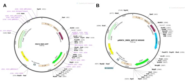
Human HOXA9-MSCV-short plasmid was a gift from Corey Largman (Addgene plasmid # 20978; http://n2t.net/addgene:20978 ; RRID:Addgene_20978)106 and MSCV‐ IRES‐GFP plasmid (Addgene), both represented in the Figure 7, and pCL-Eco retrovirus packaging vector plasmid (Addgene) were grown overnight at 37ºC in plates of Luria-Bertani medium, LB (10g/L Tryptone (BD Biosciences), 5g/L Yeast Extract (BD Biosciences), 10g/L NaCl Aldrich)), supplemented with agar (15g/L) (Sigma-Aldrich) and 100µg/mL of ampicillin (Sigma-(Sigma-Aldrich). Upon overnight incubation at 37ºC, several isolated colonies were selected from each plate and pre-inoculated in LB medium supplemented with 100µg/mL ampicillin, overnight at 37ºC with orbital agitation of 220rpm. The pre-inoculum was then inoculated in LB medium supplemented with 100µg/mL ampicillin, in bacteria to medium proportion of 1:100, for 6h at 37ºC with orbital agitation of 220rpm. To confirm the quality of the DNA, part of the pre-inoculum was used to performed DNA extraction using the GeneJET Plasmid Miniprep Kit (Thermo Scientific) according the manufacturer’s instructions. DNA concentration was measured using spectrophotometer NanoDrop 2000 (Thermo Fisher Scientific). To confirm the quality of the Human HOXA9-MSCV-short plasmid and the insert gene an enzimatic digestion was performed with SpeI (New England BioLabs) and SacII (Promega) enzymes, to confirm the pCL-Eco retrovirus packaging vector was used SacI (New England BioLabs) and the in MSCV‐IRES‐GFP plasmid, the AfeI enzyme (New England BioLabs) was used. The reaction mix with the different samples were put in a bath for 1h30 at 37ºC and the samples were run in a 1.5% agarose gel with Tris-Acetate-EDTA (TAE) buffer 1x and the plasmid DNA was revealed through Chemidoc XRS+ (BioRad).
After confirmation, one of the clones that were left growing overday (6h) was selected and left growing overnight at 37ºC with orbital agitation of 220rpm in LB medium supplemented with 150µg/mL of ampicillin (1:200 proportion of bacteria to medium). Then,