Engineering FKBP-Based Destabilizing
Domains to Build Sophisticated Protein
Regulation Systems
Wenlin An1,2, Rachel E. Jackson1, Paul Hunter1, Stefanie Gögel2, Michiel van Diepen1, Karen Liu3, Martin P. Meyer1, Britta J. Eickholt1,2*
1MRC Centre for Developmental Neurobiology, King’s College London, London, SE1 1UL, United Kingdom, 2Charité - Universitätsmedizin Berlin, Cluster of Excellence NeuroCure and Institute of Biochemistry, Berlin, 10117, Germany,3Department of Craniofacial Development, King’s College London, London, SE1 1UL, United Kingdom
Abstract
Targeting protein stability with small molecules has emerged as an effective tool to control protein abundance in a fast, scalable and reversible manner. The technique involves tag-ging a protein of interest (POI) with a destabilizing domain (DD) specifically controlled by a small molecule. The successful construction of such fusion proteins may, however, be lim-ited by functional interference of the DD epitope with electrostatic interactions required for full biological function of proteins. Another drawback of this approach is the remaining endogenous protein. Here, we combined the Cre-LoxP system with an advanced DD and generated a protein regulation system in which the loss of an endogenous protein, in our case the tumor suppressor PTEN, can be coupled directly with a conditionally fine-tunable DD-PTEN. This new system will consolidate and extend the use of DD-technology to control protein function precisely in living cells and animal models.
Introduction
Phosphatase and tensin homolog located on chromosome 10 (PTEN) is one of the most com-monly mutated or deleted tumor suppressors in human cancer that is also causally linked to autism spectrum disorder [1,2]. Changes in PTEN expression level leads to aberrant cell cycle progression as well as to alterations in cell migration [3,4], amongst other cellular responses. However, it is still a considerable challenge to ascertain the minimum alteration in PTEN level or activity that can cause the onset of tumor formation [5] or neurological changes [6], or whether disease states can be ameliorated by reinstalling PTEN expression. Our ability to address questions of this nature are currently limited by the lack of experimental systems that allow the targeted and reversible regulation of PTEN [7]. We set out to exploit FKBP DD-tech-nology to control PTEN protein function in a rapid, reversible and tunable manner. FKBP is a 12kDa FK506 binding protein, which is broadly expressed in various tissues and functions as a protein chaperone for newly synthesized polypeptides [8]. An engineered human FKBP has
OPEN ACCESS
Citation:An W, Jackson RE, Hunter P, Gögel S, van Diepen M, Liu K, et al. (2015) Engineering FKBP-Based Destabilizing Domains to Build Sophisticated Protein Regulation Systems. PLoS ONE 10(12): e0145783. doi:10.1371/journal.pone.0145783
Editor:Surinder K. Batra, University of Nebraska Medical Center, UNITED STATES
Received:September 6, 2015
Accepted:December 8, 2015
Published:December 30, 2015
Copyright:© 2015 An et al. This is an open access article distributed under the terms of theCreative Commons Attribution License, which permits unrestricted use, distribution, and reproduction in any medium, provided the original author and source are credited.
Data Availability Statement:All relevant data are within the paper.
Funding:This work was funded by a grant from the Medical Research Council (G08022289/89617), the Deutsche Forschungsgemeinschaft (SFB665, TP11; SFB958, TP16). The funders had no role in study design, data collection and analysis, decision to publish, or preparation of the manuscript.
been developed that is able to bind a small synthetic ligand, Shld1, with 1000-fold selectivity over wild-type FKBP due to an amino acid (aa) substitution at F36 (F36V) [9]. A further aa substitution at L106 (L106P) causes FKBP to degrade rapidly in the absence of Shld1 [10]. Here, we further modified FKBPF36V/L106Pby neutralizing highly charged surface amino acid residues, thereby eliminating the ability of the DD to interfere with electrostatic interactions required for full biological function of proteins. In case of PTEN, we demonstrate that
FKBPF36V/L106P/E31S/D32S-PTEN permits rapid, reversible and tunable regulation of PTEN func-tion in cellular and zebrafish models. We further eliminated a potential Cre pseudo-cleavage site within the FKBPgene sequence to render the system compatible with the Cre-LoxP
sys-tem. In conjunction, the two components will facilitate the study into protein network func-tion, as FKBP-tagged proteins and conditional mouse alleles can be combined as required by the experimental set-ups.
Material and Methods
All animal procedures were approved by the local Animal Welfare and Ethics Review Body (King’s College London) and were carried out in accordance with the Animals (Scientific Pro-cedures) Act 1986, under license from the United Kingdom Home Office.
Materials and cloning
Rabbit anti-PTEN (138G6), anti-AKT and anti-pAKT (Ser473) antibodies were from Cell Sig-naling Technology; the mouse anti-PTEN (A2B1) from Santa Cruz Biotechnology; mouse monoclonal anti GAPDH from Abcam. Phalloidin-Alexa fluorophores conjugates and anti-rabbit secondary antibody were obtained from Molecular Probes. pCDNA3+ (Invitrogen) and pCAG vector (a gift from Dr. Kobayashi Mime) were modified by addingHindIII andNotI
sites. The FKBP-PTEN cassettes for linker optimization or surface engineering were cloned
into pcDNA3+ or pCAG byHindIIIandNotIsites. The construct pCAG-FKPTEN was gener-ated by cloning a Neomycin resistance gene cassette and the FKBP-PTEN open reading
frame into the pCAG vector. The CTV vector (Addgene) was used to create constructs M3Lcta and M3Lttgby inserting FKBP-PTEN gene at theAscIsite.
Cell culture
PTEN null U87MG cells were cultured in Dulbecco’s Modified Eagle Medium (DMEM) media containing 10% FCS and 1% penicillin/streptomycin. The U87MG cell line stably expressing FKBP-PTEN was generated by G418 selection (500μg/ml) and maintained in DMEM media
containing 10% FCS and G418 (250μg/ml). Cells were maintained at 37°C, 5% CO2in a
humid atmosphere. Cerebral cortices were dissected from either CD1 or PTENflox/floxE14.5 mouse embryos, dissociated in 0.33 mg/ml trypsin (Worthington, UK) in HBSS (Gibco, UK) for 15 min at 37°C and gently triturated with a fire polished Pasteur pipette in Neurobasal medium (Gibco, UK) supplemented with 2% fetal calf serum (FCS), 2% B27, 1% Glutamax and 1% penicillin/streptomycin (P/S). Neurons seeded at a density of 2000 cell/mm2on poly-L-lysine coated culture plates.
Immuofluoresence staining
U87MG cells stably expressing FKBP-PTEN were cultured on coverslips and fixed in 4% PFA
an Alexa Fluor1
488 anti-Rabbit IgG, Alexa Fluor1
568 Phalloidin and Hoechst for 1 h at room temperature. Cells were then washed three times in PBS and mounted using Mowiol1 mount-ing medium.
Transfection and nucleofection
Transfection of plasmid DNA into U87MG cells was carried out using GeneJuice (Novagen). Nucleofection of mouse primary cortical neurons was performed by using Amaxa1Mouse Neuron Nucleofector1Kit(Lonza, UK). In order to monitor PTEN activity in transiently transfected U87MG cells, the co-transfection of AKT-GFP with FKBP-PTEN or GFP-PTEN
constructs was exploited as a fast and effective experimental protocol that allowed the analysis of PTEN activity towards PIP3-dependent signaling in transfected cell, only. This strategy was pursued in transient transfections (Figs1and2), but not in stable FKBP-PTEN expressing
U87MG cells lines (Fig 3).
Western blotting
U87MG cells or primary neuron cultures was washed with ice-cold PBS and then lysed in lyses buffer (50 mM Tris-HCl, pH7.5, 150 mM NaCl; 1% Triton X-100; 500μM NaF; 1 mM sodium
orthovanadate; 1X protease inhibitor) for 10 min. Cell debris was removed by centrifugation at 13000 rpm for 10 min. The boiled supernatants supplemented with SDS loading sample buffer were loaded and run on 8% SDS-PAGE gel. Separated proteins were transferred to a nitrocellu-lose membrane. Blots were blocked in 5% skimmed milk powder in TBST (20 mM Tris; 150 mM NaCl; 0.1% Tween) for 1 h and incubated with primary antibody in 5% skimmed milk powder/TBST overnight at 4°C or for 1h at room temperature. The expression of proteins was visualized by HRP-conjugated secondary antibody and enhanced Chemiluminescence Kit (Pierce).
Colony formation assay
A single-cell suspension was prepared in 0.35% low melting point agar and plated (100 or 200 cells per well) in 24 well plates, which were pre-coated with 0.5% agar. Cell were then cultured for 3 weeks in media containing different concentrations of Shld1. Experiments were analyzed according to the protocol published by Frankenet al. [11]. Plating efficiency was determined by the ratio of the number of colonies to the number of cells seeded.
Cell invasion assay
Cell invasion assay was performed using BioCoat™invasion chamber (BD Biosciences) with 8μm pore, according to the manufacturer’s instructions (BD Biosciences) with minor
modifica-tions. 0.5 ml of cell suspension (2.5x10⁴cells per well) prepared in serum-free media was added on rehydrated 24-well BioCoat™Matrigel™Invasion Chambers, which were placed in 24-well plate containing normal culture media (DMEM plus 10% FBS), and incubated in a humidified tissue culture incubator for 19 h, at 37°C, 5% CO2atmosphere. The cells were fixed with 4%
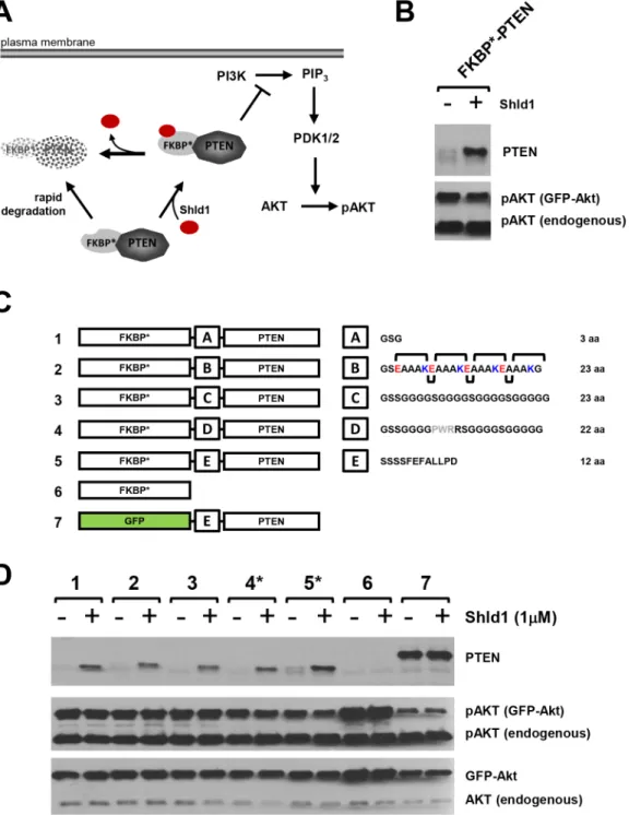
Fig 1. Consequence of linker optimization on PTEN activity of the FKBP*-PTEN fusion protein.(A)Rationale of the DD-technology: Fusion of PTEN with a modified human FKBP variant harboring the F36V and L106P point mutations (FKBP*) leads to degradation of the fusion protein. Presence of the FKBP*ligand Shld1 confers stabilisation of FKBP*-PTEN and results in inhibition of PI3K/Akt signaling. (B)The fusion protein FKBP*-PTEN shows no PTEN activity. PTEN-deficient U87MG cells were transiently co-transfected with FKBP*-PTEN and AKT-GFP. After one day, cells were treated with Shld1 for 12 hours at 1μM before cell lysis and samples analyses by western blotting using indicated antibodies.(C)Consequence of linker optimization on PTEN activity of the FKBP*-PTEN fusion protein. Schematic representation of linker optimization. FKBP*-linker-PTEN variants with different linker regions (A-E) were generated. Linker A—short flexible linker; Linker B—a helix forming linker. Linker C—long flexible linker. Linker D—long flexible linker containing the PWR motif. Linker E—the original linker present in the enzymatic active GFP-PTEN.(D)Analyses of the effect of different linkers on PTEN activity in FKBP* -PTEN fusion variants in U87MG cells. Constructs expressing FKBP*-PTEN variants or GFP-PTEN were co-transfected with AKT-GFP into a PTEN null U87MG cells. Stabilization of FKBP*-PTEN or GFP-PTEN in presence or absence of Shld1 was monitored by PTEN antibody. The phosphorylation level of AKT-GFP at Ser473 served as a fast and effective experimental protocol to control for PTEN activity towards PIP3-dependent signaling in transfected cells, only. Only constructs 4 and 5, which contain proline in the linker sequence, showed moderate PTEN activity towards PI3K signaling.
Zebrafish microinjection and visualization of FKBP
**
-PTEN expression
U87MG cells stably expressing FKBP-PTEN were transiently transfected with RFP or
pre-stained with DiI (Sigma). Approximately 100 cells were then microinjected into the perivitel-line cavity of 2 dpf (day of post fertilization) zebrafish larvae. Fish larvae were immediately imaged under an inverted fluorescence microscope (0 dpi) and then transferred into Danieau’s solution containing 4μM Shld1 or control media containing no Shld1. Fish larvae were again
imaged after 24h (1dpi) to analyze the effect of Shld1 induced FKBP-PTEN stabilization on
tumor growth and migration.
FKBP-PTEN stabilization in fish larvae was verified by immuostaining with PTEN
anti-body. Briefly, embryos were fixed in 4% PFA in PBS overnight at 4°C, and then sequentially dehydrated with 15% and 30% sucrose. Cryosections were stained by immunofluorescence staining protocols. Images were obtained by confocal microscopy.
Statistical analysis
The migration of the injected U87MG cells stably expressing FKBP-PTEN in fish embryos in
presence or absence of Shld1 were analysed in a similar way to fluorescent protein diffusion profiles [12] using the Radial Profile Plot plug-in of ImageJ. The concentric movement of cells away from a central point was quantified as a measure of increased fluorescence intensity at a distance from the central point over time. Curve fitting was carried out in Excel using the
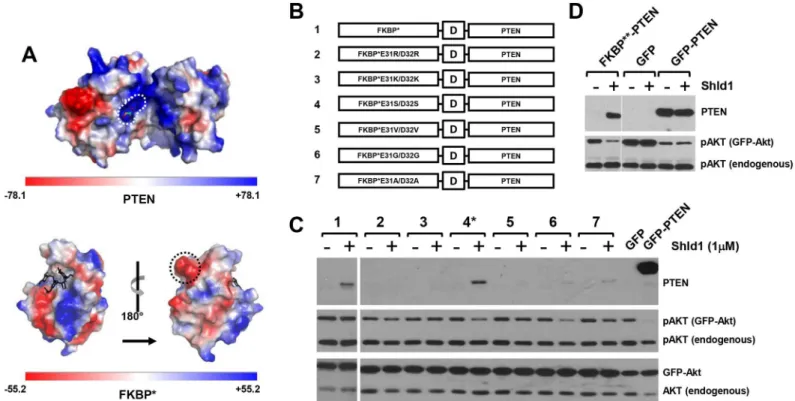
Fig 2. FKBP surface engineering restores PTEN activity of FKBP**-PTEN fusion protein. (A)Electrostatic potential map of PTEN (PDB ID: 1d5r) and FKBP (PDB ID: 1BL4). Positively charged binding pocket of the PTEN PIP3 substrate is circled with a white dashed line; the negatively charged lump formed by E31/D32 residues on the FKBP surface is circled by a black dashed line.(B)Schematic representation of amino acid residue substitutions of FKBP* protein surface at E31/D32.(C)Different FKBP*-PTEN mutants were transiently co-tansfected with AKT-GFP into U87MG cells, the expression of fusion protein and pAKT were examined by Western blotting.(D)E31S/D32S amino acid substitutions on FKBP*(FKBP**) restores PTEN activity of the FKBP** -PTEN fusion protein. FKBP**--PTEN (or GFP/GFP--PTEN) was transiently co-expressed with AKT-GFP in U87MG cells. Cells were treated with Shld1 and analyzed as before.
equation:f(t)=b0exp(-b1)(t)whereb0andb1are parameters of the curve. Migration index (Mi)
was calculated by as a ratio ofb0at time points 1dpi and 0dpi (Mi=b0(1dpi)/b0(0dpi)). The
aver-age migration index of control group (without Shld1 treatment, n = 13) is compared with that of Shld1 treated group (n = 11). The size of tumor mass was measured by ImageJ. The average ratio of tumour mass size (1 dpi/0 dpi) was calculated and analyzed by Excel1and SPSS using student two tailedt-testp<0.01;p<0.001.
Results
We fused PTEN at the C-terminus of the FKBPF36V/L106P-based DD (FKBPF36V/L106Pwill be referred to as FKBP) using a 12 aa linker sequence (-SSSSFEFALLPD-). We proposed that in
the absence of Shld1, FKBPwould be rapidly degraded by the ubiquitin proteasome system
[13], resulting in the loss of FKBP-PTEN fusion protein, whereas addition of Shld1 will
stabi-lize the protein and inhibit PI3K/Akt signaling (Fig 1a). Although expression levels of the FKBP-linker-PTEN fusion demonstrated Shld1 dependency, the protein did not alter PI3K/
Akt signaling (Fig 1b). FKBPhas been successfully used to regulate a number of different
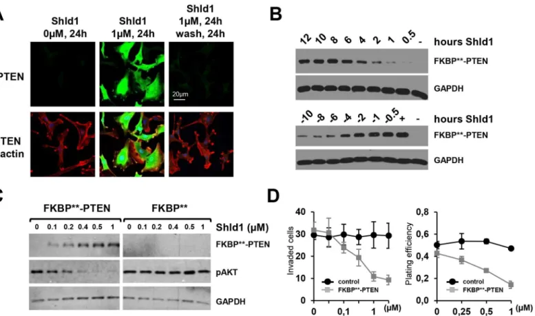
Fig 3. Engineered FKBP**permits rapid, reversible and tunable regulation of PTEN function in cellular models. (A)FKBP**-PTEN is reversibly regulated in response to Shld1 treatment in cells. U87MG cells stably expressing FKBP**-PTEN were treated with vehicle or with Shld1 (1μM) for 24 h. One set of cells was then washed thoroughly with DMEM and cultured in Shld1-free culture media for further 24hs. The expression level of FKBP**-PTEN was examined by immunofluorescence staining.(B)upper panel, U87MG cells stably expressing FKBP**-PTEN were treated with Shld1 (1μM) for different time periods; lower panel, Shld1 (1μM) was removed after 24 h treatment, and cultured in Shld1 free culture media for different time periods as indicated.(C) PTEN protein levels and activity can be maintained at specific concentrations over a prolonged period of time in dependence of Shld1. U87MG cells stably expressing FKBP**-PTEN were cultured in media containing indicated concentrations of Shld1 for 24 h.(D)Stable FKBP**-PTEN expressing U87MG cells were analyzed in cell invasion (left) and colony formation (right) assays. In the cell invasion assay, data are shown as the mean±SEM of the number of cells
migrated in three independent experiments. Plating efficiency refers to the ratio of the number of colonies to the number of cells seeded, shown as mean
±SEM of three independent experiments.
proteins, including cdc42 [14], RhoA [14], and MLYCD [15], with no reported interference in their activity. We therefore surmised that the linker might not be suitable for generating biolog-ical active FKBP-PTEN. Based on available knowledge regarding linker optimization [16,17],
we composed a number of different peptide sequences with the aim of restoring PTEN activity in the FKBP-linker-PTEN fusion protein (Fig 1c). However, the most theoretically effective
linkers [17], a helix forming linker or a long flexible linker, did not restore PTEN activity as verified by analyzing pAkt levels in PTEN-null U87MG glioblastoma cells transfected with linker variants (Fig 1d). In contrast, a linker containing the unique cyclic imino acid proline, which is unable to form hydrogen bonds to surrounding amino acids, was able to slightly improve PTEN activity (linker sequence 4,Fig 1d). This limited success in restoring PTEN activity using linker optimization suggest that other mechanisms impede biological function of the phosphatase when presented in the FKBP-PTEN fusion protein.
FKBP surface engineering restores PTEN activity of the FKBP-PTEN
fusion protein
We considered the possibility that FKBPmight directly interfere with PTEN activity by
block-ing substrate access to the active site of the enzyme, which consists of a large pocket with posi-tive charges that accommodates the PTEN phosphoinositide substrates (Fig 2a). Indeed, the electrostatic potential maps of the PTEN surface (PDB ID: 1d5r) and the FKBPsurface (PDB
ID: 1BL4) revealed complementary charge distributions of the PTEN substrate binding region and large negatively charged protrusions present on the FKBPsurface (Fig 2a). Therefore, we
engineered the FKBPsurface such that the charged regions were neutralized by replacing two
acidic amino acid residues (E31 and D32) with basic amino acid residues (Arg or Lys), with polar uncharged residues (Ser), with hydrophobic residues (Val or Ala), or with the small amino acid Gly (Fig 2b). Transfection of FKBP-PTEN surface engineered variants into
U87MG cells demonstrated that FKBPmodifications containing the aa substitutions E31S/
D32S, E31G/D32G or E31A/D32A (Fig 2b, PTEN variants 4, 6, 7) resulted in Shld1 dependent regulation of PTEN activity towards PI3K/Akt signaling. However, only the E31S/D32S FKBP
mutant was efficiently stabilized by Shld1 (Fig 2c). Hereafter, we refer to FKBPE31S/D32S
-PTEN as FKBP-PTEN. As the expression level of stabilized FKBP-PTEN defines the
dynamic range of the scalability of the FKBP-PTEN regulation system, we exploited the
strong CAG promoter sequence, a combination of the CMV early enhancer element and the chicken beta-actin promoter to drive FKBP-PTEN expression. Subcloning of the FKBP
-PTEN cassette into a pCAG vector increased the level of protein expression and activity of FKBP-PTEN to a level comparable to GFP-PTEN (Fig 2d).
Engineered FKBP
**
permits rapid, reversible and tunable regulation of
PTEN function in cellular and zebrafish models
To further characterize the scalability, speed and reversibility of FKBP-PTEN in response to
Shld1, we generated a U87MG cell line stably expressing FKBP-PTEN under the control of
the CAG promoter (Fig 3). Immunofluorescence labeling of these cells revealed that FKBP
-PTEN was robustly stabilized following the addition of Shld1, and this stabilization could be abolished by Shld1 washout (Fig 3a and 3b). FKBP-PTEN protein levels were gradually
up-regulated with increasing Shld1 concentration, resulting in a concomitant decrease in phos-phorylated Akt, indicating that PI3K signaling can be fine-tuned by altering the level of FKBP-PTEN (Fig 3c). The time-dependent stabilization profile of FKBP-PTEN showed
that the engineered FKBPresponded within a few hours of addition of Shld1 and reached a
faster than that of FKBP[14]. We also discovered a rapid turnover of FKBP-PTEN within a
few hours after the removal of Shld1 (Fig 3b). The half-life of FKBP-PTEN fusion protein is
as short as 2-4h, which is comparable to FKBP[15] and to approximately 5% of the 48–72 h
half-life of endogenous PTEN [18]. Thus, our engineered FKBPprovides an efficient
molecu-lar tool to manipulate PTEN activity in a scalable, rapid and reversible manner. To characterize whether our engineered FKBPcould finely tune PTEN activity and regulate biological
responses, we carried outin vitroinvasion and colony formation assays [11,19] using the FKBP-PTEN U87MG cell line. Cell invasion and colony formation in FKBP-PTEN
U87MG cells, but not wild type U87MG cells, could be inhibited by Shld1 in a dose-dependent manner (Fig 3d). Thus, Shld1 can be used to fine tune PTEN activity to inhibit tumor growth and tumor cell division.
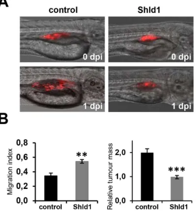
To investigate whether Shld1 can induce FKBP-PTEN stabilization and control biological
responsesin vivo, we used an established zebrafish metastasis model [20], and microinjected the FKBP-PTEN U87MG cells, either pre-stained with DiI or transfected with RFP, into the
perivitelline cavity of 2 days post fertilization (dpf) zebrafish larvae. Larvae were then trans-ferred into Danieau’s solution with or without 4μM Shld1. Images of larvae were taken
imme-diately after injections and 24h post-injection (Fig 4a). The migration indices and the increase in tumor mass size of transplanted cells were quantified over this time-course. These experi-ments demonstrated that Shld1 is effective in stabilizing FKBP-PTEN in zebrafish larvae,
resulting in inhibition of invasion and growth of FKBP-PTEN U87MG cells (Fig 4b). They
also show for the first time that DD-systems can be used effectively to manipulate protein levels on demand in the zebrafish.
Codon optimization of FKBP
**
establishes a conditional fine-tuning
protein regulation system
In order to achieve advanced regulation of protein function, it would be desirable to combine tunable FKBP-PTEN with established systems enabling precise control of gene expression,
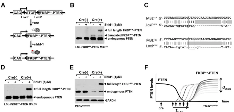
such as the Cre/LoxP system (Fig 5a). For this purpose, FKBP-PTEN was subcloned
immedi-ately downstream of the second LoxP site of the LoxP-Stop-LoxP (LSL) cassette. We tested the pCAG-LSL-FKBP-PTEN construct in mouse cortical neuronal primary cultures by transient
co-transfection with Cre-IresRFP (or, in control experiments, RFP). The FKBP-PTEN fusion
protein was only detectable by western blotting in the presence of Cre, and following applica-tion of the synthetic ligand Shld1 (Fig 5b). Surprisingly, when Cre and FKBP-PTEN were
co-expressed, the anti-PTEN antibody also identified a distinct protein band with lower molecular weight than full-length FKBP-PTEN, which was stable even in the absence of Shld1. We
rea-soned that this protein could represent a form of FKBP-PTEN with a truncation in the
FKBPdomain which allows it to escape Shld1 regulation. A mechanism involving
Cre-dependent truncation of FKBP-PTEN suggests that these two regulation systems could be
incompatible. Due to the sequence similarity of FKBPand the engineered FKBPdomain it
is likely that this might also be true of the parental DD system. We therefore investigated the potential sites of Cre action within the DD domain.
Cre recombinase is an enzyme known to catalyze site specific recombination events between two DNA recognition sites (LoxP sites) of 34 base pairs, which form a stem-loop-stem struc-ture. We aligned the LoxP sequence with the human FKBPDNA sequence and found an
88.9% similarity between the loop region of LoxP and the sequence surrounding the third ATG of the human FKBPgene (M3Lctaand LoxPrev) (Fig 5c). Consequentially, the third ATG
region (ATGCTA) of the human FKBPgene might be erroneously recognized as a Cre
the fourth ATG. In this case, although the destabilizing site L106P on FKBPprotein still
remains, the truncated FKBPmight lose its destabilizing property as shown by the
Shld1-in-dependent band (Fig 5b). To test this idea, we replaced the high similarity region ATGCTA (M3Lcta) of the human FKBPgene with silent mutations ATGTTG (M3Lttg). We found that
the truncated FKBP-PTEN band (Fig 5b) disappeared following mutations of Lctato Lttg,
whilst the expression of full-length FKBP-PTEN remained both Cre- and Shld1-dependent
(Fig 5d). Thus, we engineered FKBPfurther to implement a functioning system that offers
dual control of FKBP-PTEN expression at transcription and posttranslational levels.
In order to test if PTEN loss can be directly coupled with the expression of tunable FKBP
-PTEN, we co-transfected M3LttgpCAG-LSL-FKBP-PTEN with Cre-RFP (or RFP alone in
control experiments) into PTENflox/floxmouse neurons by nucleofection. Cre expression led to substantial decreases in endogenous PTEN levels, and created Shld1 depended regulation of exogenous FKBP-PTEN (Fig 5e). Thus, insertion of the LSL cassette ensures that loss of
endogenous PTEN and gain of tunable FKBP-PTEN takes place in the same cells.
Discussion
The targeting of protein stability with small molecules has emerged as a strategy in synthetic biology to ascertain protein function. The technique involves genetically endowing the protein
Fig 4. Engineered FKBP**permits regulation of PTEN function in the zebrafish. (A)The migration and growth of stable FKBP**-PTEN expressing U87MG cell clones injected into zebrafish embryos can be inhibited by Shld1. Prior to microinjection into the perivitelline cavity of 2 days post fertilization (dpf) zebrafish embryos, cells were pre-stained with DiI. Embryos were then transferred into fish water with (n = 11) or without (n = 13) Shld1 (4μM) and maintained for 24h. Images were taken immediately after injections (0 days post injections, dpi) and after 24 h (1dpi).(B)The migration indices and the increase in tumor mass size of transplanted cells were quantified.**p<0.01;***p<0.001.
of interest with a protein destabilizing domain specifically and reversibly controlled by a small molecule, so that the stability of the fusion protein is dependent upon the sequence of the DDs rather than that of the POI. An established DD sequence is based on a modified FKBP (FKBPF36V/L106P); however, one of the recurring problems associated is the possible interfer-ence of the FKBPF36V/L106Ptag with electrostatic interactions, as well as the remaining unaf-fected endogenous protein, which interfered here with the phosphatase activity of PTEN.
We modified FKBPF36V/L106Pby neutralizing highly charged surface amino acid residues. This modification resolved the problem of electrostatic interference by ensuring the full biolog-ical activity of PTEN, which retains highly charged amino acids within the active site essential for function. We demonstrate that our newly generated fusion protein (FKBP-PTEN)
per-mits rapid, reversible and tunable regulation of protein function in cellular as well as, for the first time, in zebrafish models. Furthermore, we have rendered the system compatible with the Cre-LoxP system. We identified and eliminated a potential Cre pseudo-cleavage site within the FKBP gene. Following Cre-mediated recombination, expression of the FKBP-PTEN gene is
Fig 5. Codon optimization of FKBP**establishes a conditional fine-tuning protein regulation system. (A)The transcription of the destabilization cassette FKBP**-PTEN was subcloned downstream of LoxP-Stop-LoxP; transcription was driven by the ubiquitous CMV-enhanced chicken beta actin promoter (CAG). Presence of Cre induces FKBP**-PTEN gene transcription; however, the translated FKBP**-PTEN will be rapidly degraded. Addition of Shld1 confers FKBP**-PTEN stabilization in a tunable and reversible manner.(B)Cre-mediated cleavage of LoxP-Stop-LoxP (LSL) created a truncated FKBP**-PTEN fusion protein. Mouse forebrain neurons were nucleofected with LSL-FKBP**-PTEN and Cre-IresRFP (Cre (+)), or RFP (Cre (-)). Cells were treated with Shld1 (or control vehicle) overnight, before analyses of cell lysates using indicated antibodies.(C)Sequence alignment of FKBP**and LoxP. The third ATG codon region sequence (TATGCTA) of FKBP**(M3Lcta) is identical to the linker region of the LoxP stem-loop sequence (TATGCTA). Codon optimization of CTA with TTG (both code for amino acid Leu) will destroy the potential pseudo-cleavage site of Cre on FKBP**.(D)Codon optimization of M3Lctato M3Lttgin FKBP
**abolished the Cre-dependent FKBP**-PTEN truncation and produced a Shld1 dependent PTEN fusion protein. The M3Lttg modified LSL-FKBP**-PTEN construct was co-expressed with Cre-IresRFP (Cre (+)), or RFP (Cre (-)) in mouse forebrain neurons as before.(E) Combinatorial use of the Cre-LoxP system with the FKBP**-PTEN/Shld1 chemical-genetic protein control system in PTENflox/floxcells. Constructs were nucleofected intoPTENloxp/loxp
mouse primary neurons as before. Note that nucleofection of primary cells occurs with efficiencies at approx. 80%, and result in a residual endogenous PTEN signals detected by western blotting.(F)The generated system is able to couple PTEN-loss directly with the expression of tunable FKBP**-PTEN. Upon Cre-mediated recombination,PTENloxp/loxpcells will lose PTEN and, at the same time, activate expression of FKBP**-PTEN. In essence, endogenous PTEN is replaced by tuneable PTEN, which will enable to test whether PTEN-loss induced phenotypes can be rescued at different time-points (tShld1) and/or at different PTEN-concentrations (dShld1).
achieved. The system now provides two layers of control over protein levels and activity: the first operating at transcriptional and the second at posttranslational level.
The development of the FKBP-Shield1/Cre-LoxP system offers unique possibilities to manipulate protein functionin vitroandin vivo, by combining the loss of endogenous proteins directly with the expression of tunable versions. This system is compatible with the building of sophisticated protein regulation networks as FKBP-tagged proteins, as well as conditional
mouse alleles can be selected and combined as required. Additionally, the system allows precise and effortless control over the re-installment of protein function following molecular deficits, thus offering a powerful means with which to address the reversibility of a number disease states, in particular those associated with developmental disorders. Therefore, this study con-solidates and extends the use of DD-technology to manipulate protein function in a specific, fast, reversible and tunable manner.
Acknowledgments
This work was funded by a grant from the Medical Research Council (G08022289/89617), the Deutsche Forschungsgemeinschaft (SFB665, TP11; SFB958, TP16). We would like to thank Robert Hindges for providing the Cre cDNA and Lloyd Trotman for PTEN flox/flox mice. We also thank Tom Wandless for providing the original FKBP cDNA, as well as providing feed-back on the work.
Author Contributions
Conceived and designed the experiments: WA REJ MvD MPM KL BJE. Performed the experi-ments: WA REJ PH MvD SG. Analyzed the data: WA REJ PH MvD. Contributed reagents/ materials/analysis tools: KL MPM. Wrote the paper: WA REJ MPM BJE.
References
1. Dahia PL (2000) PTEN, a unique tumor suppressor gene. Endocr Relat Cancer 7: 115–129. PMID:
10903528
2. van Diepen MT, Eickholt BJ (2008) Function of PTEN during the formation and maintenance of neuro-nal circuits in the brain. Dev Neurosci 30: 59–64. PMID:18075255
3. Sun H, Lesche R, Li DM, Liliental J, Zhang H, Gao J, et al. (1999) PTEN modulates cell cycle progres-sion and cell survival by regulating phosphatidylinositol 3,4,5,-trisphosphate and Akt/protein kinase B signaling pathway. Proc Natl Acad Sci U S A 96: 6199–6204. PMID:10339565
4. Yamada KM, Araki M (2001) Tumor suppressor PTEN: modulator of cell signaling, growth, migration and apoptosis. J Cell Sci 114: 2375–2382. PMID:11559746
5. Carracedo A, Alimonti A, Pandolfi PP (2011) PTEN level in tumor suppression: how much is too little? Cancer Res 71: 629–633. doi:10.1158/0008-5472.CAN-10-2488PMID:21266353
6. Kwon CH, Luikart BW, Powell CM, Zhou J, Matheny SA, Zhang W, et al. (2006) Pten regulates neuronal arborization and social interaction in mice. Neuron 50: 377–388. PMID:16675393
7. Luo L, Callaway EM, Svoboda K (2008) Genetic dissection of neural circuits. Neuron 57: 634–660. doi:
10.1016/j.neuron.2008.01.002PMID:18341986
8. Kay JE (1996) Structure-function relationships in the FK506-binding protein (FKBP) family of peptidyl-prolyl cis-trans isomerases. Biochem J 314 (Pt 2): 361–385. PMID:8670043
9. Clackson T, Yang W, Rozamus LW, Hatada M, Amara JF, Rollins CT, et al. (1998) Redesigning an FKBP-ligand interface to generate chemical dimerizers with novel specificity. Proc Natl Acad Sci U S A 95: 10437–10442. PMID:9724721
10. Egeler EL, Urner LM, Rakhit R, Liu CW, Wandless TJ (2011) Ligand-switchable substrates for a ubiqui-tin-proteasome system. J Biol Chem 286: 31328–31336. doi:10.1074/jbc.M111.264101PMID:
21768107
12. Runions J, Brach T, Kuhner S, Hawes C (2006) Photoactivation of GFP reveals protein dynamics within the endoplasmic reticulum membrane. J Exp Bot 57: 43–50. PMID:16207749
13. Iwamoto M, Bjorklund T, Lundberg C, Kirik D, Wandless TJ (2010) A general chemical method to regu-late protein stability in the mammalian central nervous system. Chem Biol 17: 981–988. doi:10.1016/j. chembiol.2010.07.009PMID:20851347
14. Banaszynski LA, Chen LC, Maynard-Smith LA, Ooi AG, Wandless TJ (2006) A rapid, reversible, and tunable method to regulate protein function in living cells using synthetic small molecules. Cell 126: 995–1004. PMID:16959577
15. Rodriguez S, Wolfgang MJ (2012) Targeted chemical-genetic regulation of protein stability in vivo. Chem Biol 19: 391–398. doi:10.1016/j.chembiol.2011.12.022PMID:22444594
16. George RA, Heringa J (2002) An analysis of protein domain linkers: their classification and role in pro-tein folding. Propro-tein Eng 15: 871–879. PMID:12538906
17. Arai R, Ueda H, Kitayama A, Kamiya N, Nagamune T (2001) Design of the linkers which effectively sep-arate domains of a bifunctional fusion protein. Protein Eng 14: 529–532. PMID:11579220
18. Wu X, Hepner K, Castelino-Prabhu S, Do D, Kaye MB, Yuan XJ, et al. (2000) Evidence for regulation of the PTEN tumor suppressor by a membrane-localized multi-PDZ domain containing scaffold protein MAGI-2. Proc Natl Acad Sci U S A 97: 4233–4238. PMID:10760291
19. Davidson L, Maccario H, Perera NM, Yang X, Spinelli L, Tibarewal P, et al. (2010) Suppression of cellu-lar proliferation and invasion by the concerted lipid and protein phosphatase activities of PTEN. Onco-gene 29: 687–697. doi:10.1038/onc.2009.384PMID:19915616
20. Rouhi P, Jensen LD, Cao Z, Hosaka K, Lanne T, Wahlberg E, et al. (2010) Hypoxia-induced metastasis model in embryonic zebrafish. Nat Protoc 5: 1911–1918. doi:10.1038/nprot.2010.150PMID: